

Evaluation of a polarization-enhanced laparoscopy prototype for improved intra-operative visualization of peritoneal metastases
- PMID: 37689765
- PMCID: PMC10492843
- DOI: 10.1038/s41598-023-41361-5
Evaluation of a polarization-enhanced laparoscopy prototype for improved intra-operative visualization of peritoneal metastases
Abstract
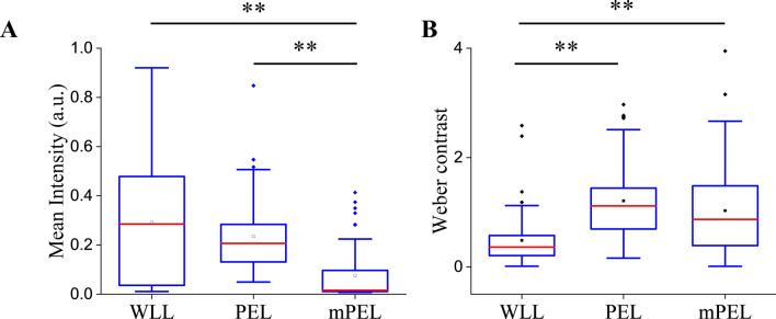
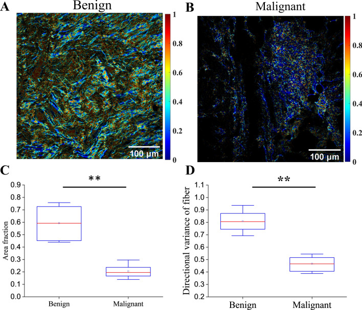
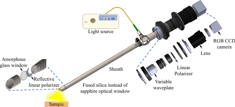
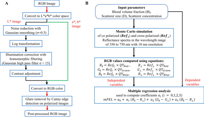
Despite careful staging, the accuracy for preoperative detection of small distant metastases remains poor, creating a clinical need for enhanced operative staging to detect occult peritoneal metastases. This study evaluates a polarization-enhanced laparoscopy (PEL) prototype and assesses its potential for label-free contrast enhancement of peritoneal metastases. This is a first-in-human feasibility study, including 10 adult patients who underwent standard staging laparoscopy (SSL) for gastrointestinal malignancy along with PEL. Image frames of all detectable peritoneal lesions underwent analysis. Using Monte Carlo simulations, contrast enhancement based on the color dependence of PEL (mPEL) was assessed. The prototype performed safely, yet with limitations in illumination, fogging of the distal window, and image co-registration. Sixty-five lesions (56 presumed benign and 9 presumed malignant) from 3 patients represented the study sample. While most lesions were visible under human examination of both SSL and PEL videos, more lesions were apparent using SSL. However, this was likely due to reduced illumination under PEL. When controlling for such effects through direct comparisons of integrated (WLL) vs differential (PEL) polarization laparoscopy images, we found that PEL imaging yielded an over twofold Weber contrast enhancement over WLL. Further, enhancements in the discrimination between malignant and benign lesions were achieved by exploiting the PEL color contrast to enhance sensitivity to tissue scattering, influenced primarily by collagen. In conclusion, PEL appears safe and easy to integrate into the operating room. When controlling for the degree of illumination, image analysis suggested a potential for mPEL to provide improved visualization of metastases.
© 2023. Springer Nature Limited.
Conflict of interest statement
The authors declare no competing interests.
Figures


References
-
- National Cancer Registration and Analysis Service. https://www.cancerdata.nhs.uk/treatments (2022).
-
- Le AT, Tzeng CW. Does finding early recurrence improve outcomes, and at what cost? J. Surg. Oncol. 2016;114:329–335. - PubMed
-
- Spagnolo E, et al. Role of fluorescence imaging for intraoperative intestinal assessment in gynecological surgery: A systematic review. Minim. Invasive Ther. Allied Technol. 2022;31:992–999. - PubMed
-
- Strigalev M, et al. Intra-operative indocyanine green fluorescence imaging in hepatobiliary surgery: A narrative review of the literature as a useful guide for the surgeon. Updates Surg. 2023;75:23–29. - PubMed
-
- Vogell A, et al. Novel imaging technologies in laparoscopic gynecologic surgery: A systematic review. J. Eng. Sci. Med. Diagn. Ther. 2018;1:010801.
Publication types
MeSH terms
Grants and funding
LinkOut - more resources
Full Text Sources
Medical
