. 2023 Nov;37(11):2197-2208.
doi: 10.1038/s41375-023-02020-w. Epub 2023 Sep 9.

FBXO21 mediated degradation of p85α regulates proliferation and survival of acute myeloid leukemia

Affiliations

FBXO21 mediated degradation of p85α regulates proliferation and survival of acute myeloid leukemia

Kasidy K Dobish et al. Leukemia. 2023 Nov.

Abstract

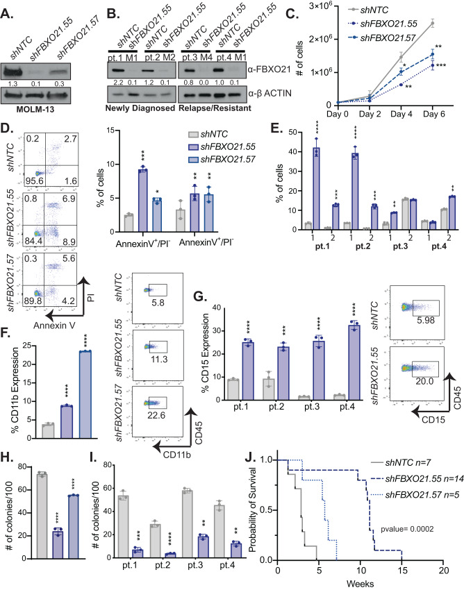
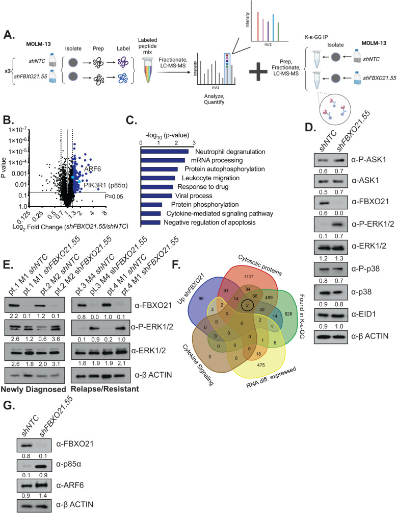
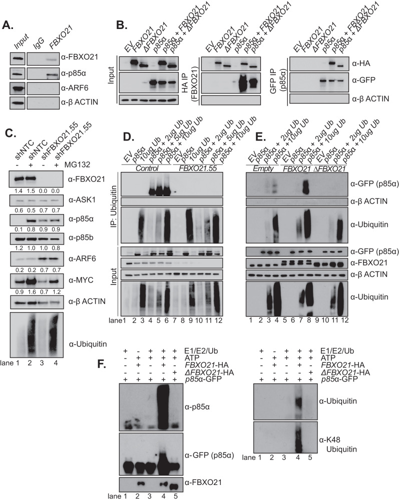
Acute myeloid leukemia (AML) is a heterogeneous disease characterized by clonal expansion of myeloid blasts in the bone marrow (BM). Despite advances in therapy, the prognosis for AML patients remains poor, and there is a need to identify novel molecular pathways regulating tumor cell survival and proliferation. F-box ubiquitin E3 ligase, FBXO21, has low expression in AML, but expression correlates with survival in AML patients and patients with higher expression have poorer outcomes. Silencing FBXO21 in human-derived AML cell lines and primary patient samples leads to differentiation, inhibition of tumor progression, and sensitization to chemotherapy agents. Additionally, knockdown of FBXO21 leads to up-regulation of cytokine signaling pathways. Through a mass spectrometry-based proteomic analysis of FBXO21 in AML, we identified that FBXO21 ubiquitylates p85α, a regulatory subunit of the phosphoinositide 3-kinase (PI3K) pathway, for degradation resulting in decreased PI3K signaling, dimerization of free p85α and ERK activation. These findings reveal the ubiquitin E3 ligase, FBXO21, plays a critical role in regulating AML pathogenesis, specifically through alterations in PI3K via regulation of p85α protein stability.


Conflict of interest statement

The authors declare no competing interests.

Figures

Fig. 1
Fig. 1. Expression of FBXO21 in AML patients.
A FBXO21 gene expression analysis of patient samples with different karyotypes from the Leukemia MILE study. B Overall patient survival from TCGA AML patient dataset. C Western blot of FBXO21 from two independent human BM (HBM) samples, two healthy CD34+ samples isolated from mobilized PB, and mononuclear cells from PB of nine AML patients (Supplementary Table 1). D Western blot probing for FBXO21 in two independent HBM samples compared to patient-derived AML (MOLM-13, THP-1, HL-60, and KASUMI-1) and ALL (MOLT-4, CCRF-CEM, and RS4(11)) cell lines. (ns non-significant, *p ≤ 0.05, ****p ≤ 0.0001).
Fig. 2
Fig. 2. Loss of FBXO21 alters cell proliferation, differentiation, and survival of AML cells.
AJ (MOLM-13: n = 3 biological replicates, AML Patients: n = 3 technical replicates) MOLM-13 cells and 4 AML primary samples (2 de novo, 2 relapse) were infected with lentiviral shRNAs against shFBXO21 and shNTC were analyzed at 72 h post puromycin selection by (A, B) western blot for knockdown in (A) AML patient derived cell line, MOLM13, and (B) 4 AML primary samples (2 de novo, 2 relapse), (C) proliferative ability of MOLM-13 cells by cell count. Cells were stained with (D, E) (left) representative flowcytometry plot and (right) bar graph of Annexin V and propidium iodide (PI) for percent of (1) AnnexinV+/PI and (2) AnnexinV+/PI+ apoptotic cells in (D) MOLM-13 and (E) AML primary cells. Cells were analyzed by flowcytometry for (F) (right) representative flowcytometry plot and (left) bar graph of CD11b (MOLM-13) and (G) (right) representative flowcytometry plot and (left) bar graph of CD15 (AML primary cells) expression. Colony forming ability by CFU assay in (H) MOLM-13 and (I) AML primary cells. J Survival of sub-lethally irradiated NSG mice transplanted with 5 × 105 MOLM-13 cells infected with shRNAs against shFBXO21 and shNTC. (*p ≤ 0.05, **p ≤ 0.01, ***p ≤ 0.001, ****p ≤ 0.0001).
Fig. 3
Fig. 3. Overexpression of FBXO21 alters cell proliferation, differentiation, and survival of AML cells.
AF (n = 6, 2 biological, 3 technical replicates) MOLM-13 cells were infected with retrovirus expressing FBXO21, ∆FBXO21, and Empty control were analyzed after sorting via FACS for (A) protein expression by western blot, (B) proliferative ability by cell count, (C) colony forming ability by CFU assay, (D) for percent of Annexin V+/PI- and Annexin V+/PI+ apoptotic cells, (E) (left) representative flowcytometry plot and (right) bar graph of CD15 expression by flow cytometry, and (F) survival of sub-lethally irradiated NSG mice transplanted with 5 × 105 cells infected with an Empty control or a plasmid overexpressing FBXO21. G MTT assay in MOLM-13 NTC and shFBXO21.55 following treatment with between 0.1-1000 nM cytarabine for 48 h. H MOLM-13, and (I) AML primary cells with FBXO21 KD and shNTC, and (J) MOLM-13 FBXO21 and Empty were treated with 50 nM cytarabine for 48 h, stained with Annexin V and PI for Annexin V+/PI and Annexin V+/PI+ apoptotic cells, and analyzed by flow cytometry. (*p ≤ 0.05, **p ≤ 0.01, ***p ≤ 0.001, ****p ≤ 0.0001).
Fig. 4
Fig. 4. RNASeq data reveals significant increase in cytokine/chemokine levels when FBXO21 is knocked down.
A Volcano plot showing fold change of expressed genes from MOLM-13 cells with silenced shFBXO21 and shNTC. B Gene ontology analysis showing pathways known to be associated with significantly upregulated (p < 0.05, ≥1-fold change) and downregulated (p < 0.05, ≤1-fold change) genes using DAVID bioinformatics database. C Localization of significantly upregulated genes (405). D Cytokine array for supernatant of MOLM-13 cells with shFBXO21 KD and shNTC, membranes showing the change in inflammatory cytokines and quantified intensities relative to internal standards. CXCL10 secretion was evaluated by enzyme-linked immunosorbent assay (ELISA) in (E) MOLM-13 cells with silenced shFBXO21 and shNTC, (F) AML primary cells with silenced shFBXO21 and shNTC, and (G) MOLM-13 cells expressing FBXO21 and Empty control.
Fig. 5
Fig. 5. Mass Spectrometry (MS) identifies potential substrate of FBXO21.
A Schematic of TMT MS and K-ε-GG IP/MS using MOLM-13 cells infected with shRNAs against shFBXO21 and shNTC. B Volcano plots showing fold change of expressed proteins from shFBXO21 compared to shNTC cells. C Gene ontology analysis showing pathways known to be associated with significantly upregulated (p < 0.05, ≥1.3-fold change) proteins using DAVID bioinformatics database. Western blot for previously known FBXO21 substrates (ASK1, EID1) and other MAPK pathway proteins (D) in MOLM-13 cells and (E) AML primary cells. F Venn Diagram of combined TMT MS, K-ε-GG IP/MS, cytosolic proteins, proteins involved in cytokine signaling pathways, and RNA-seq data comparing overlap of differentially expressed proteins and genes. G Western blot in MOLM-13 cells for validation of upregulated proteins of interest identified via the combination of proteomic and genomic analysis.
Fig. 6
Fig. 6. FBXO21 regulates p85α through ubiquitination.
A Western blot of endogenous immunoprecipitation of FBXO21 in MOLM-13 cell line. B Western blot of GFP and HA immunoprecipitation in HEK293T cells transiently transfected with plasmids expressing GFP-tagged p85α and/or HA-tagged FBXO21/∆FBXO21. C Western blot of shNTC/shFBXO21.55 MOLM-13 cells treated with 20 μM MG132 or DMSO. D Western blot of Ubiquitin immunoprecipitation in shNTC/shFBXO21.55 HEK293T cells transiently transfected with p85α and 2, 5, or 10 μg Ubiquitin. E Western blot of Ubiquitin immunoprecipitation in Empty/FBXO21/∆FBXO21 HEK293T cells transiently transfected with p85α and 5 or 10 μg Ubiquitin. F HEK293T cells were transfected with GFP-tagged p85α, HA-tagged FBXO21, HA-tagged ΔFBXO21 as indicated. After immunopurification with anti-GFP/anti-HA, in vitro ubiquitylation of p85α was performed in the presence of E1, E2, and ubiquitin (Ub). Samples were analyzed by western blot with the indicated antibodies. n = 3 for all experiments.
Fig. 7
Fig. 7. Overexpression of p85α mimics FBXO21 KD phenotype in AML cells and leads to altered PI3K pathway activation.
(n = 6, 2 biological, 3 technical replicates) MOLM-13 cells were infected with retrovirus expressing p85α and Empty control were analyzed after sorting via FACS by (A) western blot, (B) proliferative ability by cell count, (C) for percent of Annexin V+/PI- and Annexin V+/PI+ apoptotic cells, (D) colony forming ability by CFU assay, and (E) CXCL10 secretion by ELISA. Western blot in MOLM-13 cells with silenced shFBXO21 and shNTC for (F) PI3K pathway proteins and (G) native gel for PI3K complex proteins. H Schematic highlighting FBXO21 mediated alterations within PI3K signaling pathway.

References

    1. King B, Trimarchi T, Reavie L, Xu L, Mullenders J, Ntziachristos P, et al. The ubiquitin ligase FBXW7 modulates leukemia-initiating cell activity by regulating MYC stability. Cell. 2013;153:1552–66. doi: 10.1016/j.cell.2013.05.041. - DOI - PMC - PubMed
    1. Reavie L, Buckley SM, Loizou E, Takeishi S, Aranda-Orgilles B, Ndiaye-Lobry D, et al. Regulation of c-Myc ubiquitination controls chronic myelogenous leukemia initiation and progression. Cancer Cell. 2013;23:362–75. doi: 10.1016/j.ccr.2013.01.025. - DOI - PMC - PubMed
    1. Cardozo T, Pagano M. The SCF ubiquitin ligase: insights into a molecular machine. Nat Rev Mol Cell Biol. 2004;5:739–51. doi: 10.1038/nrm1471. - DOI - PubMed
    1. Kipreos ET, Pagano M. The F-box protein family. Genome Biol. 2000;1:REVIEWS3002. doi: 10.1186/gb-2000-1-5-reviews3002. - DOI - PMC - PubMed
    1. Hynes-Smith RW, Wittorf KJ, Buckley SM. Regulation of normal and malignant hematopoiesis by FBOX ubiquitin E3 ligases. Trends Immunol. 2020;41:1128–40. doi: 10.1016/j.it.2020.10.003. - DOI - PMC - PubMed

Publication types

MeSH terms