

Enhanced Detection of Suspicious Breast Lesions: A Comparative Study of Full-Field Digital Mammography and Automated Breast Ultrasound in 117 Patients with Core Needle Biopsy
- PMID: 37689969
- PMCID: PMC10501319
- DOI: 10.12659/MSM.941072
Enhanced Detection of Suspicious Breast Lesions: A Comparative Study of Full-Field Digital Mammography and Automated Breast Ultrasound in 117 Patients with Core Needle Biopsy
Abstract
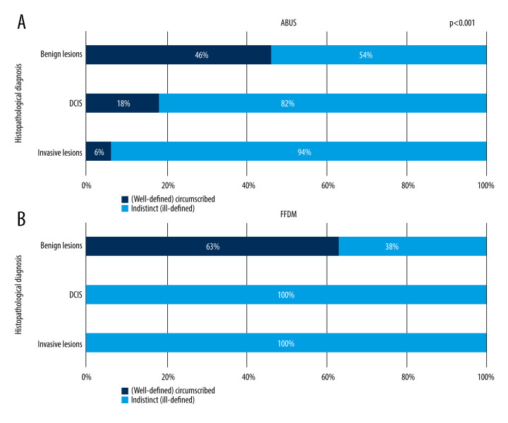
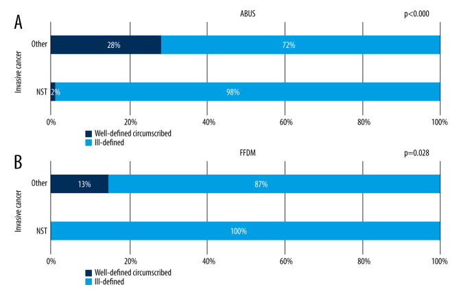
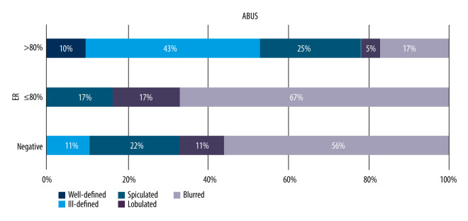
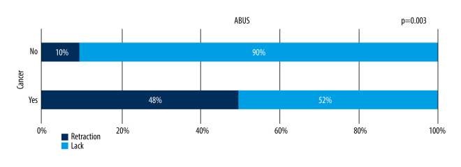
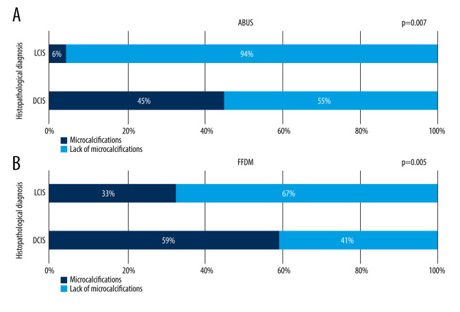
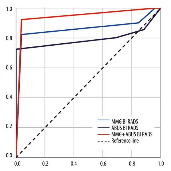
BACKGROUND This retrospective study from a single center aimed to compare the performance of full-field digital mammography (FFDM) vs automated breast ultrasound (ABUS) in the identification and characterization of suspicious breast lesions in 117 patients who underwent core-needle biopsy (CNB) of the breast. MATERIAL AND METHODS The study involved a group of 301 women. Every patient underwent FFDM followed by ABUS, which were assessed in concordance with BI-RADS (Breast Imaging Reporting and Data System) classification. RESULTS No focal lesions were found in 168 patients. In 133 patients, 117 histopathologically verified focal lesions were found. Among them, 78% appeared to be malignant and 22% benign. ABUS detected 246 focal lesions, including 115 classified as BI-RADS 4 or 5 and submitted to verification, while FFDM revealed 122 lesions, including 75 submitted to verification. The analysis revealed that combined application of both methods caused sensitivity to increase to 100, and improved accuracy improvement. Margin assessments in these examinations are consistent (P<0.00), the lesion's margin type with both methods depends on its malignant or benign character (P<0.03), lesion margins distribution on ABUS depends on estrogen receptor presence (P=0.033), and there was significant correlation between malignant character of the lesion and retraction phenomenon sign (P=0.033). ABUS obtained higher compliance between the size of the lesion in histopathology compared to FFDM (P>0.05). CONCLUSIONS The results shows that ABUS is comparable to FFDM, and even outperforms it in a few of the analyzed categories, suggesting that the combination of these 2 methods may have an important role in breast cancer detection.
Conflict of interest statement
Figures
References
-
- Kocarnik JM, Compton K, Dean FE, et al. Cancer incidence, mortality, years of life lost, years lived with disability, and disability-adjusted life years for 29 cancer groups from 2010 to 2019: A systematic analysis for the global burden of disease study 2019. JAMA Oncol. 2022;8(3):420–44. - PMC - PubMed
-
- Strategia walki z rakiem w Polsce 2015–2025 [Internet] czerwca 14, 2014. [cited 2015, November 16]. Available from: http://walkazrakiem.pl/sites/default/files/library/files/strategia_walki... [on Polish]
-
- Kolak A, Kamińska M, Sygit K, et al. Primary and secondary prevention of breast cancer. Ann Agric Environ Med. 2017;24(4):549–53. - PubMed
-
- Niell BL, Freer PE, Weinfurtner RJ, et al. Screening for breast cancer. Radiol Clin North Am. 2017;55(6):1145–62. - PubMed
MeSH terms
LinkOut - more resources
Full Text Sources
Medical
