. 2023 Nov;299(11):105239.
doi: 10.1016/j.jbc.2023.105239. Epub 2023 Sep 9.

Insulin-like growth factor binding protein-3 mediates hyperosmolar stress-induced mitophagy through the mechanistic target of rapamycin

Affiliations

Insulin-like growth factor binding protein-3 mediates hyperosmolar stress-induced mitophagy through the mechanistic target of rapamycin

Whitney Stuard Sambhariya et al. J Biol Chem. 2023 Nov.

Abstract

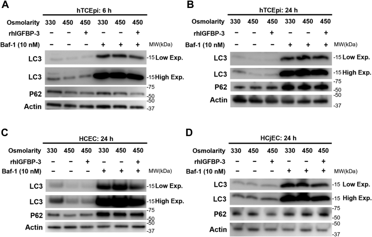
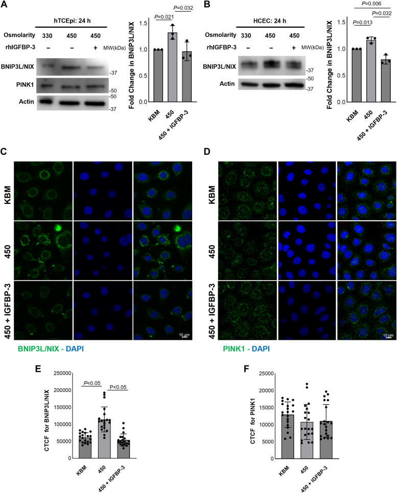
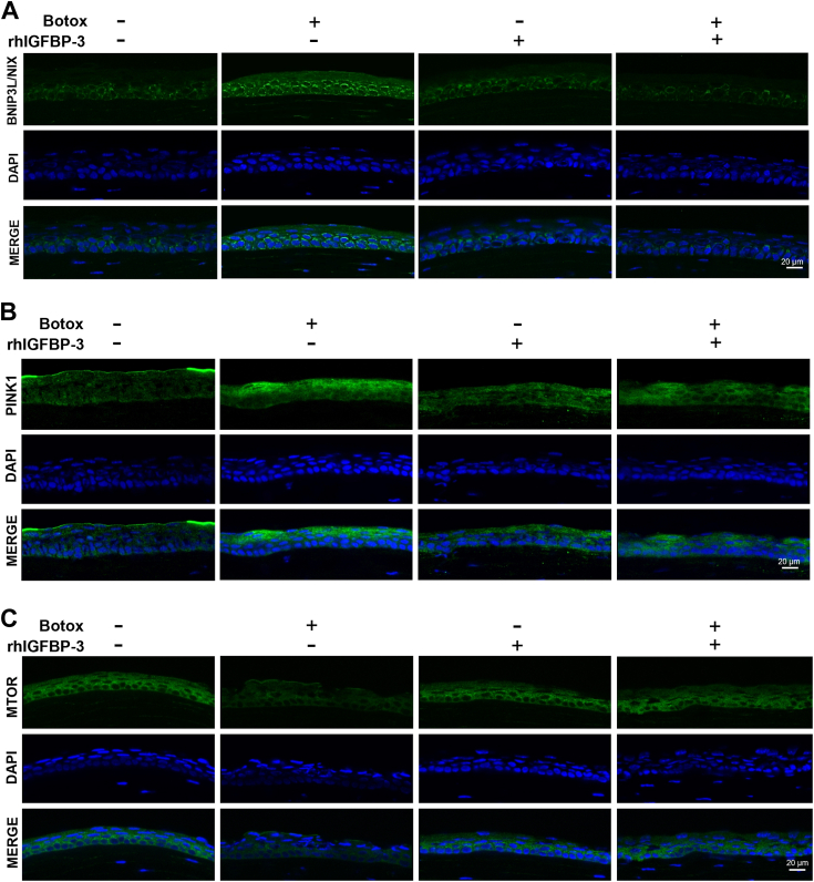
Hyperosmolarity of the ocular surface triggers inflammation and pathological damage in dry eye disease (DED). In addition to a reduction in quality of life, DED causes vision loss and when severe, blindness. Mitochondrial dysfunction occurs as a consequence of hyperosmolar stress. We have previously reported on a role for the insulin-like growth factor binding protein-3 (IGFBP-3) in the regulation of mitochondrial ultrastructure and metabolism in mucosal surface epithelial cells; however, this appears to be context-specific. Due to the finding that IGFBP-3 expression is decreased in response to hyperosmolar stress in vitro and in an animal model of DED, we next sought to determine whether the hyperosmolar stress-mediated decrease in IGFBP-3 alters mitophagy, a key mitochondrial quality control mechanism. Here we show that hyperosmolar stress induces mitophagy through differential regulation of BNIP3L/NIX and PINK1-mediated pathways. In corneal epithelial cells, this was independent of p62. The addition of exogenous IGFBP-3 abrogated the increase in mitophagy. This occurred through regulation of mTOR, highlighting the existence of a new IGFBP-3-mTOR signaling pathway. Together, these findings support a novel role for IGFBP-3 in mediating mitochondrial quality control in DED and have broad implications for epithelial tissues subject to hyperosmolar stress and other mitochondrial diseases.

Keywords: cornea; hyperosmolar stress; mTOR; mitochondria; mitophagy.


Conflict of interest statement

Conflict of interest The authors declare that they have no conflicts of interest with the contents of this article.

Figures

Figure 1
Figure 1
Hyperosmolarity promotes autophagy in corneal and conjunctival epithelial cells.AL, cells were treated with isotonic KBM or KBM with an osmolarity of 450 mOsm or 500 mOsm for 24 h. Ten nanomolars of bafilomycin (Baf-1) was used to block lysosomal fusion in each condition. A, hTCEpi cell lysates were immunoblotted for P62 and LC3-II. Membranes were imaged using low (low exp) and high (high exp) exposure times to visualize the accumulation of LC3-II in cells treated with and without Baf-1, respectively. In the absence of Baf-1, no differences were observed in LC3-II. Hyperosmolar stress increased expression of LC3-II in Baf-1–treated cells at both 450 and 500 mOsm (p < 0.05). BD, quantification of immunoblots in (A). E, in HCECs, there was a similar increase in LC3-II in cells exposed to hyperosmolar stress in the presence of Baf-1 (p = 0.008 and p = 0.012 for 500 mOsm and 450 mOsm compared to isotonic KBM, respectively). FH, quantification of immunoblots in (E). I, in HCjECs, LC3-II levels were increased in cells exposed to 500 mOsm when treated with Baf-1 (p = 0.041). JL, quantification of immunoblots shown in (I). β-actin was used as a loading control. Data expressed as mean ± SD from three independent experiments. One-way ANOVA with Student-Newman-Keuls post hoc multiple comparison test. HCECs, human corneal epithelial cells; KBM, keratinocyte basal media.
Figure 2
Figure 2
IGFBP-3 inhibits autophagy in ocular surface epithelial cells exposed to hyperosmolar stress.AD, cells were treated with isotonic KBM or 450 mOsm KBM with or without 500 ng/ml rhIGFBP-3 for 6 or 24 h. Ten nanomolars of bafilomycin (Baf-1) was used to block lysosomal fusion. Cell lysates were blotted for P62 and LC3-II. A, after 6 h, cotreatment with rhIGFBP-3 decreased LC3-II levels in the absence of Baf-1. In the presence of Baf-1, hyperosmolar stress increased LC3-II levels; cotreatment with rhIGFBP-3 decreased LC3-II compared to 450 mOsm without rhIGFBP-3. B, after 24 h of culture, cells exposed to 450 mOsm showed an increase in LC3-II levels in Baf-1–treated cells; cotreatment with rhIGFBP-3 decreased LC3-II compared to 450 mOsm without rhIGFBP-3. C, in the presence of Baf-1, HCECs cotreated with rhIGFBP-3 had less LC3-II. D, HCjECs similarly showed no changes in LC3-II in the absence of Baf-1. Cells treated with Baf-1 had higher LC3-II expression in 450 mOsm KBM than isotonic KBM. Expression of LC3-II in HCjECs cotreated with rhIGFBP-3 was lower than KBM and 450 mOsm. β-actin was used as a loading control. low exp (low exposure); high exp (high exposure). Blots are representative of three repeated experiments. HCECs, human corneal epithelial cells; IGFBP-3, insulin-like growth factor binding protein-3; KBM, keratinocyte basal media; rh, recombinant human.
Figure 3
Figure 3
IGFBP-3 inhibits autophagic flux in corneal epithelial cells exposed to hyperosmolar stress. hTCEpi cells were transfected with a GFP-mCherry-LC3 expression plasmid. Cells were then cultured in either isotonic KBM or 450 mOsm KBM with or without 500 ng/ml rhIGFBP-3 for 6 or 24 h. A, at 6 h, there was an increase in the accumulation of autophagosomes (yellow puncta) and autophagolysosomes (red puncta) in KBM compared to the KGM control. Culture in 450 mOsm KBM increased the number of autophagolysosomes. Cells in 450 mOsm cotreated with rhIGFBP-3 had fewer autophagolysosomes than 450 mOsm without rhIGFBP-3. B, in 450 mOsm KBM, there was an increase in the ratio of red to green puncta compared to KBM (p < 0.05). In contrast, treatment with rhIGFBP-3 decreased the ratio of red to green puncta (p < 0.05). C, after 24 h in 450 mOsm KBM, there was a similar increase in autophagolysosomes. Again, cotreatment with rhIGFBP-3 led to an increase in autophagosomes, further suggesting a block in autophagic flux. D, similar to the 6 h time point, the ratio of red to green puncta was increased in cells cultured in 450 mOsm KBM at 24 h (p < 0.05). This was again decreased by treatment with rhIGFBP-3 (p < 0.05). Scale bar represents 10 μm. Images are representative of three repeated experiments. IGFBP-3, insulin-like growth factor binding protein-3; KBM, keratinocyte basal media; KGM, keratinocyte growth media; rh, recombinant human.
Figure 4
Figure 4
IGFBP-3 mediates mitophagy induced during acute hyperosmolar stress through BNIP3L/NIX and PINK1 pathways. Cells were cultured in isotonic KBM or 450 mOsm KBM with or without 500 ng/ml rhIGFBP-3 for 6 h. A, immunoblotting for mitochondrial proteins involved in mitophagy, BNIP3L/NIX and PINK1, in hTCEpi cells. ß-actin was used as a loading control. B, there was an increase in BNIP3L/NIX in 450 mOsm (p = 0.04), whereas cotreatment with rhIGFBP-3 decreased BNIP3L/NIX compared to 450 mOsm alone (p = 0.008). C, PINK1 was also decreased in rhIGFBP-3 cotreated cells (p = 0.03). D and E, immunofluorescent labeling of hTCEpi cells for BNIP3L/NIX (D) and PINK1 (E). Nuclei were labeled with DAPI. Scale bar represents 10 μm. F, live-cell imaging using the mitochondrial polarization probe JC-1. JC-1 monomers (green) show mitochondria. Aggregates of JC-1 (red) represent areas of mitochondrial polarization. Scale bar represents 10 μm. G, live-cell imaging using the mitochondrial polarization probe TMRE and the mitochondrial probe MitoTracker Green. Scale bar represents 10 μm. Data presented as mean ± SD from three repeated experiments. One-way ANOVA with Student-Newman-Keuls post hoc multiple comparison test. IGFBP-3, insulin-like growth factor binding protein-3; KBM, keratinocyte basal media; rh, recombinant human; TMRE, tetramethylrhodamine ethyl ester.
Figure 5
Figure 5
IGFBP-3 blocks mitophagy through BNIP3L/NIX at 24 h. Cells were cultured in isotonic KBM or 450 mOsm KBM with or without 500 ng/ml rhIGFBP-3 for 24 h. A, immunoblotting for BNIP3L/NIX and PINK1 in hTCEpi cells. There was an increase in BNIP3L/NIX in 450 mOsm (p = 0.021), whereas cotreatment with rhIGFBP-3 decreased BNIP3L/NIX compared to 450 mOsm (p = 0.032). PINK1 was unchanged. ß-actin was used as a loading control. B, immunoblotting of HCECs showed a similar pattern of expression for BNIP3L/NIX, with increased expression at 450 mOsm compared to the isotonic control (p = 0.013), which was then decreased by cotreatment with rhIGFBP-3 (p = 0.006 and p = 0.032; KBM and 450 mOsm compared to 450 with IGFBP-3, respectively). C and D, immunofluorescent labeling of hTCEpi cells for (C) BNIP3L/NIX (green) was increased in cells treated with 450 mOsm compared to KBM (p < 0.05). This effect was blocked by cotreatment with rhIGFBP-3 (p < 0.05). D, there was no significant difference in PINK1 (green) expression at 24 h E and F, quantification of immunofluorescence in (C) and (D), respectively. Nuclei were labeled with DAPI. Scale bar represents 10 μm. Data presented as mean ± SD from three independent experiments. Images are representative of three repeated experiments. One-way ANOVA with Student-Newman-Keuls post hoc multiple comparison test. HCECs, human corneal epithelial cells; IGFBP-3, insulin-like growth factor binding protein-3; KBM, keratinocyte basal media; rh, recombinant human.
Figure 6
Figure 6
IGFBP-3 mediates the mTOR pathway to affect mitochondrial homeostasis.A, hTCEpi cells were cultured in KBM with increasing osmolarity (450 mOsm or 500 mOsm) for 24 h. Whole cell lysates were immunoblotted for mTOR and Ser(2448)-p-mTOR. As osmolarity increased, both mTOR (p < 0.001) and p-mTOR (p = 0.011 and p = 0.022) were sequentially decreased. B and C, quantification of immunoblots in (A). D, cells were cultured in isotonic KBM or 450 mOsm KBM with or without 500 ng/ml rhIGFBP-3 for 24 h. Cell lysates were again immunoblotted for mTOR and Ser(2448)-p-mTOR. In 450 mOsm KBM, mTOR was decreased (p = 0.014). This decrease was blocked by cotreatment with rhIGFBP-3 (p = 0.049). Despite a similar trend, changes in p-mTOR were not significant. Total mTOR was used as a loading control for Ser(2448)-p-mTOR. ß-actin was used as a loading control for mTOR. E and F, quantification of immunoblots in (B). G, immunofluorescent labeling of hTCEpi cells for mTOR (green) at 24 h. Nuclei were labeled with DAPI. Scale bar represents 10 μm. H, quantification of immunofluorescence in (G). I and J, hTCEpi cells were transfected with siRNA oligonucleotides targeting TSC1 or a nontargeting control. Cells were treated with isotonic KBM, 450 mOsm KBM, or 450 mOsm KBM with 500 ng/ml rhIGFBP-3. I, knockdown efficiency was confirmed using immunoblotting for TSC1. ß-actin was used as a loading control. J, mitochondrial oxygen consumption was significantly decreased in cells treated with 450 mOsm (p < 0.05) and restored to basal levels upon cotreatment with rhIGFBP-3 (p < 0.05). TSC1 knockdown cells showed no differences in OCR. K, cells were cultured in 450 mOsm KBM with or without rhIGFBP-3. Cells treated with rapamycin, an mTOR inhibitor, were cultured in parallel. Treatment with rapamycin showed significant decreases in OCR compared to nonrapamycin-treated cells (p < 0.05). Data expressed as mean ± SD from one representative experiment, n = 3. One-way ANOVA with Student-Newman-Keuls post hoc multiple comparison test. IGFBP-3, insulin-like growth factor binding protein-3; KBM, keratinocyte basal media; mTOR, mechanistic target of rapamycin; OCR, oxygen consumption rate; rh, recombinant human; TSC1, tuberous sclerosis complex 1.
Figure 7
Figure 7
Inhibition of mTOR blocks IGFBP-3–mediated mitochondrial hyperfusion. hTCEpi cells were cultured in isotonic KBM or 450 mOsm KBM with or without 500 ng/ml rhIGFBP-3 for 24 h. TEM shows distinct changes in mitochondrial morphology and lamellar structure across treatment groups. A, treatment with rapamycin blocked IGFBP-3–mediated hyperfusion. B, TEM demonstrating mitophagy in hTCEpi cells subject to hyperosmolar stress. C, similar to rapamycin, treatment with KU-0063794 blocked IGFBP-3–mediated hyperfusion. Scale bar represents 1 μm. IGFBP-3, insulin-like growth factor binding protein-3; KBM, keratinocyte basal media; mTOR, mechanistic target of rapamycin; rh, recombinant human.
Figure 8
Figure 8
IGFBP-3 regulates mitophagy and mTOR in the corneal epithelium in dry eye disease. The mouse lacrimal gland was injected with botulinum toxin or a vehicle control. Twenty-one days postinjection, mice were treated topically twice a day every other day for 7 days with 10 μl of sterile saline with or without 500 ng/ml rhIGFBP-3. Immunostaining of cryo-sectioned corneas for (A) BNIP3L/NIX (green), (B) PINK1 (green), and (C) mTOR (green). Nuclei were counterstained with DAPI (blue). Scale bar represents 20 μm. Images are representative of three or more mice per group. IGFBP-3, insulin-like growth factor binding protein-3; mTOR, mechanistic target of rapamycin; rh, recombinant human.
Supporting Figure S1
Supporting Figure S2
Supporting Figure S3
Supporting Figure S4

References

    1. Craig J.P., Nichols K.K., Akpek E.K., Caffery B., Dua H.S., Joo C.K., et al. TFOS DEWS II definition and classification report. Ocul. Surf. 2017;15:276–283. - PubMed
    1. Research in dry eye: report of the research subcommittee of the International dry eye WorkShop (2007) Ocul. Surf. 2007;5:179–193. - PubMed
    1. Png E., Samivelu G.K., Yeo S.H., Chew J., Chaurasia S.S., Tong L. Hyperosmolarity-mediated mitochondrial dysfunction requires transglutaminase-2 in human corneal epithelial cells. J. Cell. Physiol. 2011;226:693–699. - PubMed
    1. Stookey J.D., Barclay D., Arieff A., Popkin B.M. The altered fluid distribution in obesity may reflect plasma hypertonicity. Eur. J. Clin. Nutr. 2007;61:190–199. - PubMed
    1. Vernia P., Gnaedinger A., Hauck W., Breuer R.I. Organic anions and the diarrhea of inflammatory bowel disease. Dig. Dis. Sci. 1988;33:1353–1358. - PubMed

Publication types

LinkOut - more resources