. 2023 Sep 11;30(1):77.
doi: 10.1186/s12929-023-00962-y.

Platelet-derived biomaterial with hyaluronic acid alleviates temporal-mandibular joint osteoarthritis: clinical trial from dish to human

Affiliations

Platelet-derived biomaterial with hyaluronic acid alleviates temporal-mandibular joint osteoarthritis: clinical trial from dish to human

Bou-Yue Peng et al. J Biomed Sci. .

Abstract

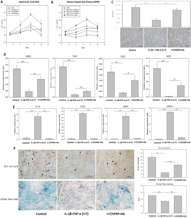
Background: Bioactive materials have now raised considerable attention for the treatment of osteoarthritis (OA), such as knee OA, rheumatoid OA, and temporomandibular joint (TMJ) OA. TMJ-OA is a common disease associated with an imbalance of cartilage regeneration, tissue inflammation, and disability in mouth movement. Recently, biological materials or molecules have been developed for TMJ-OA therapy; however, ideal treatment is still lacking. In this study, we used the combination of a human platelet rich plasma with hyaluronic acid (hPRP/HA) for TMJ-OA therapy to perform a clinical trial in dish to humans.

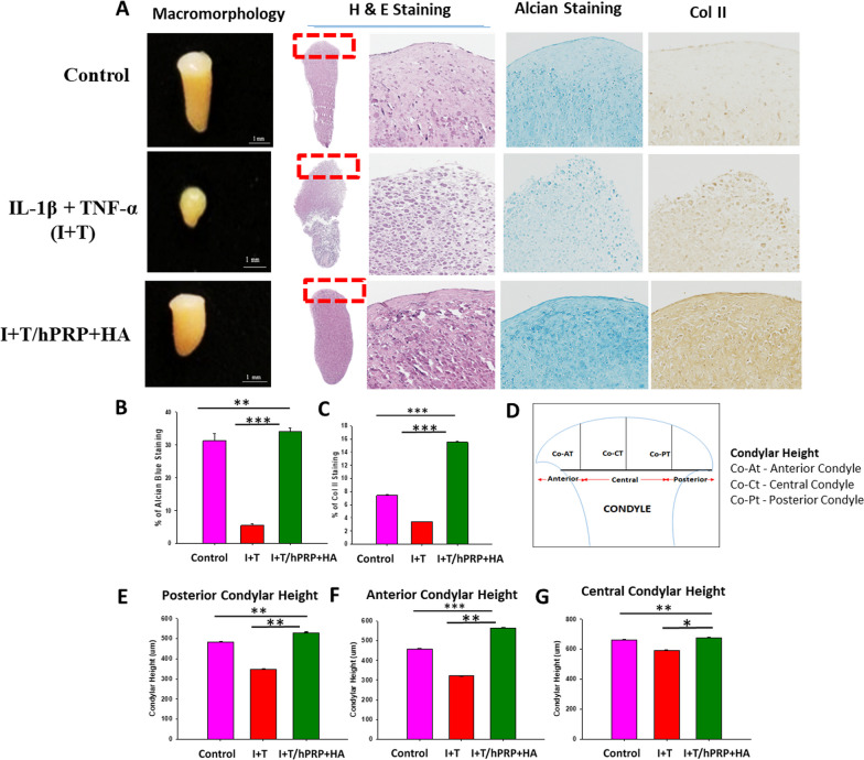
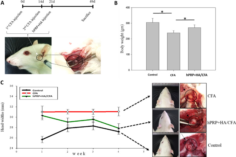
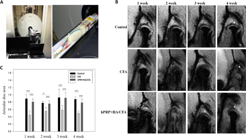
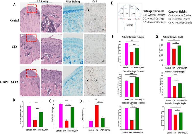
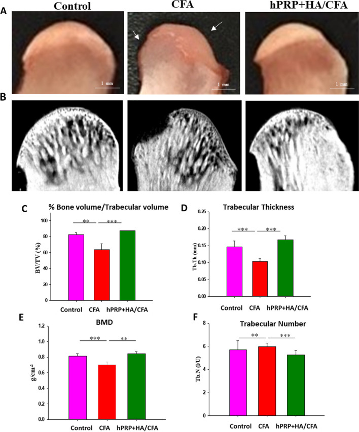
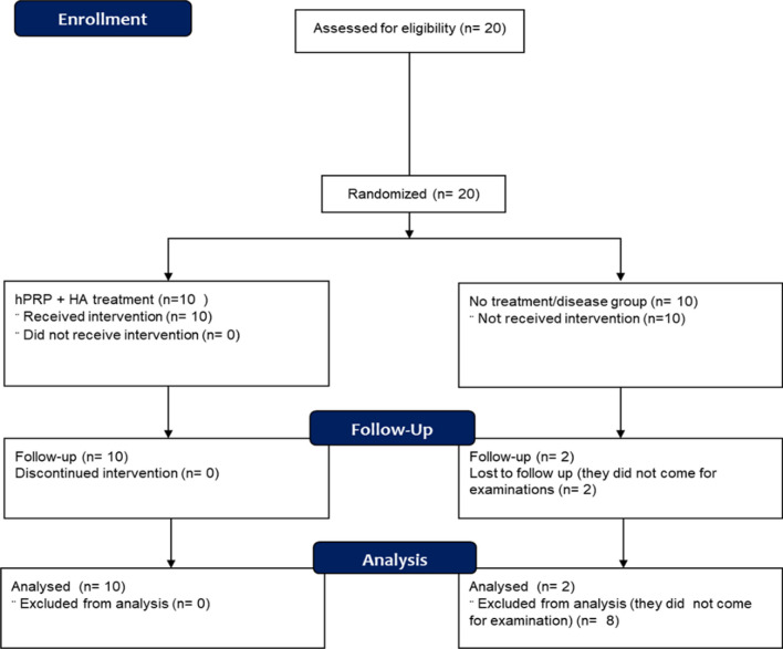
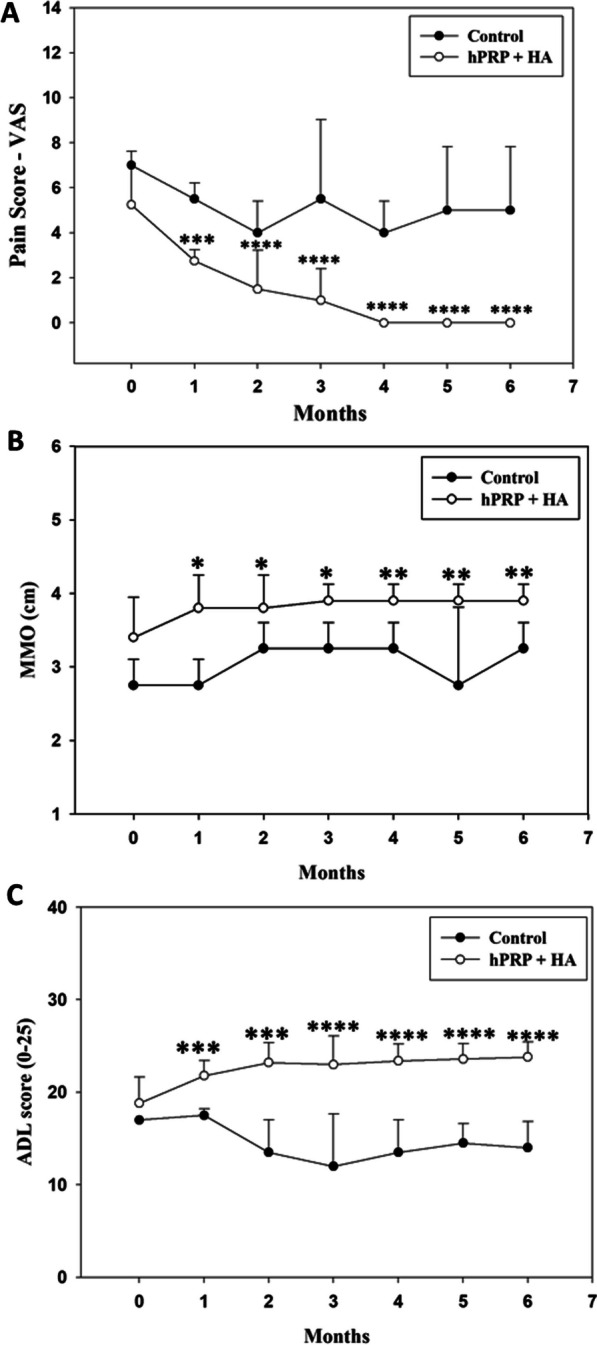
Method: Herein, hPRP was prepared, and the hPRP/HA combined concentration was optimized by MTT assay. For the clinical trial in dish, pro-inflammatory-induced in-vitro and in-vivo mimic 3D TMJ-OA models were created, and proliferation, gene expression, alcian blue staining, and IHC were used to evaluate chondrocyte regeneration. For the animal studies, complete Freund's adjuvant (CFA) was used to induce the TMJ-OA rat model, and condyle and disc regeneration were investigated through MRI. For the clinical trial in humans, 12 patients with TMJ-OA who had disc displacement and pain were enrolled. The disc displacement and pain at baseline and six months were measured by MRI, and clinical assessment, respectively.

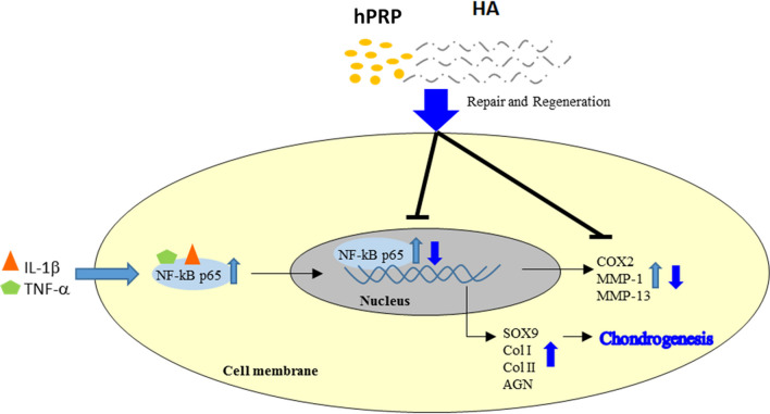
Results: Combined hPRP/HA treatment ameliorated the proinflammatory-induced TMJ-OA model and promoted chondrocyte proliferation by activating SOX9, collagen type I/II, and aggrecan. TMJ-OA pathology-related inflammatory factors were efficiently downregulated with hPRP/HA treatment. Moreover, condylar cartilage was regenerated by hPRP/HA treatment in a proinflammatory-induced 3D neocartilage TMJ-OA-like model. During the animal studies, hPRP/HA treatment strongly repaired the condyle and disc in a CFA-induced TMJ-OA rat model. Furthermore, we performed a clinical trial in humans, and the MRI data demonstrated that after 6 months of treatment, hPRP/HA regenerated the condylar cartilage, reduced disc displacement, alleviated pain, and increased the maximum mouth opening (MMO). Overall, clinical trials in dish to human results revealed that hPRP/HA promoted cartilage regeneration, inhibited inflammation, reduced pain, and increased joint function in TMJ-OA.

Conclusion: Conclusively, this study highlighted the therapeutic potential of the hPRP and HA combination for TMJ-OA therapy, with detailed evidence from bench to bedside. Trial registration Taipei Medical University Hospital (TMU-JIRB No. N201711041). Registered 24 November 2017. https://tmujcrc.tmu.edu.tw/inquiry_general.php .

Keywords: Clinical trial; Hyaluronic acid; Platelet-derived biomaterial; TMJ-OA.


Conflict of interest statement

The authors declare no conflict of interest.

Figures

Fig. 1
Fig. 1
Schematic of hPRP/HA treatment in TMJ-OA disease from bench to beside
Fig. 2
Fig. 2
In-vitro 2D hyaluronic acid (HA) and human platelet-rich plasma (hPRP) promote cell activity and anti-inflammatory effects of rat TMJ chondrocyte. Optimal concentration of (A) HA and (B) hPRP dosage in rat TMJ chondrocytes that were analyzed using the MTT assay. C The effect of HA and PRP on the cell numbers and morphological changes (200 ×) of rat TMJ chondrocytes after 2-day treatment with IL1β + TNF-α (I + T)-conditioned medium. I (10 ng/mL) + T (20 ng/mL) were added to the medium to create an in-vitro proinflammatory cytokine-induced arthritic cell model. Real-time PCR analyses of monolayer cultures of rat TMJ chondrocytes after 2-day treatment with I + T-conditioned medium. The results revealed that the combination of HA and hPRP (D) significantly increased cartilage-specific gene expression, including SOX9, collagen type I (Col-I), collagen type II (Col-II), and aggrecan, and E significantly inhibited I + T stimulated inflammatory genes including IL-1β, COX2, MMP-3, and MMP-13. F Immunocytochemistry of Col-II (black arrow indicates presence of Col-II) and its quantification (upper panel) and Alcian blue stain and its quantification (lower panel). *p < 0.05, **p < 0.01, and ***p < 0.001 compared with I + T and control group using paired t test. The results are shown as mean ± SD for three replicate
Fig. 3
Fig. 3
In-vitro 3D hyaluronic acid (HA) and platelet-rich plasma (hPRP) promote cell activity of rat TMJ chondrocytes. For the TMJ-OA model, neocartilage of TMJ chondrocytes/collagen constructs were cultured for 4 weeks in basal control, I + T, and I + T/hPRP/HA -conditioned medium. Constructs were then histologically examined using (A) macromorphology, hematoxylin and eosin (H&E) staining, IHC staining for Col-II, and Alcian blue staining. (B) Percentage areal deposition of s-GAG by quantification of TB-stained area, and (C) percentages of Col-II deposition in TMJ condylar cartilage. (D) Schematic of the anterior, central, and posterior regions of the rat TMJ condylar head. Results (E) posterior, (F) anterior, and (G) central condylar height. *p < 0.05, **p < 0.01, and ***p < 0.001 compared with I + T and control group using paired t test. The results are shown as mean ± SD for three replicate
Fig. 4
Fig. 4
Experimental design and behavioral evaluation of combined hPRP/HA treatment in CFA-induced TMJ osteoarthritis. A Time course and experimental design. TMJ-OA was induced by two injections of CFA at 0 and 14th day, followed by combined hPRP/HA treatment in the 21st day. CFA and hPRP/HA were administered through the lateral puncture technique. B Body weight changes after treatment with hPRP/HA. C Representative photographs demonstrate that the head width of rats is increased after receiving CFA induction. Both the synovium and disc became thickened, opaque, and head width increased in the CFA-induced TMJ-OA group (marked in black circle) that recovered after treatment in hPRP/HA group. Each group n = 3 rats. *p < 0.05, **p < 0.01, and ***p < 0.001, the results are shown as mean ± SD for three replicates
Fig. 5
Fig. 5
High magnetic field magnetic resonance imaging (MRI) applied to the TMJ in the rat. A Overview of the 7 T micro-imaging system. B MRI scan of control, CFA, and hPRP/HA-treated groups at 1–4 weeks posttreatment. C Measurement of the articular spaces in the coronal view. *p < 0.05, **p < 0.01, and ***p < 0.001 and the results are shown as mean ± SD for three replicates
Fig. 6
Fig. 6
hPRP/HA promote TMJ repair and regeneration in OA. Histopathological evaluation indicated that hPRP/HA restored TMJ matrix synthesis in OA. A Hematoxylin and eosin (H&E), Alcian blue staining, and immunohistochemical staining for Col-II at 4 weeks (black arrow indicate the presence of Col-II). Representative images (n = 3). Scale bars: 500 or 100 μm. B Percentage areal deposition of proteoglycan by quantification of Alcian-stained area, and C percentages of Col-II cells in TMJ condylar cartilage. D Mankin scores of samples at 4 weeks. E Schematic of the anterior, central, and posterior regions of the rat TMJ condylar head. The condylar height was measured at each region, including co-At: anterior height; co-Ct: central height; and co–Pt: posterior height of condyle. The cartilage thickness was also measured at each region, including C-At: anterior thickness; C-Ct: central thickness; and C-Pt: posterior thickness of cartilage. Measurement of F cartilage thickness and G condylar height at the anterior, posterior, and central regions. *p < 0.05, **p < 0.01,, and ***p < 0.001 and the results are shown as mean ± SD for three replicates
Fig. 7
Fig. 7
hPRP/HA treatment restored TMJ subchondral bone volume and architecture in OA. Rat TMJs were harvested for micro-CT analysis at 4 weeks posttreatment of hPRP/HA. A Macromorphological view (white arrow indicates damage area of subchondral bone) and B sagittal view. C BV/TV, D trabecular thickness, E BMD, and E trabecular number. Data represent mean ± SD. *p < 0.05, **p < 0.01, and ***p < 0.001 compared with control and CFA group and the results are shown as mean ± SD for three replicates. White arrow indicate damage area of TMJ
Fig. 8
Fig. 8
Consort flowchart for clinical trial of hPRP + HA in TMJ-OA disease
Fig. 9
Fig. 9
Therapeutic potential of the combine hPRP/HA treatment in the clinical study. A Schematic of HA and PRP administration into the superior space of TMJ-OA patients. B Disease group without treatment at 0 and 6 months of oblique sagittal MRI images represent disk displacement and osteoarthritis become worse. Quantification of the disc displacement distance from the condyle (C) and condylar thickness were measured at 0 and 6 months (cases 1 and 2, n = 1) (D). In the treatment group (E), patients of disc displacement with reduction (DDR), disc displacement without reduction (DDWR) are designated as case 1, and case 2, respectively, that were associated with disk degeneration, and osteoarthritis of the condyle at 6 months post-treatment. Oblique sagittal MRI images indicated disc recapture and condylar remodeling after using the hPRP/HA treatment compared with the disease or untreated group. In the treatment group disc displacement distance from the condyle (F) and condylar thickness (G) was measured at 0 and 6 months. *p < 0.05, **p < 0.01, and ***p < 0.001 compared with I + T and control group using paired t test
Fig. 10
Fig. 10
Clinical findings of hPRP/HA treatment. A Comparison of pain (VAS score) during the follow-up period with the preoperative level of pain. A greater decrease in pain was observed in the hPRP/HA-treated group. B Comparison of MMO during the follow-up period with preoperative MMO. A progressive increase in MMO was observed in the hPRP/HA-treated group. C Comparison of ADL during the follow-up period with the preoperative level of ADL was also increased in hPRP/HA-treated group. Clinically, the MMO and ADL scores were significantly increased in the hPRP/HA-treated group compared to the control (non-treated/disease) group, and the pain (VAS) score was significantly decreased. *p < 0.05, **p < 0.01, ***p < 0.001, and ****p < 0.0001 compared within hPRP + HA group using paired t test
Fig. 11
Fig. 11
Schematic of therapeutic mechanism action of hPRP/HA therapy in TMJ-OA disease. hPRP (human platelet rich plasma) and HA (hyaluronic acid) ameliorates the bone degradation by upregulation chondrogenic genes in inflammatory (IL-1β and TNF-α) induced TMJ-OA model

References

    1. Zhang S, Teo KYW, Chuah SJ, Lai RC, Lim SK, Toh WS. MSC exosomes alleviate temporomandibular joint osteoarthritis by attenuating inflammation and restoring matrix homeostasis. Biomaterials. 2019;200:35–47. - PubMed
    1. Li B, Guan G, Mei L, Jiao K, Li H. Pathological mechanism of chondrocytes and the surrounding environment during osteoarthritis of temporomandibular joint. J Cell Mol Med. 2021;25(11):4902–4911. - PMC - PubMed
    1. Lu K, Ma F, Yi D, Yu H, Tong L, Chen D. Molecular signaling in temporomandibular joint osteoarthritis. J Orthop Transl. 2022;32:21–27. - PMC - PubMed
    1. Wang XD, Zhang JN, Gan YH, Zhou YH. Current understanding of pathogenesis and treatment of TMJ osteoarthritis. J Dent Res. 2015;94(5):666–673. - PubMed
    1. Le ADK, Enweze L, DeBaun MR, Dragoo JL. Platelet-rich plasma. Clin Sports Med. 2019;38(1):17–44. - PubMed