

K. pneumoniae and M. smegmatis infect epithelial cells via different strategies
- PMID: 37691650
- PMCID: PMC10482649
- DOI: 10.21037/jtd-23-493
K. pneumoniae and M. smegmatis infect epithelial cells via different strategies
Abstract
Background: As the first line of defense, epithelial cells play a vital role in the initiation and control of both innate and adaptive immunity, which participate in the development of disease. Despite its therapeutic significance, little is understood about the specific interaction between pathogenic microorganisms and lung epithelial cells.
Methods: In this study, we performed a head-to-head comparison of the virulence and infection mechanisms of Klebsiella pneumoniae (K. pneumoniae) and Mycobacterium smegmatis (M. smegmatis), which represent Gram-negative/positive respiratory pathogens, respectively, in lung epithelial cell models for the first time.
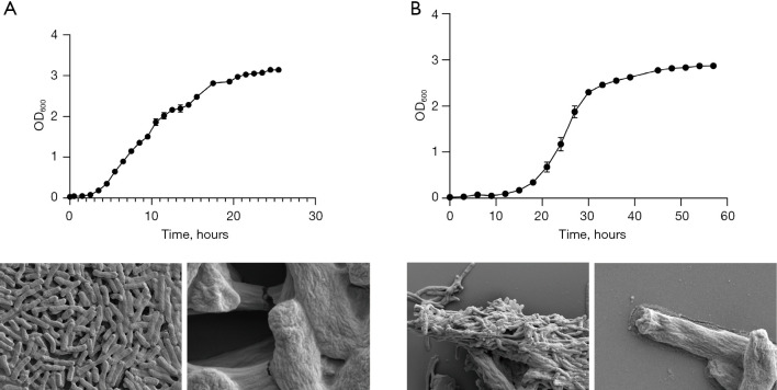
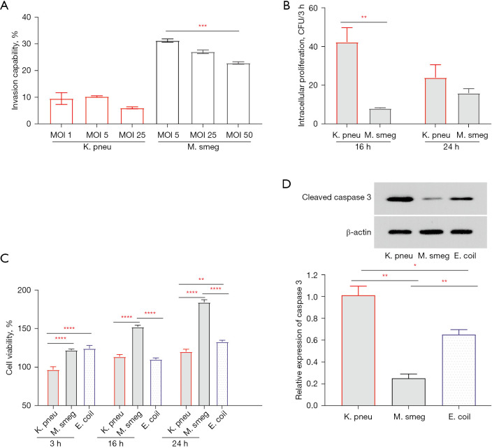
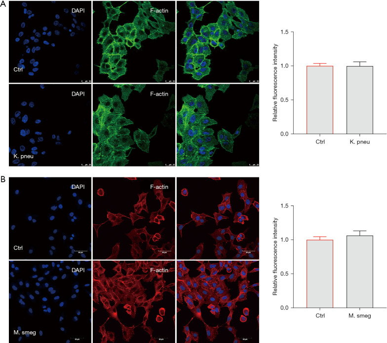
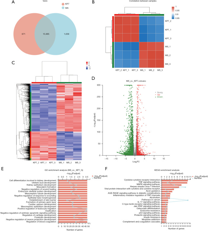
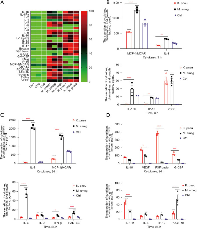
Results: Through scanning electron microscopy combined with bacterial infection experiments, we confirmed the ability of K. pneumoniae and M. smegmatis strains to form biofilm and cord factor out of the cell wall. M. smegmatis has stronger adhesion and intracellular retention ability, while K. pneumoniae is more likely to induce acute infection. These pathogens could stay and proliferate in lung epithelial cells and stimulate the secretion of specific cytokines and chemokines through a gene transcription regulator. M. smegmatis infection can promote crosstalk among epithelial cells and other immune cells in the lung from a very early stage by prompting the secretion of pro-inflammatory cytokines. Meanwhile, there were significant correlations between K. pneumonia infection and higher levels of interleukin-15 (IL-15), interleukin-1Rα (IL-1Rα), fibroblast growth factor (FGF) basic, and granulocyte colony-stimulating factor (G-CSF). At the same time, K. pneumonia infection also led to changes in the expression of cytoskeletal proteins in epithelial cells.
Conclusions: Our results emphasized the immunoprotection and immunomodulation of lung epithelial cells against exogenous pathogenic microorganisms, indicating that different pathogens damaged the host through different strategies and induced varying innate immune responses. At the same time, they provided important clues and key immune factors for dealing with complicated pulmonary infections.
Keywords: A549; Klebsiella pneumoniae (K. pneumoniae); Mycobacterium smegmatis (M. smegmatis); RNA-seq; cytokines.
2023 Journal of Thoracic Disease. All rights reserved.
Conflict of interest statement
Conflicts of Interest: All authors have completed the ICMJE uniform disclosure form (available at https://jtd.amegroups.com/article/view/10.21037/jtd-23-493/coif). The authors have no conflicts of interest to declare.
Figures
Similar articles
-
[Frontier of mycobacterium research--host vs. mycobacterium].Kekkaku. 2005 Sep;80(9):613-29. Kekkaku. 2005. PMID: 16245793 Japanese.
-
Klebsiella pneumoniae Siderophores Induce Inflammation, Bacterial Dissemination, and HIF-1α Stabilization during Pneumonia.mBio. 2016 Sep 13;7(5):e01397-16. doi: 10.1128/mBio.01397-16. mBio. 2016. PMID: 27624128 Free PMC article.
-
Structural Variability of Lipoarabinomannan Modulates Innate Immune Responses within Infected Alveolar Epithelial Cells.Cells. 2022 Jan 21;11(3):361. doi: 10.3390/cells11030361. Cells. 2022. PMID: 35159170 Free PMC article.
-
PE17 protein from Mycobacterium tuberculosis enhances Mycobacterium smegmatis survival in macrophages and pathogenicity in mice.Microb Pathog. 2019 Jan;126:63-73. doi: 10.1016/j.micpath.2018.10.030. Epub 2018 Oct 23. Microb Pathog. 2019. PMID: 30366126
-
Exploring the pathogenetic mechanisms of Mycoplasmapneumoniae (Review).Exp Ther Med. 2024 Apr 30;28(1):271. doi: 10.3892/etm.2024.12559. eCollection 2024 Jul. Exp Ther Med. 2024. PMID: 38765654 Free PMC article. Review.
Cited by
-
Miliary Tuberculosis due to Mycobacterium tuberculosis and Mycobacterium smegmatis associated with invasive aspergillosis in a renal transplant recipient.BMC Nephrol. 2025 May 7;26(1):229. doi: 10.1186/s12882-025-04159-3. BMC Nephrol. 2025. PMID: 40336084 Free PMC article.
-
Antimycobacterial and Antifungal Activities of Leaf Extracts From Trichilia emetica.Scientifica (Cairo). 2024 Dec 9;2024:8784390. doi: 10.1155/sci5/8784390. eCollection 2024. Scientifica (Cairo). 2024. PMID: 39885899 Free PMC article.
References
LinkOut - more resources
Full Text Sources
Research Materials