. 2023 Aug 24:11:1225628.
doi: 10.3389/fcell.2023.1225628. eCollection 2023.

TRiC/CCT chaperonin is required for the folding and inhibitory effect of WDTC1 on adipogenesis

Affiliations

TRiC/CCT chaperonin is required for the folding and inhibitory effect of WDTC1 on adipogenesis

Wen-Shuai Tang et al. Front Cell Dev Biol. .

Abstract

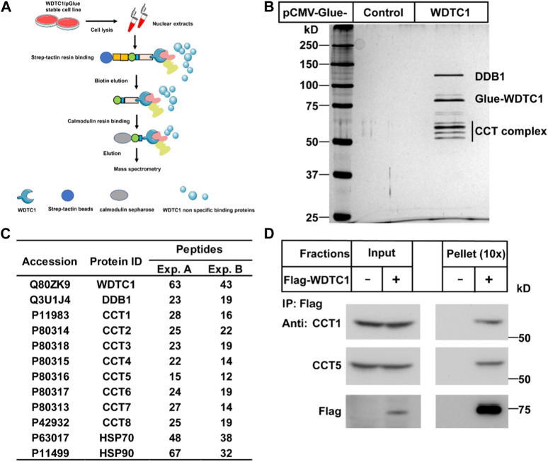
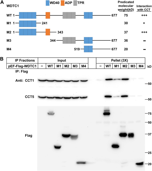
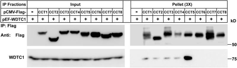
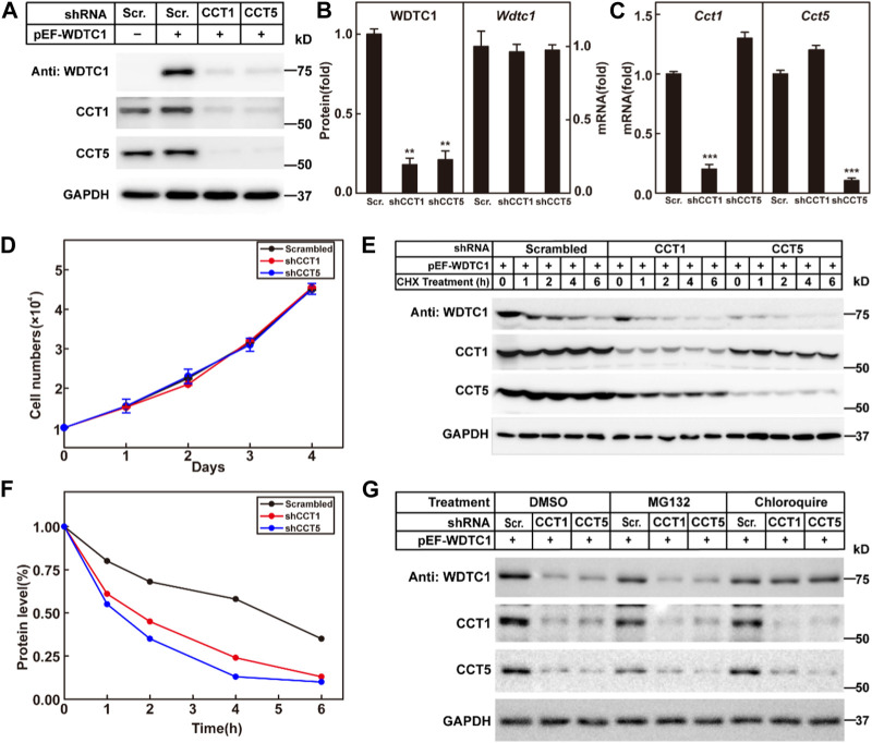
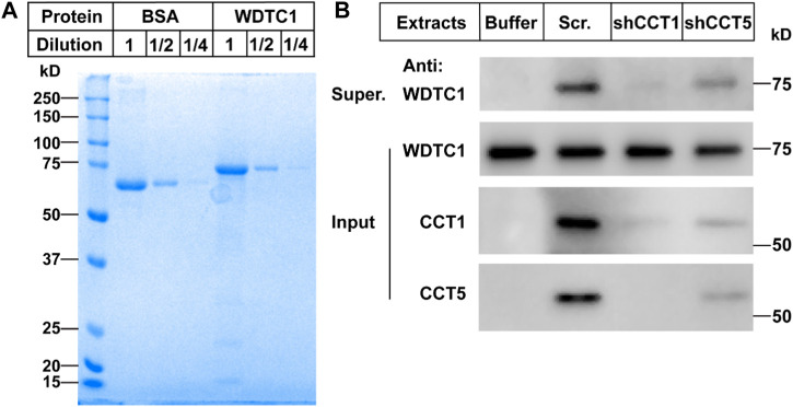
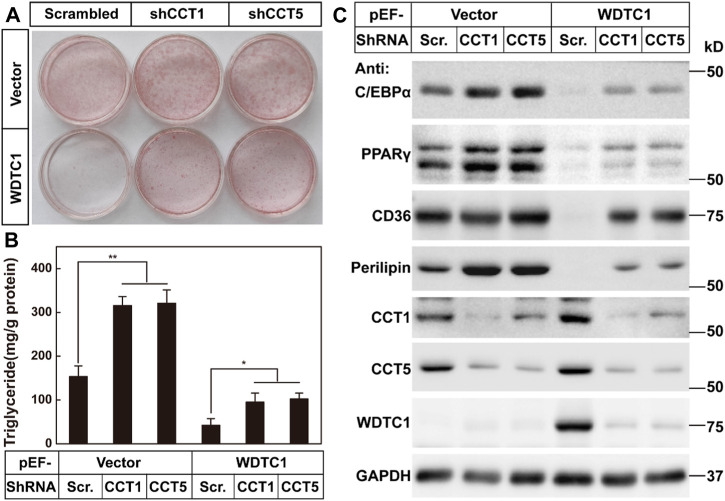
Obesity has become a global pandemic. WDTC1 is a WD40-containing protein that functions as an anti-obesity factor. WDTC1 inhibits adipogenesis by working as an adaptor of the CUL4-DDB1 E3 ligase complex. It remains unclear about how WDTC1 is regulated. Here, we show that the TRiC/CCT functions as a chaperone to facilitate the protein folding of WDTC1 and proper function in adipogenesis. Through tandem purification, we identified the molecular chaperone TRiC/CCT as WDTC1-interacting proteins. WDTC1 bound the TRiC/CCT through its ADP domain, and the TRiC/CCT recognized WDTC1 through the CCT5 subunit. Disruption of the TRiC/CCT by knocking down CCT1 or CCT5 led to misfolding and lysosomal degradation of WDTC1. Furthermore, the knockdown of CCT1 or CCT5 eliminated the inhibitory effect of WDTC1 on adipogenesis. Our studies uncovered a critical role of the TRiC/CCT in the folding of WDTC1 and expanded our knowledge on the regulation of adipogenesis.

Keywords: TRiC/CCT; WDTC1; adipogenesis; obesity; protein folding.


Conflict of interest statement

The authors declare that the research was conducted in the absence of any commercial or financial relationships that could be construed as a potential conflict of interest.

Figures

FIGURE 1
FIGURE 1
Identification of the CCT complex as WDTC1-interacting proteins. (A) Strategy to purify WDTC1-interacting proteins in HEK293T cells using Glue-WDTC1 as a bait. (B,C) On day 0, nuclear fractions were prepared from stable HEK293T cells expressing WDTC1/pGlue or vector alone and subjected to purification with Strep-Tactin beads as described. The eluted fractions were subjected to silver staining (B) and mass spectrometry for protein ID analysis. Potential candidates are listed in (C). (D) On day 0, HEK293T cells were set up at 250,000 cells/60-mm dish. On day 2, cells were transfected with pEF-Flag-WDTC1 or vector alone. On day 3, immunoprecipitation was performed using anti-Flag M2 beads and endogenous CCT1 and CCT5 were detected.
FIGURE 2
FIGURE 2
WDTC1 interacts with the CCT through its ADP domain. (A) Illustration of the domain structures of WT and different truncates of WDTC1. (B) HEK293T cells were set up, transfected with indicated plasmids, and subjected to immunoprecipitation as in Figure 1D. Input and pellet fractions were immunoblotted with indicated antibodies.
FIGURE 3
FIGURE 3
TRiC/CCT binds WDTC1 through CCT5. On day 0, HEK293T cells were set up at 250,000 cells/60-mm dish. On day 2, cells were transfected with pEF-Flag-CCT1-8 or vector alone. On day 3, immunoprecipitation was performed using anti-Flag M2 beads and endogenous WDTC1 were detected.
FIGURE 4
FIGURE 4
TRiC/CCT is required for the stability of WDTC1. (A–C) On day 0, 3T3-L1 preadipocytes infected with lentivirus expressing WDTC1 and the indicated shRNAs were set up at 1 × 10⁵ cells per 35-mm dish. On day 2, cells were harvested to detect the protein (A) and RNA (B, C) levels of WDTC1, CCT1, and CCT5. Each value represents mean ± SEM of three replicates. (D) Control, CCT1, and CCT5 knockdown 3T3-L1 cells were set up at 1 × 10⁴ cells/35-mm dish. Cells were counted every day for 4 days. Cell numbers are plotted in (D). (E,F) On day 0, 3T3-L1 preadipocytes infected with lentivirus expressing the indicated shRNAs and WDTC1 were set up at 1 × 10⁵ cells per 35-mm dish. On day 2, cells were treated with CHX (100 μg/ml) for 0, 1, 2, 4, and 6 h, then cells were harvested, and total cell lysate was immunoblotted with indicated antibodies (E); band intensities of WDTC1 are quantified and plotted in (F). (G) On day 0, 3T3-L1 preadipocytes infected with lentivirus expressing WDTC1 and the indicated shRNAs were set up at 1 × 10⁵ cells per 35-mm dish. On day 2, cells were treated with MG132(10 μg/ml) or chloroquine (50 μM) for 6 h, then cells were harvested, and total cell lysate was immunoblotted with indicated antibodies. For all panels, asterisks (*) denote the level of statistical significance (Student’s t-test). **p < 0.01 and ***p < 0.001.
FIGURE 5
FIGURE 5
TRiC/CCT is required for the protein folding of WDTC1. (A) On day 0, the WDTC1 colony was incubated in the LB medium overnight. On day 1, the bacteria were induced with 1 mM IPTG for 3 h. Cells were then harvested and lysed in PBS containing 1 mg/ml lysozyme supplemented with protease inhibitors. The final product was subjected to SDS-PAGE and Coomassie Blue Staining. (B) On day 0, the purified WDTC1 was then denatured in 6 M urea in 10 mM HEPES pH 8.0. On day 1, 100 ng denatured WDTC1 protein was diluted into cytosolic fractions prepared from 3T3-L1 cell medium buffer or cells infected with scrambled shRNA, shCC1, or shCCT5 and incubated at 25°C for 1 h. The supernatant and pellet fractions of samples were immunoblotted with indicated antibodies.
FIGURE 6
FIGURE 6
TRiC/CCT is required for the inhibitory effect of WDTC1 on adipogenesis. (A–C) 3T3-L1 preadipocytes infected with lentivirus expressing WDTC1 and the indicated shRNAs on pLKO.1 vector were set up at 1 × 10⁵ cells per 35-mm dish and subjected to differentiation. On day 8 of differentiation, cells were harvested for Oil Red O staining (A), measurement of intracellular triglyceride content (B), and Western blot tests (C). Each value represents means ± SEM of three samples. Asterisks (*) denote the level of statistical significance (Student’s t-test) between scrambled and shCCT1 or shCCT5 cells; *p < 0.05 and **p < 0.01.

Similar articles

References

    1. Angers S., Li T., Yi X., MacCoss M. J., Moon R. T., Zheng N. (2006). Molecular architecture and assembly of the DDB1-CUL4A ubiquitin ligase machinery. Nature 443 (7111), 590–593. 10.1038/nature05175 - DOI - PubMed
    1. Cong Y., Baker M. L., Jakana J., Woolford D., Miller E. J., Reissmann S., et al. (2010). 4.0-A resolution cryo-EM structure of the mammalian chaperonin TRiC/CCT reveals its unique subunit arrangement. Proc. Natl. Acad. Sci. U. S. A. 107 (11), 4967–4972. 10.1073/pnas.0913774107 - DOI - PMC - PubMed
    1. Doane W. W. (1960a). Developmental physiology of the mutant female sterile(2)adipose of Drosophila melanogaster. I. Adult morphology, longevity, egg production, and egg lethality. J. Exp. Zool. 145, 1–21. 10.1002/jez.1401450102 - DOI - PubMed
    1. Doane W. W. (1960b). Developmental physiology of the mutant female sterile(2)adipose of Drosophila melanogaster. II. Effects of altered environment and residual genome on its expression. J. Exp. Zool. 145, 23–41. 10.1002/jez.1401450103 - DOI - PubMed
    1. Galgani J. E., Kelley D. E., Albu J. B., Krakoff J., Smith S. R., Bray G. A., et al. (2013). Adipose tissue expression of adipose (WDTC1) gene is associated with lower fat mass and enhanced insulin sensitivity in humans. Obes. (Silver Spring) 21 (11), 2244–2248. 10.1002/oby.20371 - DOI - PMC - PubMed

LinkOut - more resources