. 2023 Aug 24:11:1244954.
doi: 10.3389/fbioe.2023.1244954. eCollection 2023.

Knee instability caused by altered graft mechanical properties after anterior cruciate ligament reconstruction: the early onset of osteoarthritis?

Affiliations

Knee instability caused by altered graft mechanical properties after anterior cruciate ligament reconstruction: the early onset of osteoarthritis?

Janne Spierings et al. Front Bioeng Biotechnol. .

Abstract

Anterior cruciate ligament (ACL) rupture is a very common knee joint injury. Torn ACLs are currently reconstructed using tendon autografts. However, half of the patients develop osteoarthritis (OA) within 10 to 14 years postoperatively. Proposedly, this is caused by altered knee kine(ma)tics originating from changes in graft mechanical properties during the in vivo remodeling response. Therefore, the main aim was to use subject-specific finite element knee models and investigate the influence of decreasing graft stiffness and/or increasing graft laxity on knee kine(ma)tics and cartilage loading. In this research, 4 subject-specific knee geometries were used, and the material properties of the ACL were altered to either match currently used grafts or mimic in vivo graft remodeling, i.e., decreasing graft stiffness and/or increasing graft laxity. The results confirm that the in vivo graft remodeling process increases the knee range of motion, up to >300 percent, and relocates the cartilage contact pressures, up to 4.3 mm. The effect of remodeling-induced graft mechanical properties on knee stability exceeded that of graft mechanical properties at the time of surgery. This indicates that altered mechanical properties of ACL grafts, caused by in vivo remodeling, can initiate the early onset of osteoarthritis, as observed in many patients clinically.

Keywords: anterior cruciate ligament reconstruction; graft remodeling; knee biomechanics; knee instability; osteoarthritis.


Conflict of interest statement

The authors declare that the research was conducted in the absence of any commercial or financial relationships that could be construed as a potential conflict of interest.

Figures

FIGURE 1
FIGURE 1
Schematic overview of the workflow used in this study. Subject-specific knee geometries were obtained from the Open Knee project. The material behavior of the native ACL was changed by changing the material properties of the constitutive law of the ACL to create tendon grafts or grafts with a decreasing stiffness and/or increasing transition strain to mimic graft remodeling. Validation was done by simulating an anterior drawer and a valgus stress test. The influence of graft remodeling was evaluated by simulating a gait cycle and recording the knee kinematics and tibial cartilage loading.
FIGURE 2
FIGURE 2
Schematic overview of the stress-strain relations of the grafts created and used in this research.
FIGURE 3
FIGURE 3
Overview of the joint coordinate system and the applied gait cycle loads. Three rigid cylindrical joints are connected through a four-link kinematic chain to create 6 DoFs. The imaginary links and rigid bodies are shown in green. The stance phase of the gait cycle was simulated by applying fractions of the forces, moment, and rotation obtained from the OrthoLoads database (Bergmann et al., 2014). Flexion (dashed line) is portrayed on the right y-axis.
FIGURE 4
FIGURE 4
Altering graft mechanical properties results in changed anterior tibial translation. Difference in anterior tibial translation (ATT) for tendon autografts, grafts with a decreasing stiffness, grafts with an increasing transition strain, and a combination of both.
FIGURE 5
FIGURE 5
Altering graft mechanical properties results in changed internal tibial rotation. Difference in internal tibial rotation (IR) for tendon autografts, grafts with a decreasing stiffness, grafts with an increasing transition strain, and a combination of both.
FIGURE 6
FIGURE 6
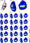
Altering graft mechanical properties results in a relocation of tibial cartilage contact pressure [MPa]. Visual representation of the tibial cartilage contact pressure distribution of model 1 for the native ACL, the tendon grafts (PT. ST, and GT), grafts with a decreasing stiffness (E1-4), grafts with an increasing transition strain (T1-3), and a combination of both (C1-5). Left: medial tibial cartilage; right: lateral tibial cartilage.
FIGURE 7
FIGURE 7
Altering graft mechanical properties results in a relocation of tibial cartilage contact pressure [MPa]. Visual representation of the tibial cartilage contact pressure distribution of model 3 for the native ACL, the tendon grafts (PT, ST, and GT), grafts with a decreasing stiffness (E1-4), grafts with an increasing transition strain (T1-3), and a combination of both (C1-5). Left: lateral tibial cartilage; right: medial tibial cartilage.
FIGURE 8
FIGURE 8
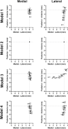
Graft mechanical properties influence the location of the tibial cartilage contact pressure in a subject-specific direction. The quantified weighted center of mass of the contact pressure on the tibial cartilage for all models.

Similar articles

Cited by

References

    1. Aronson P. A., Gieck J. H., Hertel J., Rijke A. M., Ingersoll C. D. (2010). Tibiofemoral joint positioning for the valgus stress test. J. Athl. Train. 45, 357–363. 10.4085/1062-6050-45.4.357 - DOI - PMC - PubMed
    1. Belbasis A., Fuss F. K., Sidhu J. (2015). Estimation of cruciate ligament forces via smart compression garments. Procedia Eng. 112, 169–174. 10.1016/j.proeng.2015.07.194 - DOI
    1. Bennetts C. J., Chokhandre S., Donnola S. B., Flask C. A., Bonner T. F., Colbrunn R. W., et al. (2015). “Open knee(s): magnetic resonance imaging for specimen-specific next generation knee models,” in SB3C2015, Summer Biomechanics, Bioengineering and Biotransport Conference, Snowbird Resort, Utah, USA, June 17-20, 2015.
    1. Benos L., Stanev D., Spyrou L., Moustakas K., Tsaopoulos D. E. (2020). A review on finite element modeling and simulation of the anterior cruciate ligament reconstruction. Front. Bioeng. Biotechnol. 8, 967. 10.3389/fbioe.2020.00967 - DOI - PMC - PubMed
    1. Bergmann G., Bender A., Graichen F., Dymke J., Rohlmann A., Trepczynski A., et al. (2014). Standardized loads acting in knee implants. PLoS One 9, e86035. 10.1371/journal.pone.0086035 - DOI - PMC - PubMed

LinkOut - more resources