. 2023 Aug 24;8(35):31998-32016.
doi: 10.1021/acsomega.3c03824. eCollection 2023 Sep 5.

5-Methoxyisatin N(4)-Pyrrolidinyl Thiosemicarbazone (MeOIstPyrd) Restores Mutant p53 and Inhibits the Growth of Skin Cancer Cells, In Vitro

Affiliations

5-Methoxyisatin N(4)-Pyrrolidinyl Thiosemicarbazone (MeOIstPyrd) Restores Mutant p53 and Inhibits the Growth of Skin Cancer Cells, In Vitro

Nerina Shahi et al. ACS Omega. .

Abstract

A series of novel thiosemicarbazone derivatives containing 5-methoxy isatin were designed and synthesized with modification on N(4) position. Derivatives considering structure-activity relationship have been designed and synthesized by condensing thiosemicarbazide with 5-methoxy isatin. The synthesized compounds were characterized by elemental analysis, FT-IR spectroscopy, UV-visible spectroscopy, NMR (1H, 13C) spectroscopy, mass spectrometry, and a single-crystal study. Biological evaluation of the synthesized compounds revealed that MeOIstPyrd is the most promising compound against skin cancer cell line, A431, with an IC50 value of 0.9 μM. In addition, MeOIstPyrd also exhibited low toxicity against the normal human fibroblast and the human embryonic kidney 293 cell line, HLF-1, and HEK293, respectively. Furthermore, the mechanistic study revealed that MeOIstPyrd efficiently inhibited cell proliferation, migration, and spheroid formation by activating the mitochondrial intrinsic apoptotic pathway. MeOIstPyrd also induces DNA damage and activates p53 irrespective of the p53 status. It increases the half-life of p53 and stabilizes p53 by phosphorylating it at ser15. Moreover, MeOIstPyrd was found to bind to MDM2 in the p53 sub-pocket and, therefore, block p53-MDM2 interaction. Our result exhibited potential anticancer activity of MeOIstPyrd in the A431 cell line and its ability in restoring mutant p53, which is an interesting and promising strategy for cancer therapeutics.


Conflict of interest statement

The authors declare no competing financial interest.

Figures

Scheme 1
Scheme 1. Synthetic Route of 5-Methoxyisatin TSCs and Structures of TSC Derivatives
Figure 1
Figure 1
ORTEP diagram of MeOIstHex drawn in 30% thermal probability ellipsoids showing an atomic numbering scheme. The asymmetric unit contains two crystallographically independent units and two water molecules in the crystal lattice. One of the azepane rings consists of atoms (N1 C11 C12 C13 C14 C15 C16) found to be disordered over two positions with an occupancy ratio of 53:47. The second disordered part has been omitted for the sake of clarity.
Figure 2
Figure 2
ORTEP diagram of MeOIstPipe drawn in 30% thermal probability ellipsoids showing an atomic numbering scheme.
Figure 3
Figure 3
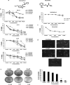
Effect of 3-methoxy isatin derivatives in different cancer cell lines. (a) shows the chemical structure of MeOIstPyrd and triapine (as a control compound). In (b), the cell growth inhibitory activity was evaluated using the CV assay, briefly. The cells were seeded and treated with compounds for 72 h, followed by staining with crystal violet. OD was taken at 450 nm. The graph shows the change in percentage viability upon treatment with compounds. (c) shows the viability of triapine in A431 cells. (d) shows the phase contrast picture of A431 cells when treated with MeOIstPyrd. Data are represented as mean ± SD (n = 3). (e) Colony formation was evaluated by the clonogenic assay where cells were treated with indicated concentrations of MeOIstPyrd, and the colonies were quantified using ImageJ; data are represented as mean ± SD (n = 2).
Figure 4
Figure 4
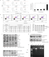
Effect of MeOIstPyrd on apoptosis. (a) A431 cells were treated either with media alone, media + DMSO, and media + MeOIstPyrd for 72 h. Cell death was accessed by PI staining, histogram represents live cells and necrotic cells, bar graph represents the percentage of dead cells in different concentrations, and experiments were done in triplicates. (b) Apoptosis was examined using annexin V/PI staining by flow cytometry. The histogram shows the percentage of early and late apoptosis. The table shows the mean standard deviation (SD) percentage of live, total apoptotic, and dead cells from three independent experiments. The graph shows the percentage of cells in early and late apoptosis for one of the experiments. Data are presented as the mean ± the SD (n = 3). (c) shows the expression of different apoptotic markers involved in mitochondrial intrinsic apoptotic pathway. (d) shows the expression of cytochrome c in cytosolic and mitochondrial fractions of cell lysate. (e) shows that MeOIstPyrd causes DNA damage; all the results were obtained in triplicates.
Figure 5
Figure 5
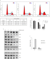
Effect of MeOIstPyrd on the cell cycle. A431 cells were treated with different concentrations of MeOIstPyrd for 48 h. The cells were fixed, stained, and processed for cell cycle analysis. Histogram shows the percentage of cells in each phase (a), data are represented as mean ± SD (n = 2), and the table shows the mean SD and percentage of cells in each phase of cell cycle. Protein and RNA were extracted for western blotting and real-time PCR, respectively. Western blots show the expression of cell cycle marker proteins treated with different concentrations of MeOIstPyrd (b). Data represented are mean ± SD (n = 2). Bar graph shows the relative expression level of RNA in control and cells treated with MeOIstPyrd (c).
Figure 6
Figure 6
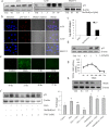
Effect of MeOIstPyrd in the expression of p53 and ROS. (a) Cells were treated with MeOIstPyrd, and p53 expression was checked by western blotting; the figure shows the effect of MeOIstPyrd on the expression of p53 in A431 (R273H), A549 (wt), and SKOV-3 (null) cell lines. (b) Immunofluorescence staining of cells with anti-p53 antibody in different cell lines. (c) Cells were treated with MeOIstPyrd or 3-AP, RNA was isolated, and the expression of p53 was checked by qRT-PCR. (d) p53 protein expression was accessed in A431 cells after 3-AP treatment. (e) Cells were treated with MeOIstPyrd at indicated time points, stained with CM-H2DCFDA for 30 min, and observed in a fluorescence microscope. Representative images are shown for the CM-H2DCFDA-stained A431 cells treated with MeOIstPyrd. (f) A431 cells were pretreated with NAC for 30 min, followed by treatment with either MeOIstPyrd or H2O2 for 24 h. The western blot result shows the expression of p53 in different conditions; the bar graph shows the data represented in mean ± SD (n = 2). (g) Total glutathione levels were measured for different time points, and the graph shows the data represented in mean ± SD (n = 2). (h) Western blot analysis for SOD at 48 h in the A431 cell line.
Figure 7
Figure 7
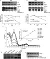
Effect of MeOIstPyrd on p53 stability and DNA damage. A431 and A549 cells were treated with MeOIstPyrd for 24 h, followed by treatment with cycloheximide for different time points. (a,b) show the expression of p53 in the presence of CHX or CHX + MeOIstPyrd. Data represented as mean ± SD (n = 3). (c) Absorption spectra of MeOIstPyrd (3 μM) with increasing concentration of calf thymus DNA (ct-DNA) (0, 20, 40, 60, 80, 100, and 120 μM). Inset: plot of [DNA]/(εa – εf) vs DNA shows a binding constant of ct-DNA in the presence of MeOIstPyrd. (d) shows the representative figure for the DNA cleavage assay where the PUC18 plasmid DNA was incubated with either doxorubicin or MeOIstPyrd. (e) MeOIstPyrd downregulates the protein expression of casein kinase 2α in the A431 cell line.
Figure 8
Figure 8
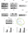
Effect of MeOIstPyrd and MG132 in p53–MDM2 interaction. A431, A549, and SKOV-3 cells were treated either with MeOIstPyrd or idasanutlin for 24 h, followed by MG132 addition for another 4 h. (a–c) show western blot analysis of p53, p-p53, and MDM2 in these cell lines. (d–f) show western blots of p53 and MDM2 treated with idasanutlin. (g) shows the expression of p53 and its downstream target MDM2 and p21 after siRNA knockdown. (h) Docking analysis of MeOIstPyrd with MDM2. (i,j) ChIP analysis shows that MeOIstPyrd treatment restores the transactivation function of p53 in mutant R273H cells. Cells were either left untreated or treated with MeOIstPyrd (3 μM) and crosslinked. Sheared chromatin was immunoprecipitated with the p53 antibody and linked with Dynabeads. DNA was eluted and examined by quantitative real-time PCR using primers that specifically target the p53 binding site in the respective promotor of the target gene. Data shown are mean ± SD (n = 3).
Figure 9
Figure 9
Effect of MeOIstPyrd on cancer migration and spheroid formation. A431 cells were grown in a monolayer, and mechanical scratch was given. (a) shows the wound healing capacity of A431 cells in the presence of MeOIstPyrd for the indicated time points. (b) Western blot pictures show the changes in the levels of the associated proteins in migration. 1000 cells per drop were seeded, and after 72 h, cell aggregates were transferred in agar media in the presence of MeOIstPyrd. (c) shows the representative figure of 3D spheroid in the presence of increasing concentration of MeOIstPyrd.

References

    1. El-Sharief M. A. M. S.; Abbas S. Y.; El-Bayouki K. A. M.; El-Gammal E. W. Synthesis of thiosemicarbazones derived from N-(4-hippuric acid)thiosemicarbazide and different carbonyl compounds as antimicrobial agents. Eur. J. Med. Chem. 2013, 67, 263–268. 10.1016/j.ejmech.2013.06.031. - DOI - PubMed
    1. Oliveira C. G.; Maia P. I. d. S.; Souza P. C.; Pavan F. R.; Leite C. Q.; Viana R. B.; Batista A. A.; Nascimento O. R.; Deflon V. M. Manganese(II) complexes with thiosemicarbazones as potential anti-Mycobacterium tuberculosis agents. J. Inorg. Biochem. 2014, 132, 21–29. 10.1016/j.jinorgbio.2013.10.011. - DOI - PubMed
    1. Sevinçli Z. Ş.; Duran G. N.; Özbil M.; Karalı N. Synthesis, molecular modeling and antiviral activity of novel 5-fluoro-1H-indole-2,3-dione 3-thiosemicarbazones. Bioorg. Chem. 2020, 104, 104202.10.1016/j.bioorg.2020.104202. - DOI - PubMed
    1. Soraires Santacruz M. C.; Fabiani M.; Castro E. F.; Cavallaro L. V.; Finkielsztein L. M. Synthesis, antiviral evaluation and molecular docking studies of N4-aryl substituted/unsubstituted thiosemicarbazones derived from 1-indanones as potent anti-bovine viral diarrhea virus agents. Bioorg. Med. Chem. 2017, 25, 4055–4063. 10.1016/j.bmc.2017.05.056. - DOI - PubMed
    1. Shakya B.; Yadav P. N. Thiosemicarbazones as Potent Anticancer Agents and their Modes of Action. Mini-Rev. Med. Chem. 2020, 20, 638–661. 10.2174/1389557519666191029130310. - DOI - PubMed