. 2023 Aug 23:14:1221436.
doi: 10.3389/fpls.2023.1221436. eCollection 2023.

Chloroplast magnesium transporters play essential but differential roles in maintaining magnesium homeostasis

Affiliations

Chloroplast magnesium transporters play essential but differential roles in maintaining magnesium homeostasis

Emilija Dukic et al. Front Plant Sci. .

Abstract

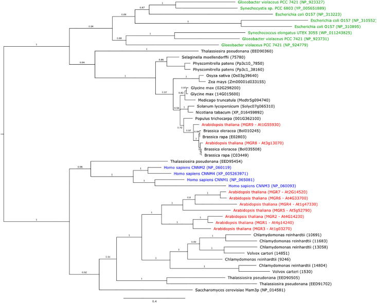
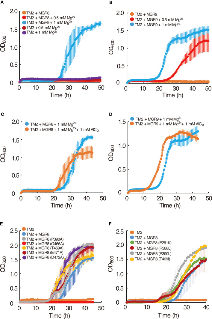
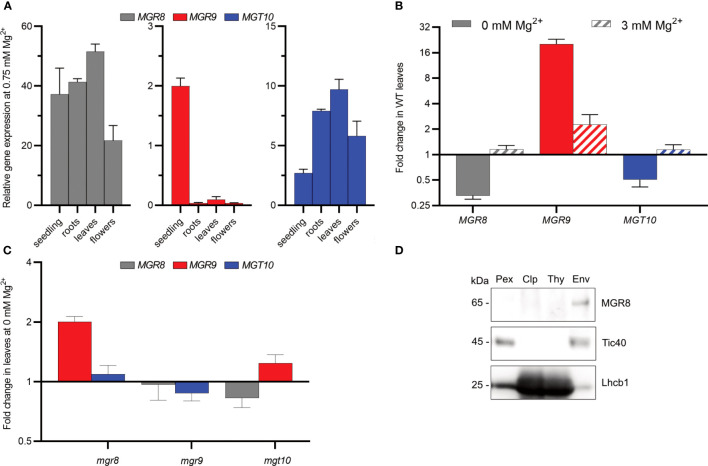
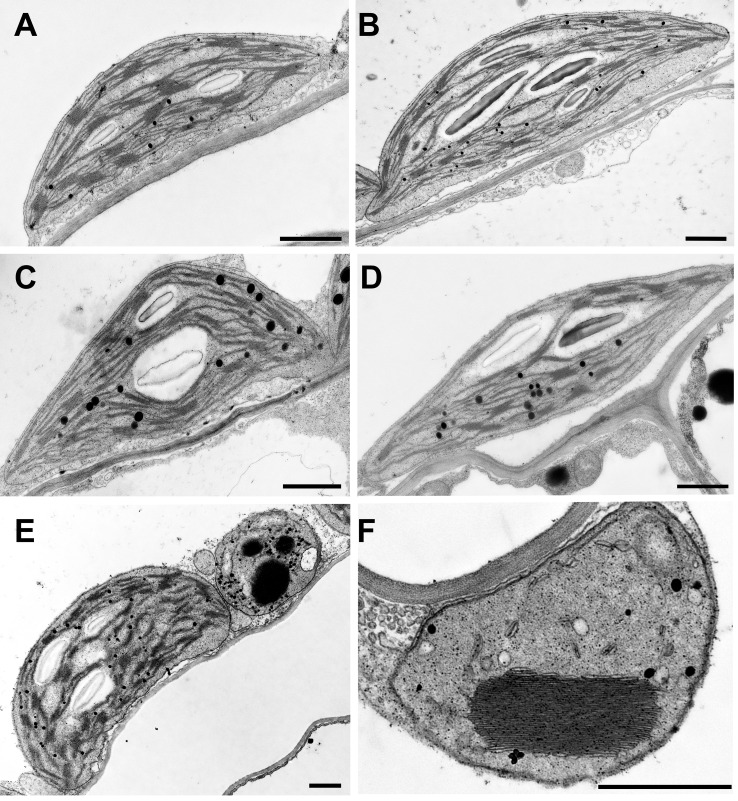
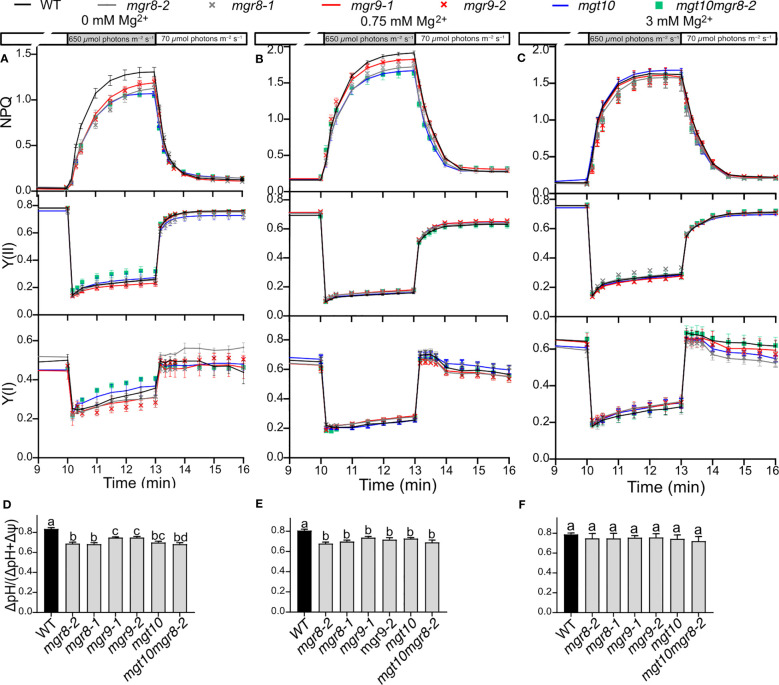
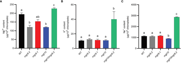
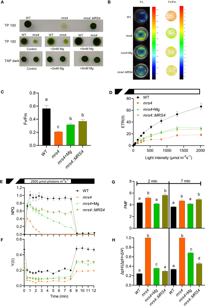
Magnesium (Mg2+) is essential for photosynthesis in the chloroplasts of land plants and algae. Being the central ion of chlorophyll, cofactor and activator of many photosynthetic enzymes including RuBisCO, magnesium-deficient plants may suffer from leaf chlorosis symptoms and retarded growth. Therefore, the chloroplast Mg2+ concentration is tightly controlled by magnesium transport proteins. Recently, three different transporters from two distinct families have been identified in the chloroplast inner envelope of the model plant Arabidopsis thaliana: MGT10, MGR8, and MGR9. Here, we assess the individual roles of these three proteins in maintaining chloroplast Mg2+ homeostasis and regulating photosynthesis, and if their role is conserved in the model green alga Chlamydomonas reinhardtii. Phylogenetic analysis and heterologous expression revealed that the CorC-like MGR8 and MGR9 transport Mg2+ by a different mechanism than the CorA-like MGT10. MGR8 and MGT10 genes are highest expressed in leaves, indicating a function in chloroplast Mg2+ transport. MGR9 is important for chloroplast function and plant adaptation in conditions of deficiency or excess of Mg2+. Transmission electron microscopy indicated that MGT10 plays a differential role in thylakoid stacking than MGR8 and MGR9. Furthermore, we report that MGR8, MGR9, and MGT10 are involved in building up the pH gradient across the thylakoid membrane and activating photoprotection in conditions of excess light, however the mechanism has not been resolved yet. While there are no chloroplast MGR-like transporters in Chlamydomonas, we show that MRS4 is a homolog of MGT10, that is required for photosynthesis and cell growth. Taken together, our findings reveal that the studied Mg2+ transporters play essential but differential roles in maintaining chloroplast Mg2+ homeostasis.

Keywords: Arabidopsis thaliana; Chlamydomonas reinhardtii; chlorophyll fluorescence; chloroplast; magnesium homeostasis; magnesium transporter; photosynthesis.


Conflict of interest statement

The authors declare that the research was conducted in the absence of any commercial or financial relationships that could be construed as a potential conflict of interest.

Figures

Figure 1
Figure 1
Phylogenetic tree of protein sequences from plants, bacteria, green algae, diatoms, yeast, and humans, that are homologs of the Arabidopsis MGR8 and MGR9. The tree was constructed using MrBayes v3.2.6. Bacteria are indicated in green text, the human CNNM1–4 homologs in blue, and all Arabidopsis MGRs in red. Numbers above branches indicate posterior probability values and the expected number of changes per site along the branches is indicated by the scale bar.
Figure 2
Figure 2
Complementation of the E. coli TM2 Mg2+ auxotrophy with MGR8 and MGR9 and Al3+ inhibition. (A) Growth curves of TM2 transformed with the pTV118N vector containing MGR8 cDNA and with the empty vector. (B) Growth curves of TM2 transformed with the plasmid containing MGR9 cDNA. Cells were grown at 37°C on LB medium supplemented with different concentrations of MgSO4. LB medium without added MgSO4 contained 0.17 mM Mg2+ (Ishijima et al., 2015). (C, D) Effect of Al3+ on the growth curves of TM2 transformed with the plasmid containing MGR8 (C) and MGR9 (D) cDNA. Cells were grown at 37°C on LB medium supplemented with 1 mM MgSO4. AlCl3 was added at 0 mM and 1 mM concentration. (E, F) Growth curves of TM2 transformed with the plasmid containing MGR8 wild type, P390A, G466A, T469A, E471A, and D472A (E), E261K, R388L, P390L, and T469I (F) mutant cDNA and with the empty pTV118N vector. Cells were grown at 37°C on LB medium supplemented with 1 mM MgSO4. The OD600 was measured every 0.5 h. Data are average values of three or more independent experiments, and bars indicate means ± S.E.M.
Figure 3
Figure 3
Expression pattern and chloroplast localization in Arabidopsis. (A) Relative expression in 2-week-old seedlings and roots, leaves, and flowers of 6-week-old wild type plants grown hydroponically at 0.75 mM Mg2+ was determined using quantitative RT-PCR. ACTIN8 and PEX4 were used as internal standards. (B, C) Wild type (WT) plants (B) and mutants (C) were grown first for two weeks at 0.75 mM Mg2+ and then for three to four weeks at either 0 or 3 mM Mg2+. The plotted data represent expression fold change relative to the expression at 0.75 mM Mg2+ in the same genotype. Where two independent lines were available, the obtained data are presented as averages. The scale on the Y-axis in (B, C) is log2, whereas the fold change values are non-log2-transformed. The data presented in (A–C) are means ± S.E.M (n = 4 plants). (D) Total protein extracts (Pex), Chloroplasts (Clp), thylakoid (Thy), and envelope (Env) membranes were prepared from wild type leaves. Localization of MGR8 in chloroplast and subfractions was performed by immunoblotting with an MGR8-peptide-specific antibody. Purity of fractions was confirmed using antibodies against marker proteins for the respective compartment: inner envelope translocon complex Tic40 protein and the light harvesting Chl a/b binding thylakoid protein Lhcb1. Uncropped version of the immunoblots is shown in Supplementary Figure S8 .
Figure 4
Figure 4
Chloroplast ultrastructure in Arabidopsis. Wild type plants and mutants were grown in standard Mg2+ conditions for six weeks. Representative TEM photos of chloroplasts are shown. (A) Wild-type, (B) mgr8-2, (C) mgr9-1, (D) mgt10 green interveinal region, (E, F) mgt10 chlorotic, yellow vein region. (E) Mesophyll cell with normal chloroplast and peculiar plastid. (F) peculiar plastid with macro-granum, vesicles, and no stroma thylakoids typical for bundle sheath cells. Scale bar: 1 μm.
Figure 5
Figure 5
Dynamics of photosynthesis and photoprotection in fluctuating light. Wild type (WT) plants and mutants were grown hydroponically first for two weeks at 0.75 mM Mg2+ and then for four to five weeks at the indicated Mg2+ concentrations. Plants were dark adapted for 20 min, illuminated for 10 min with low light, then for 3 min with high light, and then again for 3 min in low light. Chl fluorescence and electrochromic shift were recorded with Dual-PAM-100. The plots in (A–C) show non-photochemical quenching (NPQ), PSII, and PSI quantum yields (Y(II) and (YI)). The plots in (D–F) show the partitioning of the proton motive force to ΔpH as determined from ECS measurements at the end of transition from low to high light. The plotted data are means ± S.E.M. (n = 4-7 plants). Different letters indicate statistically significant differences among the genotypes according to Tukey one-way ANOVA (P < 0.05).
Figure 6
Figure 6
Mineral content of Arabidopsis chloroplasts. Intact chloroplasts were prepared from shoots of plants grown at 0.75 mM Mg2+ and their mineral content was determined using ICP-MS. (A) Mg2+, (B) K+, and (C) Na+ content of Arabidopsis chloroplasts. The presented data are expressed as means ± S.E.M. (n = 3 chloroplast preparations). Different letters indicate statistically significant differences among the genotypes according to Tukey one-way ANOVA (P < 0.05).
Figure 7
Figure 7
Chlamydomonas mrs4 experiences light stress. Wild type (WT), mrs4, and complemented mrs4:MRS4 were grown in the light (100 μmol photons m−2 s−1) on TP medium (TP100) or in darkness on TAP (TAP dark), and where indicated supplemented with 5 mM Mg2+. (A) Spot tests on agar plates showing that complementation with VcMRS4 and Mg2+ supplementation improve the growth of the mutant. (B, C) Chl fluorescence imaging using FluorCam shows significantly reduced maximum quantum yield of PSII photochemistry (Fv/Fm) in mrs4 due to enhanced minimum fluorescence (F0). Complementation with VcMRS4 and Mg2+ supplementation improved the photosynthetic efficiency of the mutant. Chl fluorescence and electrochromic shift were recorded with a Dual-Pam-100. (D) Rapid light response curves of electron transport rates of PSII (ETR(II)). (E, F) Slow kinetics of Chl fluorescence induction during 8 min of illumination followed by 4 min in darkness. The data show reduced ETR(II), NPQ, and Y(II) in mrs4 and improvement by complementation with VcMRS4 and Mg2+ supplementation of cells grown in TP100. (G, H) The total proton motive force (PMF) and partitioning to ΔpH were determined from ECS decay kinetics in darkness of cells grown in TAP dark and pre-exposed at 660 μmol photons m−2 s−1 for 2 or 7 min. The data in (C–G) are means ± S.E.M. (n = 3 replicates). Different letters indicate significant differences among the genotypes according to Tukey one-way ANOVA (P < 0.05). The PMF in the mrs4 mutant consists of 100% ΔpH, indicating that the cells experience high light stress.

References

    1. Alonso J. M., Stepanova A. N., Leisse T. J., Kim C. J., Chen H., Shinn P., et al. (2003). Genome-wide insertional mutagenesis of Arabidopsis thaliana . Science 301, 653–657. doi: 10.1126/science.1086391 - DOI - PubMed
    1. Böszörményi A., Dobi A., Skribanek A., Pávai M., Solymosi K. (2020). The effect of light on plastid differentiation, chlorophyll biosynthesis, and essential oil composition in rosemary (Rosmarinus officinalis) leaves and cotyledons. Front. Plant Sci. 11, 196. doi: 10.3389/fpls.2020.00196 - DOI - PMC - PubMed
    1. Camacho C., Coulouris G., Avagyan V., Ma N., Papadopoulos J., Bealer K., et al. (2009). BLAST+: architecture and applications. BMC Bioinf. 10, 1–9. doi: 10.1186/1471-2105-10-421 - DOI - PMC - PubMed
    1. Chen A., Hansen T. H., Olsen L. I., Palmgren M., Husted S., Schjoerring J. K., et al. (2020). Towards single-cell ionomics: A novel micro-scaled method for multi-element analysis of nanogram-sized biological samples. Plant Methods 16, 1–13. doi: 10.1186/S13007-020-00566-9 - DOI - PMC - PubMed
    1. Chen Y. S., Kozlov G., Fakih R., Yang M., Zhang Z., Kovrigin E. L., et al. (2020). Mg2+-ATP sensing in CNNM, a putative magnesium transporter. Structure 28, 324–335.e4. doi: 10.1016/j.str.2019.11.016 - DOI - PubMed