. 2022 Aug 11;8(5):e10392.
doi: 10.1002/btm2.10392. eCollection 2023 Sep.

Shaping the "hot" immunogenic tumor microenvironment by nanoparticles co-delivering oncolytic peptide and TGF-β1 siRNA for boosting checkpoint blockade therapy

Affiliations

Shaping the "hot" immunogenic tumor microenvironment by nanoparticles co-delivering oncolytic peptide and TGF-β1 siRNA for boosting checkpoint blockade therapy

Cao Dai Phung et al. Bioeng Transl Med. .

Abstract

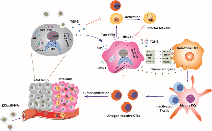
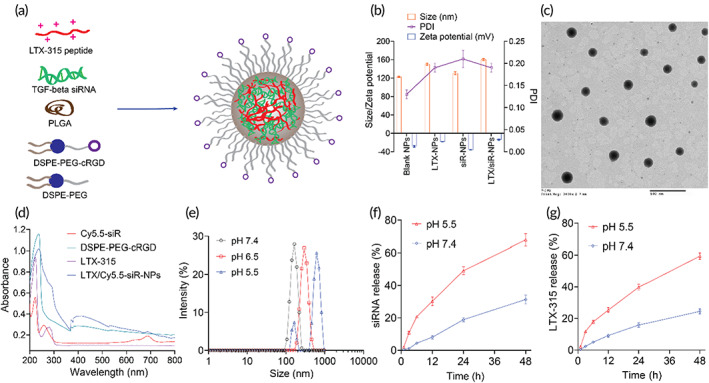
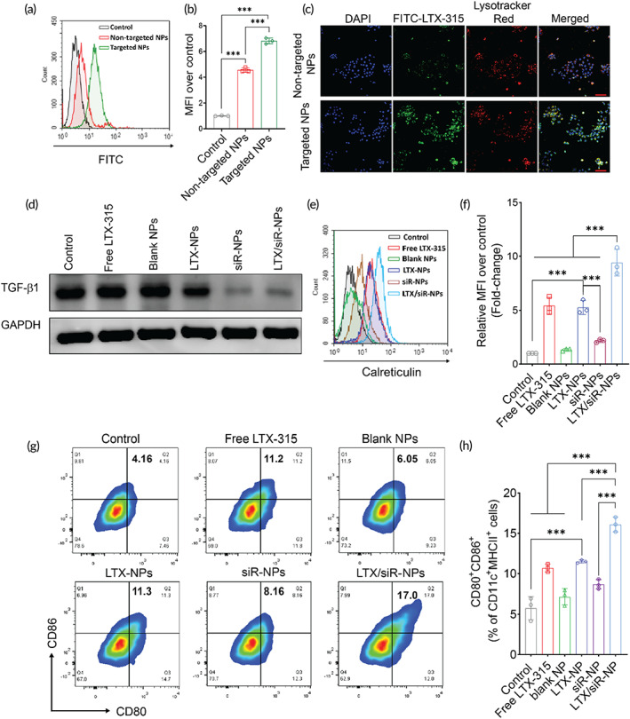
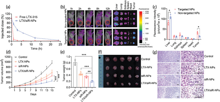
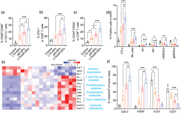
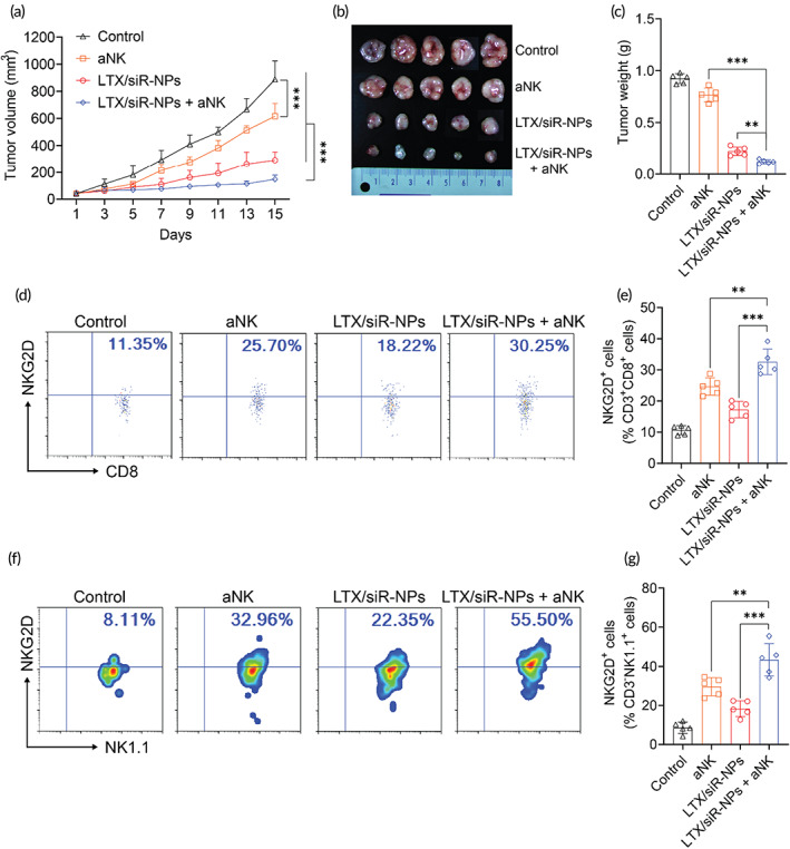
Induction of potent immune responses toward tumors remains challenging in cancer immunotherapy, in which it only showed benefits in a minority of patients with "hot" tumors, which possess pre-existing effector immune cells within the tumor. In this study, we proposed a nanoparticle-based strategy to fire up the "cold" tumor by upregulating the components associated with T and NK cell recruitment and activation and suppressing TGF-β1 secretion by tumor cells. Specifically, LTX-315, a first-in-class oncolytic cationic peptide, and TGF-β1 siRNA were co-entrapped in a polymer-lipid hybrid nanoparticle comprising PLGA, DSPE-mPEG, and DSPE-PEG-conjugated with cRGD peptide (LTX/siR-NPs). The LTX/siR-NPs showed significant inhibition of TGF-β1 expression, induction of type I interferon release, and triggering immunogenic cell death (ICD) in treated tumor cells, indicated via the increased levels of danger molecules, an in vitro setting. The in vivo data showed that the LTX/siR-NPs could effectively protect the LTX-315 peptide from degradation in serum, which highly accumulated in tumor tissue. Consequently, the LTX/siR-NPs robustly suppressed TGF-β1 production by tumor cells and created an immunologically active tumor with high infiltration of antitumor effector immune cells. As a result, the combination of LTX/siR-NP treatment with NKG2A checkpoint inhibitor therapy remarkably increased numbers of CD8+NKG2D+ and NK1.1+NKG2D+ within tumor masses, and importantly, inhibited the tumor growth and prolonged survival rate of treated mice. Taken together, this study suggests the potential of the LTX/siR-NPs for inflaming the "cold" tumor for potentiating the efficacy of cancer immunotherapy.

Keywords: LTX‐315; cancer immunotherapy; hybrid nanoparticle; tumor microenvironment.


Conflict of interest statement

The authors have no conflict of interest to declare.

Figures

SCHEME 1
SCHEME 1
Schematic illustration of the mechanism of action of LTX/siR‐NPs in shaping the “hot” immunogenic tumor microenvironment
FIGURE 1
FIGURE 1
LTX/siR‐NPs preparation and characterization. (a) The representative structure of LTX/siR‐NPs. (b) Particle size distribution and zeta potential of LTX/siR‐NPs were measured by the dynamic light scattering method. (c) The spherical morphology of LTX/siR‐NPs was observed by transmission electron microscopy (scale bar: 500 nm). (d) The UV–VIS spectra of components and LTX/siR‐NPs confirmed the successful load of LTX‐315 and TGF‐β siRNA into the hybrid nanoparticles. (e) The influence of pH conditions on the size of LTX/siR‐NPs. The in vitro accumulative drug‐release profile of (f) siRNA and (g) LTX‐315 at different pH conditions
FIGURE 2
FIGURE 2
Cellular uptake of LTX/siR‐NPs into 4T1 cells followed by the induction of the immunogenic cell death and reduced expression of TGF‐β1. The internalization of targeted LTX/siR‐NPs (with cRGD conjugation) and nontargeted LTX/siR‐NPs (without cRGD conjugation) into 4T1 cells was confirmed by (a, b) flow cytometry analysis, and (c) confocal laser scanning microscopy (scale bar: 50 nm). (d) Western Blot analysis of TGF‐β1 expression in 4T1 cells after treatment with (1) PBS (Control), (2) free LTX‐315, (3) blank NPs, (4) LTX‐NPs, (5) siR‐NPs, and (6) LTX/siR‐NPs. (e, f) The plasma membrane expression of calreticulin in 4T1 cells after treatment determined by flow cytometry. (g, h) The expression of co‐stimulatory molecules on the surface of BMDCs after the treatments with supernatant of treated cancer cells. Data were presented as mean ± SD (n = 3). **p < 0.01; ***p < 0.001
FIGURE 3
FIGURE 3
Hybrid nanoparticles prolong the circulation time, improve the accumulation at the tumor, and antitumor efficacy of loading cargoes. (a) The plasma pharmacokinetic profiles of LTX‐315 by intravenous administration of free LTX‐315 and LTX‐315 loaded NPs. (b) Fluorescent images of mice and major organs were collected from mice injected with hybrid NPs conjugated with cRGD (targeted NPs) or nontargeted NPs co‐entrapping LTX‐315 and Cyanine 5.5‐labeled siRNA. (c) Quantification of fluorescence intensities of targeted NPs and nontargeted NPs in tumors and organs of treated mice. Data were represented as mean ± SD (n = 3). Two‐tailed Student's t‐test, *p < 0.05. (d) Tumor volumes of mice treated with PBS (control), LTX‐NPs, siR‐NPs, and LTX/siR‐NPs with increasing time. Data were represented as mean ± SD (n = 5), two‐way ANOVA with Tukey's multiple comparison test of tumor sizes at Day 15, ***p < 0.001. (e) Tumor weight, (f) images of isolated tumors, and (g) histological analysis of tumors collected from treated mice. Data represented as mean ± SD (n = 5). One‐way ANOVA with Tukey's multiple comparison test, **p < 0.01; ***p < 0.001
FIGURE 4
FIGURE 4
LTX/siR‐NPs re‐shaped the immune cell profile of tumor microenvironment. Flow cytometric analysis of (a) matured DC in tdLNs, (b) IFN‐γ produced by CD8+ T cells and (c) effector memory CD8+ T cells in the spleen, and (d) intratumoral profiles of immune cells of the mice treated with indicated formulations. (e) Heatmap visualization of gene expression in tumors collected from treated mice. (f) Cell percentages (%/mm2 of tumor mass) apoptosis markers (cleaved caspse‐3; cleaved PARP), tumor proliferation (Ki‐67), and angiogenesis marker (CD31) were detected by Immunohistochemical analysis. Data represented as mean ± SD (n = 5). One‐way ANOVA with Tukey's multiple comparison test, *p < 0.05; **p < 0.01; ***p < 0.001
FIGURE 5
FIGURE 5
The synergic antitumor efficacy of LTX/siR‐NPs and NKG2A blockade. (a) Tumor volume of 4T1 tumor‐bearing mice treated with PBS (control), anti‐NKG2A antibody (aNK), LTX/siR‐NPs, and LTX/siR‐NPs + aNK with increasing time. (b) Images and (c) weight of 4T1 tumor isolated from treated mice. (d–g) Flow cytometric and data analysis of NKG2D expression in intratumoral CD8+ and NK cells. Data represented as mean ± SD (n = 5). Two‐way ANOVA (a) and one‐way ANOVA (c, e, g) with Tukey's multiple comparison test, *p < 0.05; **p < 0.01; ***p < 0.001

References

    1. Phung CD, Nguyen HT, Tran TH, Choi H‐G, Yong CS, Kim JO. Rational combination immunotherapeutic approaches for effective cancer treatment. J Control Release. 2019;294:114‐130. doi:10.1016/j.jconrel.2018.12.020 - DOI - PubMed
    1. Phung CD, Pham TT, Nguyen HT, et al. Anti‐CTLA‐4 antibody‐functionalized dendritic cell‐derived exosomes targeting tumor‐draining lymph nodes for effective induction of antitumor T‐cell responses. Acta Biomater. 2020;115:371‐382. doi:10.1016/j.actbio.2020.08.008 - DOI - PubMed
    1. Sanchez‐Correa B, Lopez‐Sejas N, Duran E, et al. Modulation of NK cells with checkpoint inhibitors in the context of cancer immunotherapy. Cancer Immunol Immunother. 2019;68(5):861‐870. doi:10.1007/s00262-019-02336-6 - DOI - PMC - PubMed
    1. Phung CD, Nguyen HT, Choi JY, et al. Reprogramming the T cell response to cancer by simultaneous, nanoparticle‐mediated PD‐L1 inhibition and immunogenic cell death. J Control Release. 2019;315:126‐138. doi:10.1016/j.jconrel.2019.10.047 - DOI - PubMed
    1. Phung CD, Tran TH, Kim JO. Engineered nanoparticles to enhance natural killer cell activity towards onco‐immunotherapy: a review. Arch Pharm Res. 2020;43(1):32‐45. doi:10.1007/s12272-020-01218-1 - DOI - PubMed