. 2023 Apr 21;8(5):e10525.
doi: 10.1002/btm2.10525. eCollection 2023 Sep.

Partial decellularization eliminates immunogenicity in tracheal allografts

Affiliations

Partial decellularization eliminates immunogenicity in tracheal allografts

Zheng Hong Tan et al. Bioeng Transl Med. .

Abstract

There is currently no suitable autologous tissue to bridge large tracheal defects. As a result, no standard of care exists for long-segment tracheal reconstruction. Tissue engineering has the potential to create a scaffold from allografts or xenografts that can support neotissue regeneration identical to the native trachea. Recent advances in tissue engineering have led to the idea of partial decellularization that allows for the creation of tracheal scaffolds that supports tracheal epithelial formation while preserving mechanical properties. However, the ability of partial decellularization to eliminate graft immunogenicity remains unknown, and understanding the immunogenic properties of partially decellularized tracheal grafts (PDTG) is a critical step toward clinical translation. Here, we determined that tracheal allograft immunogenicity results in epithelial cell sloughing and replacement with dysplastic columnar epithelium and that partial decellularization creates grafts that are able to support an epithelium without histologic signs of rejection. Moreover, allograft implantation elicits CD8+ T-cell infiltration, a mediator of rejection, while PDTG did not. Hence, we establish that partial decellularization eliminates allograft immunogenicity while creating a scaffold for implantation that can support spatially appropriate airway regeneration.

Keywords: decellularization; immunogenicity; orthotopic tracheal transplantation; regenerative medicine; tissue‐engineered tracheal graft.


Conflict of interest statement

The authors have no conflicts of interest to disclose as described by the American Journal of Transplantation.

Figures

FIGURE 1
FIGURE 1
Orthotopic tracheal transplantation resulted in similar graft patency and survival among graft types. (a) Representative images of orthotopic tracheal transplantation. (b) Orthotopic tracheal transplantation survival rates. Animals were euthanized at 10 days, 30 days, and 90 days for end‐point analysis. (c) Sagittal micro‐computed tomography of the airway demonstrating graft patency, quantification of graft cross‐sectional area (yellow brackets denote graft). (d) Representative H&E sections of native and PDTG (preimplant). (e) Representative longitudinal H&E sections of tracheal grafts (10 days). (f) Epithelial height (10 days, # represents increased epithelial height in Allograft [ATG] compared to native, * = increased epithelial height in Allografts vs other graft types [## p = 0.0063 vs. native, *p = 0.028 vs. STG, ***p = 0.0003 vs. PDTA, ***p = 0.0003 vs. PDTS]). (g) Epithelial height at 1‐month. (h) Epithelial height at 3‐month # = decreased epithelial height in Allograft versus native (p = 0.0493) * = decreased epithelial height in Allografts compared to other graft types (p = 0.0143 ATG vs. PDTA, p = 0.0211 Allograft vs. PDTS). (i) Representative IF images of tracheal epithelium and multiciliated cells (ACT+ [red], 10 days). (j) % Graft coverage with ACT+ epithelial cells (10 days, # = decreased epithelialization vs native, p = 0.0003 vs. STG, p = 0.0037 vs. PDTS, p = 0.0019 vs. PDTA, p < 0.0001 vs. Allograft, * = change in epithelialization within graft types, p = 0.0238 Allograft vs. STG, p = 0.0013 ATG vs. PDTS, p = 0.0221 Allograft vs. PDTA). (k) % ACT cell coverage at 1‐month, * = decreased epithelialization versus other graft types (p = 0.0313 Allograft vs. STG, p = 0.0178 PDTA vs. Allograft). (l)% ACT cell coverage at 3‐month, * = decreased epithelialization compared versus graft types (p = 0.0242 STG vs. Allograft, p = 0.0402 PDTA vs. ATG, p = 0.0241 PDTS vs. Allograft). H&E, hematoxylin and eosin; IF, immunofluorescent; PDTA, partially decellularized tracheal allografts; PDTG, partially decellularized tracheal graft; PDTS, partially decellularized tracheal syngrafts; STG, syngeneic tracheal grafts.
FIGURE 2
FIGURE 2
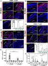
Partial decellularization attenuates CD8+ T‐cell mediated rejection. (a) Representative IF images of CD8+ T‐cells in grafts at 10 days and (b) native trachea (c) CD8+ T‐cells/mm2 (10 days) #### = increased CD8+ T‐cells vs Native (10 days, p < 0.0001) **** = decreased CD8+ T‐cells compared to Allograft (ATG) (10 days, p < 0.0001 for all graft types) * = decreased CD8+ T‐cells compared to Allograft at 10 days (p = 0.0324 STG vs. PDTA, p = 0.0184 STG vs. Allograft). (d) Representative images of CD8+ T‐cells in grafts at 1‐month and (e) 3‐months. (f) CD8+ T‐cells/mm2 at 1‐month and 3‐months # = increased CD8+ T‐cells versus STG (p < 0.0001 for STG‐1 m vs. Allograft‐1 m, p < 0.0001 for STG‐3 m vs. ATG‐3 m), ^ = increased CD8+ T‐cells versus PDTA (p < 0.0001 for PDTA‐1 m vs. Allograft‐1 m, p < 0.0001 for PDTA‐3 m vs. Allograft‐3 m), ~ = increased CD8+ T‐cells versus PDTS (p < 0.0001 for PDTS‐1 m vs. Allograft‐1 m, p < 0.0001 for PDTS‐3 m vs. Allograft‐3 m), **** = decreased CD8+ T‐cells between Allograft at 1‐ and 3‐month (p < 0.0001). (g) Representative Terminal deoxynucleotidyl transferase dUTP nick end labeling assay (TUNEL) images of the implanted grafts. red denotes apoptotic cells while blue denotes cellular nuclei. (h) Representative IF images of CD4+ T‐cells in grafts at 10 days and (i) native trachea. (j) CD4+ T‐cells/mm2 at 10 days # = increased CD4+ T‐cells versus Native at 10 days (p = 0.0242 vs. PDTS, p = 0.0007 vs. PDTA, p = 0.0004 vs. Allograft). (k) CD4+ T‐cells/mm2 at chronic time points * = increase in CD4+ T‐cells between grafts (p = 0.0312 for STG 1 m vs. PDTA 1 m, p = 0.0014 for STG 1 m vs. PDTS 1 m, p < 0.0001 for STG 1 m vs. Allograft 1 m, p = 0.0004 for STG 3 m vs. Allograft 3 m, p = 0.0011 for PDTA 3 m vs. Allograft 3 m, p = 0.0020 for PDTS 3 m vs. Allograft 3 m, p = 0.0066 for STG 1 m vs. STG 3 m, p = 0.0219 for PDTA 1 m vs. PDTA 3 m, p = 0.0226 for PDTS 1 m vs. PDTS 3 m). IF, immunofluorescent; PDTA, partially decellularized tracheal allografts; PDTS, partially decellularized tracheal syngrafts; STG, syngeneic tracheal grafts.

References

    1. Chiang T, Pepper V, Best C, Onwuka E, Breuer CK. Clinical translation of tissue engineered trachea grafts. Ann Otol Rhinol Laryngol. 2016;125(11):873‐885. doi:10.1177/0003489416656646 - DOI - PMC - PubMed
    1. Pearlman AM, Mujumdar V, McAbee KE, Terlecki RP. Outcomes of adult urethroplasty with commercially available acellularmatrix. Ther Adv Urol. 2018;10(11):351‐355. doi:10.1177/1756287218790370 - DOI - PMC - PubMed
    1. Taufique ZM, Bhatt N, Zagzag D, Lebowitz RA, Lieberman SM. Revascularization of AlloDerm used during endoscopic skull base surgery. J Neurol Surg B Skull Base. 2019;80(1):46‐50. doi:10.1055/S-0038-1666851 - DOI - PMC - PubMed
    1. Badria AF, Koutsoukos PG, Mavrilas D. Decellularized tissue‐engineered heart valves calcification: what do animal and clinical studies tell us? J Mater Sci Mater Med. 2020;31(12):132. doi:10.1007/S10856-020-06462-X - DOI - PMC - PubMed
    1. Cheng CW, Solorio LD, Alsberg E. Decellularized tissue and cell‐derived extracellular matrices as scaffolds for orthopaedic tissue engineering. Biotechnol Adv. 2014;32(2):462‐484. doi:10.1016/J.BIOTECHADV.2013.12.012 - DOI - PMC - PubMed