

This is a preprint.
Large-scale annotated dataset for cochlear hair cell detection and classification
- PMID: 37693382
- PMCID: PMC10491224
- DOI: 10.1101/2023.08.30.553559
Large-scale annotated dataset for cochlear hair cell detection and classification
Update in
-
Large-scale annotated dataset for cochlear hair cell detection and classification.Sci Data. 2024 Apr 23;11(1):416. doi: 10.1038/s41597-024-03218-y. Sci Data. 2024. PMID: 38653806 Free PMC article.
Abstract
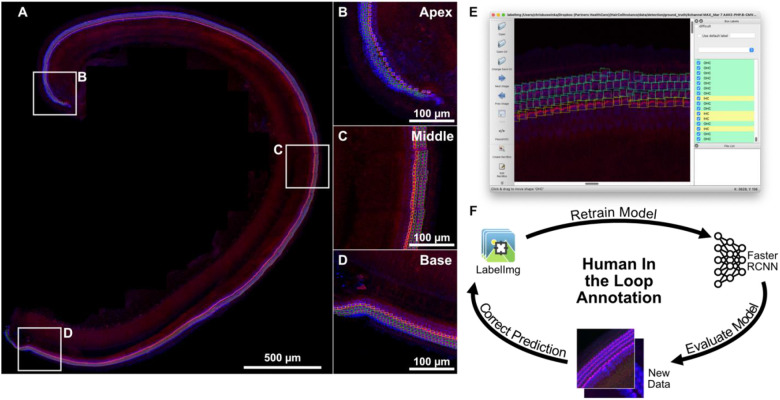
Our sense of hearing is mediated by cochlear hair cells, localized within the sensory epithelium called the organ of Corti. There are two types of hair cells in the cochlea, which are organized in one row of inner hair cells and three rows of outer hair cells. Each cochlea contains a few thousands of hair cells, and their survival is essential for our perception of sound because they are terminally differentiated and do not regenerate after insult. It is often desirable in hearing research to quantify the number of hair cells within cochlear samples, in both pathological conditions, and in response to treatment. However, the sheer number of cells along the cochlea makes manual quantification impractical. Machine learning can be used to overcome this challenge by automating the quantification process but requires a vast and diverse dataset for effective training. In this study, we present a large collection of annotated cochlear hair-cell datasets, labeled with commonly used hair-cell markers and imaged using various fluorescence microscopy techniques. The collection includes samples from mouse, human, pig and guinea pig cochlear tissue, from normal conditions and following in-vivo and in-vitro ototoxic drug application. The dataset includes over 90'000 hair cells, all of which have been manually identified and annotated as one of two cell types: inner hair cells and outer hair cells. This dataset is the result of a collaborative effort from multiple laboratories and has been carefully curated to represent a variety of imaging techniques. With suggested usage parameters and a well-described annotation procedure, this collection can facilitate the development of generalizable cochlear hair cell detection models or serve as a starting point for fine-tuning models for other analysis tasks. By providing this dataset, we aim to supply other groups within the hearing research community with the opportunity to develop their own tools with which to analyze cochlear imaging data more fully, accurately, and with greater ease.
Keywords: annotation; cochlea; detection; hair cells; inner hair cell; machine-learning-ready data; outer hair cell.
Figures
References
Publication types
Grants and funding
- R21 DC018237/DC/NIDCD NIH HHS/United States
- R21 DC020312/DC/NIDCD NIH HHS/United States
- R01 DC016365/DC/NIDCD NIH HHS/United States
- R01 DC014712/DC/NIDCD NIH HHS/United States
- R01 DC021325/DC/NIDCD NIH HHS/United States
- R01 DC020322/DC/NIDCD NIH HHS/United States
- T32 DC000038/DC/NIDCD NIH HHS/United States
- R01 DC020190/DC/NIDCD NIH HHS/United States
- ZIA DC000079/ImNIH/Intramural NIH HHS/United States
- R01 DC017166/DC/NIDCD NIH HHS/United States
- P30 CA014195/CA/NCI NIH HHS/United States
- P50 DC015857/DC/NIDCD NIH HHS/United States
- R01 DC021075/DC/NIDCD NIH HHS/United States
- R01 DC020268/DC/NIDCD NIH HHS/United States
- R01 DC015242/DC/NIDCD NIH HHS/United States
LinkOut - more resources
Full Text Sources