

This is a preprint.
Exploring the Residue-Level Interactions between the R2ab Protein and Polystyrene Nanoparticles
- PMID: 37693402
- PMCID: PMC10491123
- DOI: 10.1101/2023.08.28.554951
Exploring the Residue-Level Interactions between the R2ab Protein and Polystyrene Nanoparticles
Update in
-
Exploring Residue-Level Interactions between the Biofilm-Driving R2ab Protein and Polystyrene Nanoparticles.Langmuir. 2024 Jan 16;40(2):1213-1222. doi: 10.1021/acs.langmuir.3c02609. Epub 2024 Jan 4. Langmuir. 2024. PMID: 38174900 Free PMC article.
Abstract
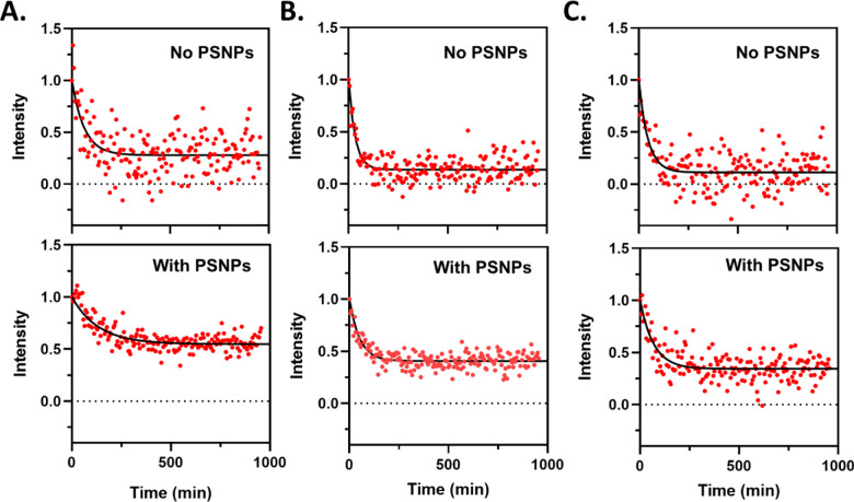
In biological systems, proteins can bind to nanoparticles to form a "corona" of adsorbed molecules. The nanoparticle corona is of high interest because it impacts the organism's response to the nanomaterial. Understanding the corona requires knowledge of protein structure, orientation, and dynamics at the surface. Ultimately, a residue-level mapping of protein behavior on nanoparticle surfaces is needed, but this mapping is difficult to obtain with traditional approaches. Here, we have investigated the interaction between R2ab and polystyrene nanoparticles (PSNPs) at the level of individual residues. R2ab is a bacterial surface protein from Staphylococcus epidermidis and is known to interact strongly with polystyrene, leading to biofilm formation. We have used mass spectrometry after lysine methylation and hydrogen-deuterium exchange (HDX) NMR spectroscopy to understand how the R2ab protein interacts with PSNPs of different sizes. Through lysine methylation, we observe subtle but statistically significant changes in methylation patterns in the presence of PSNPs, indicating altered protein surface accessibility. HDX measurements reveal that certain regions of the R2ab protein undergo faster exchange rates in the presence of PSNPs, suggesting conformational changes upon binding. Both results support a recently proposed "adsorbotope" model, wherein adsorbed proteins consist of unfolded anchor points interspersed with regions of partial structure. Our data also highlight the challenges of characterizing complex protein-nanoparticle interactions using these techniques, such as fast exchange rates. While providing insights into how proteins respond to nanoparticle surfaces, this research emphasizes the need for advanced methods to comprehend these intricate interactions fully at the residue level.
Keywords: adsorbotope; corona; interaction; nanoparticle; protein; structure.
Conflict of interest statement
Conflicts of Interest The authors declare the following competing financial interest(s): J.S.S. discloses a significant interest in GenNext Technologies, Inc., a growth-stage company seeking to commercialize technologies for protein higher-order structure analysis.
Figures


References
-
- Kokkinopoulou M., Simon J., Mailaender V., Lieberwirth I. and Landfester K., in European Microscopy Congress 2016: Proceedings, American Cancer Society, 2016, pp. 71–72.
-
- Deuker M. F. S., Mailänder V., Morsbach S. and Landfester K., Nanoscale Horiz., 2023, 10.1039.D3NH00198A. - PubMed
-
- Walkey C. D., Olsen J. B., Song F., Liu R., Guo H., Olsen D. W. H., Cohen Y., Emili A. and Chan W. C. W., ACS Nano, 2014, 8, 2439–2455. - PubMed
Publication types
Grants and funding
LinkOut - more resources
Full Text Sources