[Preprint]. 2023 Sep 6:2023.08.30.555581.
doi: 10.1101/2023.08.30.555581.

Cryptosporidium infection of human small intestinal epithelial cells induces type III interferon and impairs infectivity of Rotavirus

Affiliations

Cryptosporidium infection of human small intestinal epithelial cells induces type III interferon and impairs infectivity of Rotavirus

Valentin Greigert et al. bioRxiv. .

Update in

Abstract

Cryptosporidiosis is a major cause of severe diarrheal disease in infants from resource poor settings. The majority of infections are caused by the human-specific pathogen C. hominis and absence of in vitro growth platforms has limited our understanding of host-pathogen interactions and development of effective treatments. To address this problem, we developed a stem cell-derived culture system for C. hominis using human enterocytes differentiated under air-liquid interface (ALI) conditions. Human ALI cultures supported robust growth and complete development of C. hominis in vitro including all life cycle stages. C. hominis infection induced a strong interferon response from enterocytes, likely driven by an endogenous dsRNA virus in the parasite. Prior infection with Cryptosporidium induced type III IFN secretion and consequently blunted infection with Rotavirus, including live attenuated vaccine strains. The development of hALI provides a platform for further studies on human-specific pathogens, including clinically important coinfections that may alter vaccine efficacy.

Keywords: antiviral; co-infection; enteric infection; interferon; mucosal immunity; parasitology; vaccine.


Figures

Fig. 1 –
Fig. 1 –. Human ALI system promoted development of a highly complex epithelial tissue composed of differentiated cell types.
(A) Human air-liquid interface (hALI) was generated by seeding intestinal stem cells on a confluent layer of irradiated human intestinal myofibroblasts platted on Matrigel-coated transwells. Cells were cultured in 50% conditioned media (CM) complemented with Y-27632 (10 μM) and an inhibitor of the TGF-β pathway SB431542 (SBi, 10 μM). After 72 hr of growth, SB431542 was removed from the growth media. Seven days after seeding, medium from the top chamber was removed to generate ALI cultures, and SB431542 added back to culture medium when the purpose of the culture was parasite growth. Cells were used for infection 3 to 4 days after top medium removal. (B) Hematoxylin-eosin staining of sections of hALI showing the thickening and differentiation of the monolayer over time after removal of medium in the top chamber. Enterocytes became more columnar and goblet cells more abundant and mucus-producing over time. (C, D) Immunostaining of flat mounted (C) or transversal sections (D) 14 days after top medium removal revealed an uniform brush-border stained by anti-villin antibodies, disrupted by goblet cells secreting mucus stained with anti-Muc2 antibodies, and the presence of both enteroendocrine cells (anti-chromogranin A antibodies) and Paneth cells (anti-lysozyme antibodies). B-catenin antibodies delineate cells, similar to E-cadherin (adehrens junctions). ZO-1 (tight junctions) is expressed at the apical end of cellular junctions, on the lumen side (arrowheads). Scale bars = 50 μm.
Fig. 2 –
Fig. 2 –. Human ALI system supported Cryptosporidium hominis culture with complete life cycle development.
(A) Growth of C. hominis in hALI cultures over a 7-day period. Mean ± S.E. N = 3, n = 3 (B) Immunostaining of various stages of C. hominis cultured on hALI using a panel of previously described mAb to C. parvum (green) and a pan-Cryptosporidium polyclonal antibody (red)(12). Sporozoites and trophozoites were stained 6 hr after infection (pi), meronts 24 hr pi and microgamonts and macrogamonts 72 hr pi. (C) EdU was used in combination with Crypt-a-Glo (CaG) to differentiate newly-formed oocysts from carryover oocysts. (D) Immunostaining of newly-formed oocysts following infection of hALI with C. hominis oocysts, 72 hr pi. Scale bar = 3 μm
Fig. 3 –
Fig. 3 –. The permeability of human ALI did not increase after infection, as opposed to HCT-8 cell monolayers.
(A) Trans-epithelial electrical resistance (TEER) and Dextran flux measured across hALI of HCT-8 monolayers infected with C. parvum or C. hominis. hALI was infected 3 days after top medium removal. Mean ± S.E. N = 2, n = 4. Kruskal-Wallis test followed by Dunn’s multiple comparison test. * P < 0.05, **** P < 0.0001. (B) Immunostaining of flat mounted hALI transwell culture showed tight junction (TJ; ZO-1, green) and adherens junction (AJ; E-cadherin, green) organization, with or without infection with C. parvum (Vicia villosa lectin, VVL, red). Scale bar = 50 μm. (C) Intercellular junction organization quantification (IJOQ) of hALI and HCT-8 cells infected with C. parvum vs. not infected, for AJ (E-cadherin) and TJ (ZO-1). hALI was infected 3 days after top medium removal. One representative experiment for each cell type is showed. Mean ± S.E. N = 2, n = 6. Mann-Whitney U test. ** P < 0.01, ns = non significant.
Fig. 4 –
Fig. 4 –. Infection of human ALI with Cryptosporidium induced the expression of genes involved in inflammation, including type I, II and III interferon pathways.
(A) hALI was infected with either C. parvum or C. hominis, with a mock infection as control, for a total of 72 hr. Cells were harvested for RNA-Seq after 24 hr of infection. Bottom chamber media was harvested for cytokine quantification using a Luminex bead assay after 4 hr, 24 hr and 72 hr of infection. (B) Growth of C. hominis and C. parvum in hALI cultures used for this experiment over a 3-day period. Mean ± S.E. N = 3, n = 1. (C) Volcano plot of differentially expressed genes in hALI 24 hr after infection with C. parvum. Genes up- or down-regulated by more than 2-fold and P < 0.05 are shown in red. (D) Scatter-plot showing genes upregulated following C. hominis infection vs. C. parvum infection. (E) Pathway analysis of upregulated genes following C. hominis or C. parvum infection (MsigDB Hallmark 2020). (F) GSEA analysis showing upregulated (up) and down-regulated (down) pathways in hALI infected with C. parvum. (G) Network plot showing the involvement of highly expressed genes following C. parvum infection in several upregulated pathways (WikiPathway).
Fig. 5 –
Fig. 5 –. Infection of human ALI with C. parvum induced the production of inflammatory cytokines, including type I and III interferons.
(A, B) Induction of chemokines and cytokines following infection with C. parvum or C. hominis based on Luminex bead assay. Mean ± S.E. N = 3, n = 1. Kruskal-Wallis test followed by Dunn’s multiple comparison test. * P < 0.05. (C) CSpV1 load in each batch of Cryptosporidium oocysts was measured by RT-qPCR. Each strain was tested thrice, on 3 different batches (i.e. 3 different oocyst-producing calves for C. parvum, and 3 different oocyst-producing gnotobiotic piglets for C. hominis).
Fig. 6 –
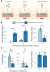
Fig. 6 –. Pre-infection of human ALI with C. parvum inhibited Rotavirus growth.
(A) hALI was infected with C. parvum for 24 hr prior infection with Rotavirus (RV). Cells were harvested 2 hr and 24 hr after infection with RV for virus quantification by RT-qPCR. H0, H24, H48: time since C. parvum infection (hr) (B) (left) IFN-λ3 gene expression as measured by RT-qPCR after hALI infection with C. parvum + RV vs. mock + RV infection. Mean ± S.E. N = 2, n = 3. One-way ANOVA followed with Tukey’s HSD test. (right) RV growth in hALI pre-infected with C. parvum as compared to uninfected hALI. N = 2, n = 3. Mann-Whitney U test. ** P < 0.01. (C) (left) RV vaccine strains growth inhibition in hALI pre-infected with C. parvum as compared to uninfected hALI. Mean ± S.E. N = 4, n = 3. After detection and removal of outliers with the 1.5xIQR rule, Mann-Whitney U test. * P < 0.05, **** P < 0.0001. (right) Percentage of inhibition of RV vaccine strains growth over 4 independent experiments. Mean ± S.E. Mann-Whitney U test. ns = non significant.

References

    1. Feng Y, Ryan UM, Xiao L. Genetic Diversity and Population Structure of Cryptosporidium. Trends Parasitol. 2018;34(11):997–1011. - PubMed
    1. Kotloff KL, et al. Burden and aetiology of diarrhoeal disease in infants and young children in developing countries (the Global Enteric Multicenter Study, GEMS): a prospective, case-control study. The Lancet. 2013;382(9888):209–222. - PubMed
    1. Kotloff KL. The Burden and Etiology of Diarrheal Illness in Developing Countries. Pediatr Clin North Am. 2017;64(4):799–814. - PubMed
    1. Checkley W, et al. A review of the global burden, novel diagnostics, therapeutics, and vaccine targets for Cryptosporidium. The Lancet Infectious Diseases. 2015;15(1):85–94. - PMC - PubMed
    1. Theodos CM, et al. Profiles of healing and nonhealing Cryptosporidium parvum infection in C57BL/6 mice with functional B and T lymphocytes: the extent of gamma interferon modulation determines the outcome of infection. Infect Immun. 1997;65(11):4761–4769. - PMC - PubMed

Publication types