[Preprint]. 2023 Aug 29:2023.08.28.555150.
doi: 10.1101/2023.08.28.555150.

Preliminary cross-sectional investigations into the human glymphatic system using multiple novel non-contrast MRI methods

Affiliations

Preliminary cross-sectional investigations into the human glymphatic system using multiple novel non-contrast MRI methods

Swati Rane Levendovszky et al. bioRxiv. .

Update in

Abstract

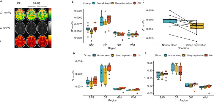
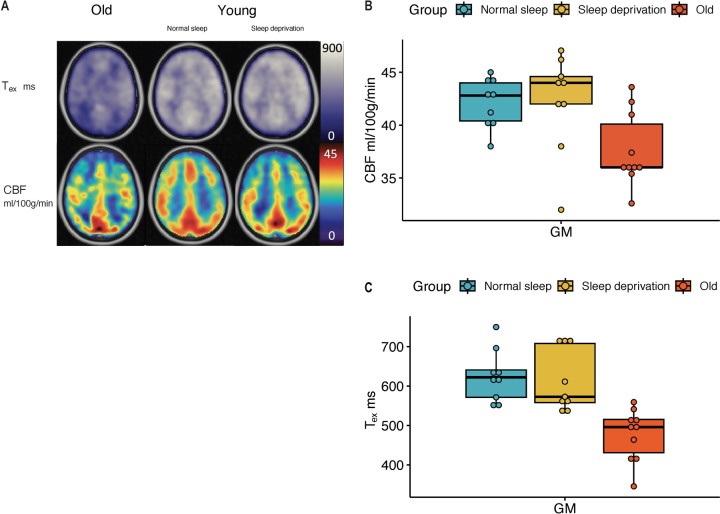
We discuss two potential non-invasive MRI methods to cross-sectionally study two distinct facets of the glymphatic system and its association with sleep and aging. We apply diffusion-based intravoxel incoherent motion (IVIM) imaging to evaluate pseudodiffusion coefficient, D*, or cerebrospinal fluid (CSF) movement across large spaces like the subarachnoid space (SAS). We also performed perfusion-based multi-echo, Hadamard encoded multi-delay arterial spin labeling (ASL) to evaluate whole brain cortical cerebral blood flow (CBF) and transendothelial exchange (Tex) of water from the vasculature into the perivascular space and parenchyma. Both methods were used in young adults (N=9, 6F, 23±3 years old) in the setting of sleep and sleep deprivation. To study aging, 10 older adults, (6F, 67±3 years old) were imaged after a night of normal sleep only and compared with the young adults. D* in SAS was significantly (p<0.05) lesser after sleep deprivation (0.014±0.001 mm2/s) than after normal sleep (0.016±0.001 mm2/s), but was unchanged with aging. Cortical CBF and Tex on the other hand, were unchanged after sleep deprivation but were significantly lower in older adults (37±3 ml/100g/min, 476±66 ms) than young adults (42±2 ml/100g/min, 624±66 ms). IVIM was thus, sensitive to sleep physiology and multi-echo, multi-delay ASL was sensitive to aging.

Keywords: ASL; IVIM; aging; glymphatics; sleep.


Conflict of interest statement

Conflicts of Interest: None.

Figures

Figure 1:
Figure 1:
A. IVIM parameters, D*, D, and f in older adults (first column), in young adults with normal sleep (middle column), and the same young adults with sleep deprivation (third column) in the SAS. B. D* values in all three groups: young adults after a night of normal sleep (blue), young adults after 24 hours of sleep deprivation (yellow), and older adults after normal sleep (red). C. Only D* was significantly different (p=0.009) after 24 hours of sleep deprivation compared to a night of normal sleep. D, E. D, and f values in all three groups: young adults after a night of normal sleep (blue), young adults after 24 hours of sleep deprivation (yellow), and older adults after normal sleep (red). No difference was observed with aging. Supplementary figure 1 shows the ROIs generated for this study.
Figure 2:
Figure 2:
A. ASL parameters, Whole-brain cortical CBF and Tex in older adults (first column), in young adults with normal sleep (middle column), and the same young adults with sleep deprivation (third column). CBF was not significantly different in young adults after sleep deprivation (yellow), when compared to those after a night of normal sleep (blue). Detailed paired comparison is shown in B. CBF was significantly different in older adults (red) compared to young adults (p=0.005). C. Tex was also not significantly different in young adults after sleep deprivation (yellow), when compared to those after a night of normal sleep (blue). Like CBF, Tex was significantly shorter in older adults (red) compared to young adults (p=0.0001).

References

    1. Iliff JJ, Lee H, Yu M, et al. Brain-wide pathway for waste clearance captured by contrast-enhanced MRI. J Clin Invest. 2013;123(3):1299–1309. doi: 10.1172/JCI67677 - DOI - PMC - PubMed
    1. Iliff JJ, Wang M, Liao Y, et al. A Paravascular Pathway Facilitates CSF Flow Through the Brain Parenchyma and the Clearance of Interstitial Solutes, Including Amyloid β. Sci Transl Med. 2012;4(147):147ra111. doi: 10.1126/scitranslmed.3003748 - DOI - PMC - PubMed
    1. Xie L, Kang H, Xu Q, et al. Sleep Drives Metabolite Clearance from the Adult Brain. Science. 2013;342(6156):373–377. doi: 10.1126/science.1241224 - DOI - PMC - PubMed
    1. Simon M, Wang MX, Ismail O, et al. Loss of perivascular aquaporin-4 localization impairs glymphatic exchange and promotes amyloid β plaque formation in mice. Alzheimers Res Ther. 2022;14(1):59. doi: 10.1186/s13195-022-00999-5 - DOI - PMC - PubMed
    1. Iliff JJ, Chen MJ, Plog BA, et al. Impairment of glymphatic pathway function promotes tau pathology after traumatic brain injury. J Neurosci. 2014;34(49):16180–16193. doi: 10.1523/JNEUROSCI.3020-14.2014 - DOI - PMC - PubMed

Publication types