[Preprint]. 2023 Sep 1:2023.09.01.555937.
doi: 10.1101/2023.09.01.555937.

Lipid scrambling is a general feature of protein insertases

Affiliations

Lipid scrambling is a general feature of protein insertases

Dazhi Li et al. bioRxiv. .

Update in

Abstract

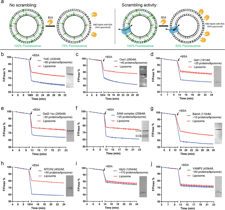
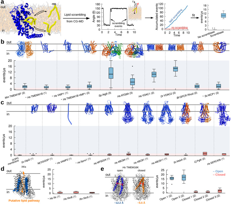
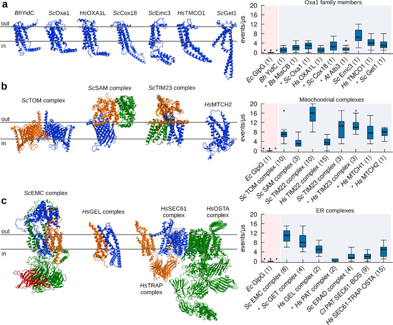
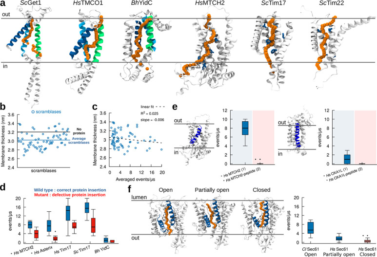
Glycerophospholipids are synthesized primarily in the cytosolic leaflet of the endoplasmic reticulum (ER) membrane and must be equilibrated between bilayer leaflets to allow the ER and membranes derived from it to grow. Lipid equilibration is facilitated by integral membrane proteins called "scramblases". These proteins feature a hydrophilic groove allowing the polar heads of lipids to traverse the hydrophobic membrane interior, similar to a credit-card moving through a reader. Nevertheless, despite their fundamental role in membrane expansion and dynamics, the identity of most scramblases has remained elusive. Here, combining biochemical reconstitution and molecular dynamics simulations, we show that lipid scrambling is a general feature of protein insertases, integral membrane proteins which insert polypeptide chains into membranes of the ER and organelles disconnected from vesicle trafficking. Our data indicate that lipid scrambling occurs in the same hydrophilic channel through which protein insertion takes place, and that scrambling is abolished in the presence of nascent polypeptide chains. We propose that protein insertases could have a so-far overlooked role in membrane dynamics as scramblases.


Conflict of interest statement

Declaration of Interest: The authors declare no competing interests.

Figures

Figure 1.
Figure 1.. Multiple protein insertases have lipid scrambling activity in vitro.
(a) Schematic of the BSA back extraction assay. (b-e) Members of the Oxa1 superfamily (YidC, Oxa1, Get1, and the Get1/2 complex) can scramble glycerophospholipids. (f-g) The β-barrel membrane protein insertase, Sam50 in complex with Sam35 and Sam37, and the bacterial ortholog of Sam50, BamA, have scrambling activity. (h) The outer mitochondrial membrane insertase MTCH2 scrambles. (i-j) Negative controls, GlpG and VAMP2, do not scramble. Proteoliposomes used in the assays were analyzed by SDS-PAGE (insets) to confirm efficient reconstitution; approximate numbers for proteins/liposome were estimated assuming 50% recovery of lipids after reconstitution. (See Methods for exact liposome compositions, details of which varied according to experimentalist.)
Figure 2.
Figure 2.. CG simulations recapitulate known activity of lipid scramblases.
a. Protocol used to quantify lipid scrambling in CG-MD simulations. b. CG-MD simulations reproduce lipid scrambling activity by known lipid scramblases of different structure and oligomerization state. c. CG-MD simulations correctly reproduce lack of lipid scrambling activity by proteins that do not have scrambling activity in vitro. d,e. CG-MD simulations recapitulate conformational-dependent lipid scrambling activity by proteins from the XK (d) and TMEM16K (e) families. AlphaFold structures are denoted by the * symbol, oligomerization state is in parenthesis. Light blue and light red shadings indicate scrambling vs non-scrambling activity, respectively. The cut-off used was 1 events/μs.
Figure 3.
Figure 3.. CG-MD simulations identify protein insertase complexes as lipid scramblases.
a. All members of the Oxa1 family of insertases have in silico lipid scrambling activity in their monomeric form. Left: 3D structure of selected members of the Oxa1 family. Right: In silico lipid scrambling quantification. The negative control EcGlpG is shown as reference. b. Mitochondrial insertase complexes have in silico lipid scrambling activity. Left: 3D structure of selected mitochondrial insertase complexes. Right: In silico lipid scrambling quantification. c. ER insertase complexes have in silico lipid scrambling activity. Left: 3D structure of selected ER insertase complexes. Right: In silico lipid scrambling quantification. AlphaFold structures are denoted by the * symbol, number of proteins in the complex is in parenthesis.
Figure 4.
Figure 4.. Lipid scrambling takes place via the same mechanistic pathway as in protein insertion.
A. In silico lipid scrambling pathway (orange) in selected protein insertases. The position of the lipid polar head at different times along the scrambling pathway is depicted with orange spheres. Regions involved in protein insertion are shown in green, blue and cyan for Oxa1 family proteins, while residues involved in protein insertion are shown in blue for HsMTCH2, ScTim17 and ScTim22. B. Protein scramblases induce limited (0.2 nm on average) membrane thinning. C. Membrane thickness has minimal correlation with lipid scrambling activity in silico. D. Mutants proposed to decrease protein insertion activity also reduce lipid scrambling. E. The presence of a nascent polypeptide inside the protein hydrophilic cavity abolishes lipid scrambling. Left: HsMCTH2. Right: HsOXA1L. f. Different conformations of Sec61 (lateral gate open, partially open and closed) have different lipid scrambling activity. AlphaFold structures are denoted by the * symbol. Number of proteins in the system is in parenthesis.

References

    1. Abraham M.J., Murtola T., Schulz R., Páll S., Smith J.C., Hess B., Lindahl E., 2015. GROMACS: High performance molecular simulations through multi-level parallelism from laptops to supercomputers. SoftwareX 1–2, 19–25. 10.1016/j.softx.2015.06.001 - DOI
    1. Adlakha J., Hong Z., Li P., Reinisch K.M., 2022. Structural and biochemical insights into lipid transport by VPS13 proteins. J. Cell Biol. 221, e202202030. 10.1083/jcb.202202030 - DOI - PMC - PubMed
    1. Anghel S.A., McGilvray P.T., Hegde R.S., Keenan R.J., 2017. Identification of Oxa1 Homologs Operating in the Eukaryotic Endoplasmic Reticulum. Cell Rep. 21, 3708–3716. 10.1016/j.celrep.2017.12.006 - DOI - PMC - PubMed
    1. Antonicka H., Lin Z.-Y., Janer A., Aaltonen M.J., Weraarpachai W., Gingras A.-C., Shoubridge E.A., 2020. A High-Density Human Mitochondrial Proximity Interaction Network. Cell Metab. 32, 479–497.e9. 10.1016/j.cmet.2020.07.017 - DOI - PubMed
    1. Bai L., You Q., Feng X., Kovach A., Li H., 2020. Structure of the ER membrane complex, a transmembrane-domain insertase. Nature 584, 475–478. 10.1038/s41586-020-2389-3 - DOI - PMC - PubMed

Publication types