Investigation of Morphometric Factors Associated With Adolescent ACL Rupture
- PMID: 37693804
- PMCID: PMC10492494
- DOI: 10.1177/23259671231194928
Investigation of Morphometric Factors Associated With Adolescent ACL Rupture
Abstract
Background: There are no definitive anatomic morphometric risk factors for adolescent anterior cruciate ligament (ACL) injury.
Purpose: To compare the parameters used to define the tibial and femoral morphometric structure of the knee between adolescent patients with and without ACL rupture.
Study design: Cross-sectional study; Level of evidence, 3.
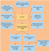
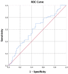
Methods: Included were magnetic resonance imaging (MRI) scans and radiographs of 115 patients aged 10 to 17 years who were evaluated for ACL rupture at a single institution between February 1, 2019, and January 31, 2022. Images from 115 patients with intact MRI scans were included as controls. We investigated the following imaging parameters: tibial slope (on lateral radiograph), lateral condylar height, tibial sulcus height, medial condylar height, condylar width, intercondylar notch with, intercondylar notch angle, notch index, eminence width, tibial plateau width, eminence width/tibial plateau width, medial/lateral/overall eminence height, medial plateau depth, and 2 different eminence angles. Parameters were compared between groups using the chi-square, Fisher exact, Student t, or Mann-Whitney U test, as appropriate. Receiver operating characteristic analysis was conducted for cutoff values of significant parameters.
Results: There were no significant differences in age, sex, or side affected between groups. Only the medial plateau depth was found to be statistically significant between the ACL rupture and ACL intact groups (2.6 vs 2.2 mm; P = .015). A statistically significant cutoff value could not be obtained for the medial plateau depth.
Conclusion: Medial plateau depth was found to be significantly greater in adolescent patients with ACL rupture compared with ACL-intact controls.
Keywords: ACL rupture; adolescents; anterior cruciate ligament; morphometric study.
© The Author(s) 2023.
Conflict of interest statement
The authors have declared that there are no conflicts of interest in the authorship and publication of this contribution. AOSSM checks author disclosures against the Open Payments Database (OPD). AOSSM has not conducted an independent investigation on the OPD and disclaims any liability or responsibility relating thereto.
Figures


References
-
- Ahmad R, Patel A, Mandalia V, Toms A. Posterior tibial slope: effect on, and interaction with, knee kinematics. JBJS Rev. 2016;4(4):e31-e36. - PubMed
-
- Alentorn-Geli E, Pelfort X, Mingo F, et al.. An evaluation of the association between radiographic intercondylar notch narrowing and anterior cruciate ligament injury in men: the notch angle is a better parameter than notch width. Arthroscopy. 2015;31(10):2004-2013. - PubMed
-
- Al-Saeed O, Brown M, Athyal R, Sheikh M. Association of femoral intercondylar notch morphology, width index and the risk of anterior cruciate ligament injury. Knee Surg Sports Traumatol Arthrosc. 2013;21(3):678-682. - PubMed
-
- Bernhardson AS, Aman ZS, Dornan GJ, et al.. Tibial slope and its effect on force in anterior cruciate ligament grafts: anterior cruciate ligament force increases linearly as posterior tibial slope increases. Am J Sports Med. 2019;47(2):296-302. - PubMed
LinkOut - more resources
Full Text Sources

