Review
. 2023 Aug 31;27(1):2677.
doi: 10.4102/sajr.v27i1.2677. eCollection 2023.

Imaging evaluation of the benign and malignant lesions of the floor of the mouth: Pictorial review

Affiliations
Review

Imaging evaluation of the benign and malignant lesions of the floor of the mouth: Pictorial review

Ashim K Lahiri et al. SA J Radiol. .

Abstract

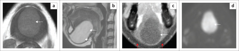
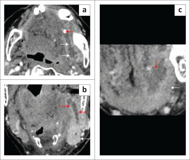
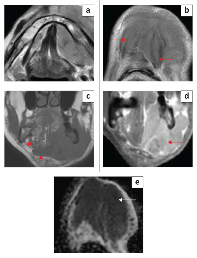
The floor of the mouth is an important anatomical region of the oral cavity where primary benign and malignant disease processes can originate or secondary pathologies can extend into adjacent spaces. Knowledge of the anatomy is crucial for accurate localisation of pathology and understanding the spread of disease. The sublingual space is the dominant component of the floor of the mouth, bounded inferiorly by the mylohyoid muscle that separates it from the submandibular space. Imaging is immensely important to characterise and map the extent of disease, considering the fact that the bulk of the disease may be submucosal and not visible on clinical inspection.

Contribution: The floor of the mouth is a complex anatomical region for radiological evaluation. The purpose of this pictorial review is to present an understanding of the relevant anatomy and to demonstrate the role and appropriate application of different imaging modalities. This article highlights the imaging spectrum of a wide range of various benign conditions including normal variants and a variety of malignant lesions at different tumour stages, with an aim to establish the correct diagnosis, avoid misinterpretation and help in treatment planning.

Keywords: CT; MRI; computed tomography; floor of mouth; mylohyoid; sublingual space.


Conflict of interest statement

The authors declare that they have no financial or personal relationships that may have inappropriately influenced them in writing this article.

Figures

FIGURE 1
FIGURE 1
MRI anatomy of the floor of the mouth on T1 weighted (T1W) images indicating the muscles of the floor of the mouth and the extrinsic muscles of the tongue. Inferior axial (a), mid axial (b), superior axial (c) and coronal (d).
FIGURE 2
FIGURE 2
Anatomy of the sublingual space. T2 fat-suppressed coronal (a), T2 axial (b), T1 contrast-enhanced fat-suppressed (c) and T1 coronal without fat-suppression (d) show the sublingual glands (red arrows), mylohyoid muscle (d, white arrow), geniohyoid (b, white arrow). T2 axial (e) demonstrates the deep part of submandibular glands (white arrows) in floor of the mouth (FOM), posterior to the mylohyoid muscles and sublingual glands (red arrows). CT coronal (f) shows the mixed fat density of sublingual glands (red arrows) between the genioglossus (white arrow) and mylohyoid muscles (black arrows).
FIGURE 3
FIGURE 3
Mylohyoid Boutonniere in a 60-year-old patient who presented with intermittent swelling of the right submandibular region. T1 axial (a), T2 axial (b), T1 coronal (c) and T2-short tau inversion recovery (STIR) coronal (d) demonstrate a deficient right mylohyoid muscle (red arrow) anteriorly and an enlarged right sublingual gland herniating inferiorly through the mylohyoid muscle defect (white arrow).
FIGURE 4
FIGURE 4
Mylohyoid Boutonniere in an adult patient who presented with a 2-year history of a fluctuating right submandibular neck lump. Ultrasound (a, b) demonstrate the enlarged inferiorly herniated right sublingual gland (white arrow) through the mylohyoid defect (red arrow). Subsequent MRI for ongoing concerns confirms the findings with a mylohyoid defect (red arrow) and herniated sublingual gland (white arrow) on the T2W axial image (c) and T1 contrast-enhanced fat-suppressed coronal image (d).
FIGURE 5
FIGURE 5
A case of a ranula in a 9-year-old child with a painless submandibular swelling. Ultrasound (a, b) showed a sonolucent cystic mass (4 cm × 2 cm; red arrows) with homogeneous low-level internal echoes, located medial to the submandibular gland (white arrow) and dissecting into the floor of the mouth. A different case of a ranula in an adult patient. Ultrasound (c) demonstrated a cystic mass, with internal debris and septations, in the left anterior submandibular region. MRI indicated the ranula (d, red arrow) on the T2W axial (d) and confirms extension to the sublingual space (e, white arrow) and communicating channel (e, red arrow).
FIGURE 6
FIGURE 6
A ranula in an 85-year-old patient who presented with a painless swelling in the left floor of the mouth. MRI neck T2-weighted (T2W) axial (a) T2-weighted short-tau inversion recovery (T2W-STIR) posterior coronal image (b) reveal the left floor of the mouth simple cystic mass dissecting through the left mylohyoid muscle (red arrow) and indenting the left geniohyoid muscle (a, white arrow). The anterior coronal T2W short-tau inversion recovery (STIR) image (c) shows the cyst limited by the anterior belly of digastric muscle inferiorly (white arrow). No enhancement of the cyst wall or intracystic enhancing component on the contrast-enhanced coronal T1 fat-suppressed image (d).
FIGURE 7
FIGURE 7
A plunging (diving) ranula in an adult patient with a history of a fluctuant soft swelling in the left submandibular region. Initial ultrasound (not included) revealed a partly imaged cystic lesion. T2 axial MRI neck (a) indicates a cystic lesion in the left submandibular region, anterior to the left submandibular gland, extending from anterior floor of the mouth. The T2-weighted short tau inversion recovery (T2W-STIR) coronal anterior image (b) shows the anterior extent of the cyst in the left sublingual space (red arrow) above the anterior belly of digastric muscle (b, white arrow). The mid part of lesion (c) shows the cyst is dissecting inferiorly, deep to mylohyoid muscle (red arrow), suggesting a tail-sign. The T2-weighted short-tau inversion recovery (T2W-STIR) coronal image (d) shows small non-cystic debris (red arrow); there was no evidence of a vascular malformation.
FIGURE 8
FIGURE 8
Epidermoid cyst in an adult female patient. T1 axial MRI neck with contrast (a) shows a large well defined, 5 cm, simple, non-enhancing cystic mass at the floor of mouth (a, white arrow) with intermediate homogeneous signal, splaying the anterior belly of the digastric muscles. T2 sagittal (b) shows homogeneous hyperintensity (white arrows). The contrast-enhanced T1 fat-saturated coronal image (c) shows a non-enhancing cyst (white arrow), limited inferiorly by an intact mylohyoid sling (red arrows). There is a marked restriction of diffusion on the high b-value diffusion-weighted image (d).
FIGURE 9
FIGURE 9
(a, c) Obstructing submandibular duct calculus with adenitis and a floor of the mouth (FOM) abscess. A 98-year-old female patient presented with a large, tense swelling in the left FOM and submandibular region, a swollen tongue and difficult breathing. Contrast-enhanced axial CT neck (a) demonstrated a calcified obstructing calculus at the distal end of the left submandibular duct (red arrow) and a markedly dilated duct (white arrows). The left submandibular gland was enlarged and markedly enhancing because of acute adenitis (b, white arrow and dilated duct, red arrows). There is a left anterior FOM collection (c, red arrow), deep to the left mylohyoid muscle (c, white arrow).
FIGURE 10
FIGURE 10
A case of Ludwig’s Angina in an adult patient who presented with acute stridor, difficulty swallowing and marked swelling in the left upper neck. The patient was intubated. CT neck with contrast was partly degraded by artefacts from dental amalgam. There was extensive soft tissue thickening of the floor of the mouth (FOM) (a, b) with the loss of the normal anatomical boundaries at the FOM and other neck spaces, marked narrowing of airspaces (b, white arrow) and evidence of intubation (a, white arrow). There was an irregular left parapharyngeal abscess (c, red arrows), which was drained. The sagittal image (d) shows the extension of cellulitis into the anterior mediastinum (red arrows).
FIGURE 11
FIGURE 11
A 69-year-old male patient presented with a painful swelling in the right sublingual space, difficulty in swallowing and pain in the submandibular region. Contrast-enhanced CT (a to d) demonstrates an irregular rim-enhancing abscess (red arrows). The axial image (b) shows mild borderline enlargement with increased enhancement of the right submandibular gland (white arrow). The abscess is located above the mylohyoid muscle (c, white arrows).
FIGURE 12
FIGURE 12
Stage T4 squamous cell carcinoma of the mandible in an adult patient. There is a large bulky mass lesion involving the left mandibular body, with full-thickness osseous destruction, and the lateral floor of the mouth, with invasion of the left mylohyoid and hyoglossus muscles (red arrows on the left and normal muscles on the right (white arrows) in (a, b; T2-axial views) and on the coronal T1 contrast-enhanced fat-suppressed image (c). There is extensive infiltration of the left masseter muscle (c) and retromolar trigone (b, red arrows). CT mandible (d) shows full-thickness cortical osseous destruction on the left.
FIGURE 13
FIGURE 13
(a, b) Locally advanced squamous cell carcinoma, seen as a right submandibular gland-based aggressive mass, invading the floor of the mouth. Axial T2 (a) shows the large tumour (white arrows) and invasion of right sublingual gland (red arrow). Axial T2 (b) demonstrates invasion of the entire anterior belly of right digastric muscle (red arrow). The T1 contrast-enhanced fat-saturated coronal image (c) shows the heterogeneously enhancing mass with necrotic areas. There is focal invasion of bony mandible (red arrow) and inferior attachment of right masseter muscle (white arrow).
FIGURE 14
FIGURE 14
Left anterior floor of the mouth bulky small cell carcinoma (stage T4) in an elderly patient, invading the extrinsic tongue muscles. T2-weighted short-tau inversion recovery (T2W-STIR) coronal (a) and sagittal T2 (b) demonstrate the mildly hyperintense, bulky tumour. The T1 contrast-enhanced fat-suppressed image (c) reveals an ill-defined, inhomogeneously enhancing tumour.
FIGURE 15
FIGURE 15
Stage T4 anterior floor of the mouth (FOM) locally advanced squamous cell carcinoma in a 58-year-old male patient who presented with recurrent swelling in the right submandibular region and fullness in the FOM. Axial T2 (a) shows the dilated right submandibular duct (red arrow) and the right FOM tumour obstructing the duct (white arrow). The coronal short tau inversion recovery (STIR) (b) shows the oedematous right submandibular gland (white arrow) and dilated duct (red arrow). The coronal contrast-enhanced fat-suppressed image (c) demonstrates the inhomogeneously enhancing tumour invading the extrinsic tongue muscles and right sublingual gland (red arrow).
FIGURE 16
FIGURE 16
Stage T2 squamous cell carcinoma of the right floor of the mouth with enlargement of the right sublingual gland, crossing the midline and obstructing the left submandibular duct in a 68-year-old male. The T1 post-contrast fat-suppressed axial image (a) shows the tumour crossing the midline (red arrows). The T1 post-contrast fat-suppressed coronal image (b) demonstrates the less enhancing tumour component (white arrow) in contrast to the normal sublingual gland and the dilated left submandibular gland duct (red arrow). The T2-weighted short tau inversion recovery (T2W-STIR) coronal image (c) shows the tumour involving the right sublingual gland (white arrows) and a dilated distal left submandibular gland duct (red arrow). The T2-STIR coronal image (d) shows the enlarged left submandibular gland with a dilated ductal system (red arrow).
FIGURE 17
FIGURE 17
Mucoepidermoid carcinoma of the salivary gland in the left anterior floor of mouth/sublingual space, in an adult patient. A tumour is obstructing the opening of left submandibular duct. T2-weighted short tau inversion recovery (T2W-STIR) axial image (a) shows the slightly hyperintense tumour projecting into left submandibular gland duct opening (red arrow). The T2 axial (b, red arrows) shows the dilated left submandibular gland duct. The T1 contrast-enhanced fat-suppressed coronal image (c) demonstrates the enhancing tumour (red arrows).
FIGURE 18
FIGURE 18
An 89-year-old male patient with aggressive diffuse large B-cell lymphoma. MRI neck; T2 axial (a, b), unenhanced T1 coronal (c) and post-contrast T1 fat sat coronal (d) demonstrate a large bulky soft tissue mass with full-thickness marrow infiltration and cortical destruction of the left mandibular body. The mass is involving the left floor of the mouth (FOM) and sublingual space with infiltration of the left muscles of the FOM, submental region and left submandibular gland. There is a marked restriction of diffusion on the apparent diffusion coefficient (ADC) map (e). T1 coronal (c) shows normal right mylohyoid and anterior belly of digastric muscles (red arrows).
FIGURE 19
FIGURE 19
An elderly patient with tumour recurrence within the graft in the left floor of the mouth (FOM). Axial T2 images (a, b) show the homogeneous signal of the graft (a, red arrows) and recurrence within the graft with a loss of the bright signal (b, red arrows).
FIGURE 20
FIGURE 20
An elderly patient with a malignant tumour in the right FOM and submandibular region as seen on the T1 coronal post contrast (a). The lesion is markedly avid on 18F-fluorodeoxyglucose positron emission tomography/computed tomography (18F-FDG PET/CT) (b). The T2 axial (c) shows the post-operative appearance after right mandibular resection and graft repair (bright signal of graft fat; red arrows). The contrast-enhanced fat-suppressed T1 weighted (T1W) coronal image (d) shows complete suppression of fat signal (red arrows) and mild oedema at the surgical site.
FIGURE 21
FIGURE 21
False-positive uptake on positron emission tomography (PET) imaging. An elderly female patient who underwent resection of an oral tongue squamous cell carcinoma and neck dissection, followed by radiotherapy. The baseline post-treatment T2 axial images (a, b) show marked oedema in the right upper neck and floor of the mouth (FOM) region with an absent right submandibular gland and oedematous left submandibular gland. A 18F-fluorodeoxyglucose positron emission tomography/computed tomography (18F-FDG PET/CT) (c) performed after 6 months for a synchronous sinister lung lesion showed non-significant uptake at the FOM. A follow-up 18F-FDG PET/CT (d) performed for a malignant lung lesion after 1 year showed significant uptake at the right FOM; however, the MRI (e, post-contrast fat-suppressed axial) and clinical examination did not reveal any mass lesion at the FOM. Persistent PET uptake after another year (f) and normal FOM on coronal contrast-enhanced CT neck (g).

References

    1. Law CP, Chandra RV, Hoang JK, Phal PM. Imaging the oral cavity: Key concepts for the radiologist. Br J Radiol. 2011;84(1006):944–957. 10.1259/bjr/70520972 - DOI - PMC - PubMed
    1. Patel S, Bhatt AA. Imaging of the sublingual and submandibular spaces. Insights Imaging. 2018;9(3):391–401. 10.1007/s13244-018-0615-4 - DOI - PMC - PubMed
    1. Agarwal AK, Kanekar SG. Submandibular and sublingual spaces: diagnostic imaging and evaluation. Otolaryngol Clin North Am. 2012;45(6):1311–1323. 10.1016/j.otc.2012.08.005 - DOI - PubMed
    1. Varghese J, Kirsch C. Magnetic resonance imaging of the oral cavity and oropharynx. Top Magn Reson Imaging. 2021;30(2):79–83. 10.1097/RMR.0000000000000282 - DOI - PubMed
    1. Tantiwongkosi B, Yu F, Kanard A, et al. . Role of 18F-FDG PET/CT in pre and post treatment evaluation in head and neck carcinoma. World J Radiol. 2014;6(5): 177–191. 10.4329/wjr.v6.i5.177 - DOI - PMC - PubMed

LinkOut - more resources