. 2023 Nov 1;208(9):930-943.
doi: 10.1164/rccm.202305-0818OC.

Inflammatory Activity of Epithelial Stem Cell Variants from Cystic Fibrosis Lungs Is Not Resolved by CFTR Modulators

Affiliations

Inflammatory Activity of Epithelial Stem Cell Variants from Cystic Fibrosis Lungs Is Not Resolved by CFTR Modulators

Shan Wang et al. Am J Respir Crit Care Med. .

Erratum in

Abstract

Rationale: CFTR (cystic fibrosis transmembrane conductance regulator) modulator drugs restore function to mutant channels in patients with cystic fibrosis (CF) and lead to improvements in body mass index and lung function. Although it is anticipated that early childhood treatment with CFTR modulators will significantly delay or even prevent the onset of advanced lung disease, lung neutrophils and inflammatory cytokines remain high in patients with CF with established lung disease despite modulator therapy, underscoring the need to identify and ultimately target the sources of this inflammation in CF lungs. Objectives: To determine whether CF lungs, like chronic obstructive pulmonary disease (COPD) lungs, harbor potentially pathogenic stem cell "variants" distinct from the normal p63/Krt5 lung stem cells devoted to alveolar fates, to identify specific variants that might contribute to the inflammatory state of CF lungs, and to assess the impact of CFTR genetic complementation or CFTR modulators on the inflammatory variants identified herein. Methods: Stem cell cloning technology developed to resolve pathogenic stem cell heterogeneity in COPD and idiopathic pulmonary fibrosis lungs was applied to end-stage lungs of patients with CF (three homozygous CFTR:F508D, one CFTR F508D/L1254X; FEV1, 14-30%) undergoing therapeutic lung transplantation. Single-cell-derived clones corresponding to the six stem cell clusters resolved by single-cell RNA sequencing of these libraries were assessed by RNA sequencing and xenografting to monitor inflammation, fibrosis, and mucin secretion. The impact of CFTR activity on these variants after CFTR gene complementation or exposure to CFTR modulators was assessed by molecular and functional studies. Measurements and Main Results: End-stage CF lungs display a stem cell heterogeneity marked by five predominant variants in addition to the normal lung stem cell, of which three are proinflammatory both at the level of gene expression and their ability to drive neutrophilic inflammation in xenografts in immunodeficient mice. The proinflammatory functions of these three variants were unallayed by genetic or pharmacological restoration of CFTR activity. Conclusions: The emergence of three proinflammatory stem cell variants in CF lungs may contribute to the persistence of lung inflammation in patients with CF with advanced disease undergoing CFTR modulator therapy.

Keywords: CFTR modulators; cystic fibrosis; lung inflammation; lung stem cells; neutrophils..


Figures
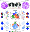
Figure 1.
Figure 1.
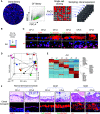
Stem cell libraries from cystic fibrosis lung. (A) Schematic of generation of libraries of epithelial stem cells from control and cystic fibrosis (CF) lungs. CF lungs show cystic bronchiectasis by computed tomography scan and a biallelic loss of phenylalanine 508 by DNA sequencing. Culture plates show colonies formed by clonogenic epithelial cells on lawns of irradiated feeder cells. (B) Single-cell RNA sequencing (scRNA-seq) cluster profiles of stem cell libraries of four CF lungs derived from transplant surgery together with pie charts quantifying the distribution of cells in the various clusters, and the merging of these profiles to detail CF epithelial variants 1–6 (CFv1–CFv6). (C) scRNA-seq analysis of three control adult lungs and one fetal lung, corresponding pie charts detailing variant clusters (TBE, blue; GCM, green; SCM, orange; iSCM, red), and a merged profile highlighting these clusters. GCM = goblet cell metaplasia; iSCM = inflammatory squamous cell metaplasia; SCM = squamous cell metaplasia; TBE = terminal bronchial epithelia; tSNE = t-distributed stochastic neighbor embedding.
Figure 2.
Figure 2.
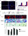
Xenografts of cystic fibrosis (CF) stem cell libraries display pathogenic features. (A) Schematic of xenograft model for assessing the pathologic properties of stem cell libraries involving the expansion of stem cell colonies (in green circles) on 3T3 feeder cells (left), the subcutaneous injection of library epithelial cells in immunodeficient mice to generate nodules (middle), and the analysis of self-assembled epithelial cysts (red asterisks) marked by the human-specific monoclonal antibody STEM123 (right). Scale bar, 200 μm.(B) Histological assessment of xenograft nodules formed by stem cell libraries from patients without obstructive disease by Muc5AC immunofluorescence (left), hematoxylin and eosin (H&E, middle), and Masson’s trichrome staining (right). Scale bar, 200 μm. (C) Histological assessment of xenograft nodules formed from CF libraries by Muc5AC and αTub immunofluorescence (left), H&E (middle) revealing cell infiltrates in epithelial cysts, and Masson’s trichrome staining showing fibrosis adjacent to cysts (arrows, right). Scale bar, 200 μm. Insets: Magnification of Muc5AC-positive goblet cells (left), immunohistochemistry of cellular infiltrates with antibodies to the neutrophil marker Ly6G (middle), and submucosal fibrosis via Masson’s trichrome staining (blue). Scale bar, 200 μm.
Figure 3.
Figure 3.
Clonogenic analysis of cystic fibrosis (CF) libraries reveals disease-linked fate profiles. (A) Schematic for isolating discrete clones representative of the cluster heterogeneity of CF stem cell libraries involving single-cell sorting to 384-well plates and clonal expansion. (B and C) In vitro differentiation of clones representative of each cluster (CF epithelial variants 1–6 [CFv1–v6]) evident in the single-cell RNA sequencing (scRNA-seq) analysis in air–liquid interface (ALI) differentiation cultures and marker immunofluorescence analysis of histological sections of differentiated epithelia by antibodies to Aqp4 (alveolar), Muc5AC (goblet cell metaplasia [GCM]), and IVL (involucrin) (squamous cell metaplasia [SCM]). (D) Principal component analysis of differential gene expression by CFv1–v6 clones as stem cells (SC) and their ALI-differentiated counterparts (ALI). (E) Heatmap of differential gene expression by epithelia derived from in vitro differentiation of CFv1–v6 clones showing typical markers of terminal bronchial epithelia (CFv1, CFv2), GCM (CFv3, CFv4), and SCM (CFv5, CFv6). Scale bar, 20 μm. (F) Histology and indicated marker immunofluorescence of sections of nodules formed by xenografting CFv1–v6 clones. Scale bar, 200 μm.
Figure 4.
Figure 4.
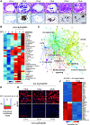
Three proinflammatory variants dominate cystic fibrosis (CF) lung. (A) Histological sections (upper panel) and CD45 immunohistochemistry (lower panel) depicting transepithelial accumulation of neutrophils in nodules of xenografted stem cell clones representative of CF epithelial variant 2 (CFv2), v4, and v6. Scale bar, 100 μm. (B) Heatmap of differential expression of inflammatory genes across CFv1–v6 clones. (C) Network analysis of inflammatory genes expressed in aggregate by CFv2, CFv4, and CFv6 stem cell clones. (D) Schematic for collecting basolateral media conditioned by air–liquid interface (ALI) differentiated CFv1–CFv6 stem cell clones for human vascular endothelial cell (HUVEC) activation assays. (E) Immunofluorescence micrographs of E-selectin expression on HUVEC cells after 48-hour exposure to conditioned media from ALI-differentiated clones representative of CFv1–v6. Scale bar, 30 μm. (F) Heatmap of differential gene expression in HUVEC cells exposed to media conditioned by differentiated CFv1 or CFv2 clones. TPM = transcripts per million.
Figure 5.
Figure 5.
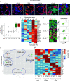
Overlap between cystic fibrosis (CF) and chronic obstructive pulmonary disease (COPD) variant epithelial cells. (A) Immunofluorescence micrographs of histology sections of CF epithelial variants 1–6 (CFv1–v6) xenograft nodules stained with the epithelial marker E-cadherin (red), the myofibroblast marker αSMA (green), and the DNA dye DAPI (blue). Scale bar, 200 μm. (B) Quantification of ratio of αSMA+ myofibroblasts bordering epithelial cysts in xenograft nodules formed by the indicated CF variant clone. (C) Heatmap of differential expression of genes linked to fibrosis by CF variant stem cell clones. (D) Immunofluorescence images and fluorescence-activated cell sorting analysis of cocultures of the indicated clones with normal HuLFs with antibodies to the epithelial marker E-cadherin (green) and the myofibroblast marker αSMA (red). Scale bar, 50 μm. (E) Principal component analysis of whole-genome expression profiles of CFv1–v6 clones with those dominating the COPD lung (21). (F) Expression heat map of selected marker genes for TBE, GCM, and SCM in CFv1–v6 and those identified in COPD lung. HuLFs = human lung fibroblasts; GCM = goblet cell metaplasia; SCM = squamous cell metaplasia; TBE = terminal bronchial epithelia; TPM = transcripts per million.
Figure 6.
Figure 6.
Inflammatory phenotype of cystic fibrosis (CF) variants independent of CFTR (cystic fibrosis transmembrane conductance regulator) activity. (A) Left: schematic of lentiviral transduction of the wild-type (WT) CFTR cDNA and the fluorescence-activated cell sorting of d-Tomato+ cells for clonal expansion. Scale bar, 100 μm. Right: short-circuit current measurements of normal terminal bronchial epithelial stem cells (WT/WT), CF epithelial variant 2 (CFv2) (ΔF508/ΔF508), and CFv2 transduced with a lentivirus driving WT CFTR (CFv2 + Lenti-CFTRWT). See Methods in online supplement. (B) Neutrophilic inflammation in xenograft nodules arising from transplanted CFv2 and CFv2 expressing transduced, WT CFTR. Scale bars, 50 μm. (C) Histogram of gene sets common to CFv2 and CFv2 + WT CFTR, CFv4 and CFv4 + WT CFTR, or CFv6 and CFv6 + WT CFTR (left), and corresponding heatmaps of differentially expressed inflammatory genes (right). Tailed inflammatory genes listed in Figure E10. DIDS = anion exchange inhibitor 4,4′-Diisothiocyano-2,2′-stilbenedisulfonic acid; F&I = forskolin and 3-isobutyl-1-methylxanthine (IBMX); FSC-H-A = forward scatter height and area; iGCM = inflammatory goblet cell metaplasia; iNM = inflammatory normal; iSCM = inflammatory squamous cell metaplasia; PE-A = phycoerythrin area.
Figure 7.
Figure 7.
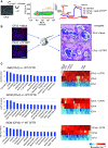
Impact of CFTR (cystic fibrosis transmembrane conductance regulator) modulators on pathologic features of cystic fibrosis (CF) library xenografts. (A) E-selectin immunofluorescence of human vascular endothelial cell (HUVEC) cultures exposed to media conditioned by CF epithelial variant 1 (CFv1) and CFv2 cells differentiated in the absence (left) and presence (right) of triple combinations of CFTR modulators (ivacaftor [3 μM ], elexacaftor [3 μM], and tezacaftor [4 μM]). Scale bar, 30 μm. (B) Histogram of expression of key genes in HUVEC activation in cells treated with media conditioned by CFv1, CFv2, or CFv2 in the presence of triple combination of CFTR modulators. (C) Schematic of CF stem cell library transplantation to immunodeficient mice and systemic treatment of mice with CFTR modulator cocktail ivacaftor, elexacaftor, and tezacaftor (30 mg/kg/d). (D) Histological sections of xenograft nodules stained with antibodies to Muc5A/C and α-Tubulin (top), stained with Masson’s trichrome to detect fibrosis (middle), and immunohistochemistry of antibodies to the murine hematopoietic marker mCD45 (brown). Scale bar, 200 μm. (E) Morphometric quantification of neutrophilic inflammation in xenograft nodules of stem cell libraries from each of four CF stem cell libraries with and without systemic treatment by CFTR modulator cocktail ivacaftor, elexacaftor, and tezacaftor (30 mg/kg/d). TPM = transcripts per million; UT = untreated.

Comment in

References

    1. Rowe SM, Miller S, Sorscher EJ. Cystic fibrosis. N Engl J Med . 2005;352:1992–2001. - PubMed
    1. Elborn JS. Cystic fibrosis. Lancet . 2016;388:2519–2531. - PubMed
    1. Riordan JR, Rommens JM, Kerem B, Alon N, Rozmahel R, Grzelczak Z, et al. Identification of the cystic fibrosis gene: cloning and characterization of complementary DNA. Science . 1989;245:1066–1073. - PubMed
    1. Matsui H, Grubb BR, Tarran R, Randell SH, Gatzy JT, Davis CW, et al. Evidence for periciliary liquid layer depletion, not abnormal ion composition, in the pathogenesis of cystic fibrosis airways disease. Cell . 1998;95:1005–1015. - PubMed
    1. Gibson-Corley KN, Meyerholz DK, Engelhardt JF. Pancreatic pathophysiology in cystic fibrosis. J Pathol . 2016;238:311–320. - PMC - PubMed

Publication types

Substances