. 2023 Sep 19;120(38):e2302489120.
doi: 10.1073/pnas.2302489120. Epub 2023 Sep 11.

Serine starvation silences estrogen receptor signaling through histone hypoacetylation

Affiliations

Serine starvation silences estrogen receptor signaling through histone hypoacetylation

Albert M Li et al. Proc Natl Acad Sci U S A. .

Abstract

Loss of estrogen receptor (ER) pathway activity promotes breast cancer progression, yet how this occurs remains poorly understood. Here, we show that serine starvation, a metabolic stress often found in breast cancer, represses estrogen receptor alpha (ERα) signaling by reprogramming glucose metabolism and epigenetics. Using isotope tracing and time-resolved metabolomic analyses, we demonstrate that serine is required to maintain glucose flux through glycolysis and the TCA cycle to support acetyl-CoA generation for histone acetylation. Consequently, limiting serine depletes histone H3 lysine 27 acetylation (H3K27ac), particularly at the promoter region of ER pathway genes including the gene encoding ERα, ESR1. Mechanistically, serine starvation impairs acetyl-CoA-dependent gene expression by inhibiting the entry of glycolytic carbon into the TCA cycle and down-regulating the mitochondrial citrate exporter SLC25A1, a critical enzyme in the production of nucleocytosolic acetyl-CoA from glucose. Consistent with this model, total H3K27ac and ERα expression are suppressed by SLC25A1 inhibition and restored by acetate, an alternate source of acetyl-CoA, in serine-free conditions. We thus uncover an unexpected role for serine in sustaining ER signaling through the regulation of acetyl-CoA metabolism.

Keywords: SLC25A1; breast cancer; estrogen receptor; histone acetylation; serine metabolism.


Conflict of interest statement

The authors declare no competing interest.

Figures

Fig. 1.
Fig. 1.
Serine starvation represses the estrogen signaling pathway and promotes endocrine resistance. (A and B) Enrichment plots via GSEA on RNA-seq performed in control vs. 24 h serine-starved MDA-MB-231 (A) or MCF7 (B) cells. NES, nuclear enrichment score. (C) Heatmap of the expression of select estrogen signaling pathway genes as measured by RNA-seq in B. (D) Quantitative real-time PCR analysis for estrogen signaling pathway genes in 24-h serine-starved MCF7 cells. (E) Immunoblot for estrogen receptor alpha (ERα) in whole cell lysates from MCF7 or T47D cells grown in control (+Ser) or serine-free (–Ser) media for 24 h. (F) Four-day proliferation assay of MCF7 and T47D cells in ±Ser media, treated with increasing doses of fulvestrant (ICI). (G) Four-day proliferation assay of MCF7 and T47D cells in ±Ser media treated with ICI or the CDK1 inhibitor RO-3306. Data represent mean ± SD (D, F, and G), N = 3 per group. Statistics: two-tailed unpaired Student’s t test; **P < 0.01, ^P < 0.001, #P < 0.0001.
Fig. 2.
Fig. 2.
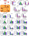
Reduced glucose flux through glycolysis and the TCA cycle under serine starvation leads to citrate and acetyl-CoA insufficiency. (A) Schematic of U-13C-glucose labeling to serine and intermediates in glycolysis and the TCA cycle. (B) Isotopomer distribution of serine from MCF7 cells cultured in ±Ser glucose-free media supplemented with U-13C-glucose for 6 h. (C) LC-MS was used to determine the NADH/NAD+ ratio at various time points of −Ser in MCF7 cells. (D) Isotopomer distribution of pyruvate, acetyl-CoA, citrate, fumarate, and malate from MCF7 cells cultured in ±Ser glucose-free media supplemented with U-13C-glucose for 6 h. (E and F) LC-MS was used to determine the levels of intermediates in glycolysis (E) and the TCA cycle (F) at various time points of −Ser in MCF7 cells. Data are mean ± SD (BF), N = 3 per group. Statistics: two-tailed unpaired Student’s t test; *P < 0.05, **P < 0.01, ^P < 0.001, #P < 0.0001. Abbreviations: G6P, glucose 6-phosphate; G1P, glucose 1-phosphate; F6P, fructose 6-phoshpate; F16BP, fructose 1,6-bisphosphate; DHAP, dihydroxyacetone phosphate; G3P, glycerol 3-phosphate; 3PG, 3-phosphoglycerate; PEP, phosphoenolpyruvate; Ac-CoA, acetyl-Coenzyme A; CoA, coenzyme A; αKG, alpha ketoglutarate; NAD+, nicotinamide adenine dinucleotide.
Fig. 3.
Fig. 3.
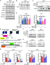
Serine starvation suppresses estrogen receptor signaling through H3K27 hypoacetylation. (A) Immunoblot for ERα and common H3K acetylation and methylation modifications, performed on nuclear lysates from MCF7 cells grown in ±Ser media at 12 h, 24 h, and 48 h. (B) Immunoblot for ERα and H3K27ac, performed on nuclear lysates isolated from T47D cells grown in ±Ser media at 12 h, 24 h, and 48 h. (C and D) Immunoblot for H3K27ac and ERα in nuclear lysates (C) and qPCR analysis for ESR1 and PGR (D) from MCF7 cells grown in ±Ser media treated with either suberanilohydroxamic acid (SAHA) or romidepsin (Rom) for 18 h. (E) Schematic of two ways acetate may contribute to nuclear Ac-CoA pools to support histone acetylation. (F) Immunoblot for H3K27ac in nuclear lysates from MCF7 and T47D cells grown in ±Ser media supplemented with glycerol triacetate (GTA) or sodium acetate (NaAc) for 24 h. (G) qPCR analysis for ESR1 and PGR in MCF7 and T47D cells grown in ±Ser media supplemented with GTA for 18 h. (H) Immunoblot for ERα in whole cell extracts from MCF7 and T47D cells grown in ±Ser media supplemented with different concentrations of GTA for 24 h. Data are mean ± SD (D and G), N = 3 per group. Statistics: two-tailed unpaired Student’s t test; *P < 0.05, **P < 0.01, ^P < 0.001.
Fig. 4.
Fig. 4.
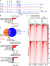
H3K27ac at promoters of down-regulated genes is lost under serine starvation and is restored by acetate. (A) Bedgraphs of H3K27ac at the ESR1 locus in MCF7 cells. Several enriched peaks around the promoter region of cells cultured in +Ser are highlighted in gray. (B) Pathway analysis via Enrichr of genes that lost at least one H3K27ac peak in −Ser. (C) Overlap of RNA-seq and ChIP-seq data. (D) Pathway analysis via ENRICHR of genes either down-regulated or up-regulated by serine starvation via RNA-seq overlapped with genes that lost at least one H3K27ac peak in −Ser. (E) Heatmaps of H3K27ac signal distribution along genes that were identified as either significantly up-regulated or down-regulated by −Ser in MCF7 cells. Abbreviations: TSS, transcription start site; TES, transcription end site.
Fig. 5.
Fig. 5.
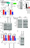
Serine starvation suppresses histone acetylation and gene expression by inhibiting the mitochondrial citrate exporter SLC25A1. (A) Schematic of enzymes involved in acetyl-CoA generation from mitochondria-derived citrate. The relative levels of SLC25A1 and ACLY mRNA were determined from RNA-seq. (B) Immunoblot for ERα, H3K27ac, SLC25A1, and ACLY in whole-cell extracts from MCF7 cells grown in ±Ser media for 24 h. (C) Ac-CoA/CoA measurements in MCF7 cells expressing shNT, shSLC25A1#1, or shSLC25A1#2. (D) Immunoblot for ERα, SLC25A1, and H3K27ac in whole cell lysates from MCF7 cells expressing shNT, shSLC25A1#1, or shSLC25A1#2 grown in media supplemented with increasing concentrations of GTA for 24 h. (E) Immunoblot for ERα, SLC25A1, ACLY, and H3K27ac in whole-cell lysates (Top) and nuclear lysates (Bottom) from MCF7 and T47D cells treated with the SLC25A1 inhibitors CNASB and CTPI-2 for 18 h. (F) Four-day proliferation assay of MCF7 cells expressing shNT, shSLC25A1#1, or shSLC25A1#2 treated with 1 nM ICI or 2 mM GTA. (G) Immunoblot for ERα, SLC25A1, and H3K27ac in whole cell lysates from MCF7 cells expressing EV or SLC25A1 OE grown in ±Ser media for 24 h. Data are mean ± SD (A, C, and F), N = 3 per group. Statistics: two-tailed unpaired Student’s t test; *P < 0.05, **P < 0.01, ^P < 0.001, #P < 0.0001. Abbreviations: TPM, transcripts per million.

References

    1. Couse J. F., Korach K. S., Estrogen receptor null mice: What have we learned and where will they lead us? Endocr. Rev. 20, 358–417 (1999). - PubMed
    1. Osborne C. K., Schiff R., Mechanisms of endocrine resistance in breast cancer. Annu. Rev. Med. 62, 233–247 (2011). - PMC - PubMed
    1. Chang M., Tamoxifen resistance in breast cancer. Biomol. Ther. (Seoul) 20, 256–267 (2012). - PMC - PubMed
    1. Lim E., et al. , Aberrant luminal progenitors as the candidate target population for basal tumor development in BRCA1 mutation carriers. Nat. Med. 15, 907–913 (2009). - PubMed
    1. Fan M., et al. , Triggering a switch from basal- to luminal-like breast cancer subtype by the small-molecule diptoindonesin G via induction of GABARAPL1. Cell Death Dis. 11, 635 (2020). - PMC - PubMed

Publication types