Predicting successful clinical candidates for fiducial-free lung tumor tracking with a deep learning binary classification model
- PMID: 37696265
- PMCID: PMC10691617
- DOI: 10.1002/acm2.14146
Predicting successful clinical candidates for fiducial-free lung tumor tracking with a deep learning binary classification model
Abstract
Objectives: The CyberKnife system is a robotic radiosurgery platform that allows the delivery of lung SBRT treatments using fiducial-free soft-tissue tracking. However, not all lung cancer patients are eligible for lung tumor tracking. Tumor size, density, and location impact the ability to successfully detect and track a lung lesion in 2D orthogonal X-ray images. The standard workflow to identify successful candidates for lung tumor tracking is called Lung Optimized Treatment (LOT) simulation, and involves multiple steps from CT acquisition to the execution of the simulation plan on CyberKnife. The aim of the study is to develop a deep learning classification model to predict which patients can be successfully treated with lung tumor tracking, thus circumventing the LOT simulation process.
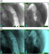
Methods: Target tracking is achieved by matching orthogonal X-ray images with a library of digital radiographs reconstructed from the simulation CT scan (DRRs). We developed a deep learning model to create a binary classification of lung lesions as being trackable or untrackable based on tumor template DRR extracted from the CyberKnife system, and tested five different network architectures. The study included a total of 271 images (230 trackable, 41 untrackable) from 129 patients with one or multiple lung lesions. Eighty percent of the images were used for training, 10% for validation, and the remaining 10% for testing.
Results: For all five convolutional neural networks, the binary classification accuracy reached 100% after training, both in the validation and the test set, without any false classifications.
Conclusions: A deep learning model can distinguish features of trackable and untrackable lesions in DRR images, and can predict successful candidates for fiducial-free lung tumor tracking.
Keywords: 4DCT; CyberKnife; LOT simulation; SBRT; accuray; artificial intelligence; binary classification; deep learning; digitally reconstructed radiograph (DRR); lung cancer; lung tumors; motion modeling; radiation therapy; respiratory motion; respiratory motion modeling; transfer learning.
© 2023 The Authors. Journal of Applied Clinical Medical Physics published by Wiley Periodicals, LLC on behalf of The American Association of Physicists in Medicine.
Conflict of interest statement
The authors declare no conflicts of interest.
Figures


Similar articles
-
Deep match: A zero-shot framework for improved fiducial-free respiratory motion tracking.Radiother Oncol. 2024 May;194:110179. doi: 10.1016/j.radonc.2024.110179. Epub 2024 Feb 24. Radiother Oncol. 2024. PMID: 38403025
-
Simultaneous object detection and segmentation for patient-specific markerless lung tumor tracking in simulated radiographs with deep learning.Med Phys. 2024 Mar;51(3):1957-1973. doi: 10.1002/mp.16705. Epub 2023 Sep 8. Med Phys. 2024. PMID: 37683107
-
Robust localization of poorly visible tumor in fiducial free stereotactic body radiation therapy.Radiother Oncol. 2024 Nov;200:110514. doi: 10.1016/j.radonc.2024.110514. Epub 2024 Aug 29. Radiother Oncol. 2024. PMID: 39214256
-
Deep learning-based target decomposition for markerless lung tumor tracking in radiotherapy.Med Phys. 2024 Jun;51(6):4271-4282. doi: 10.1002/mp.17039. Epub 2024 Mar 20. Med Phys. 2024. PMID: 38507259 Free PMC article.
-
Deep learning-based target tracking with X-ray images for radiotherapy: a narrative review.Quant Imaging Med Surg. 2024 Mar 15;14(3):2671-2692. doi: 10.21037/qims-23-1489. Epub 2024 Mar 7. Quant Imaging Med Surg. 2024. PMID: 38545053 Free PMC article. Review.
Cited by
-
Comparison of Various GAN-Based Bone Suppression Imaging for High-Accurate Markerless Motion Tracking of Lung Tumors in CyberKnife Treatment.Thorac Cancer. 2025 Feb;16(4):e70014. doi: 10.1111/1759-7714.70014. Thorac Cancer. 2025. PMID: 39994000 Free PMC article.
-
A Thorough Review of the Clinical Applications of Artificial Intelligence in Lung Cancer.Cancers (Basel). 2025 Mar 4;17(5):882. doi: 10.3390/cancers17050882. Cancers (Basel). 2025. PMID: 40075729 Free PMC article. Review.
References
-
- Kilby W, Naylor M, Dooley JR, Maurer CR, Sayeh S. Handbook of Robotic and Image‐Guided Surgery. Elsevier; 2020:15‐38.
-
- Bahig H, Campeau M‐P, Vu T, et al. Predictive parameters of CyberKnife fiducial‐less (XSight Lung) applicability for treatment of early non‐small cell lung cancer: a single‐center experience. Int J Radiat Oncol Biol Phys. 2013;87:583‐589. - PubMed
MeSH terms
LinkOut - more resources
Full Text Sources
Medical

