. 2023 Sep 12;8(1):344.
doi: 10.1038/s41392-023-01621-2.

Oit3, a promising hallmark gene for targeting liver sinusoidal endothelial cells

Affiliations

Oit3, a promising hallmark gene for targeting liver sinusoidal endothelial cells

Zhi-Wen Li et al. Signal Transduct Target Ther. .

Abstract

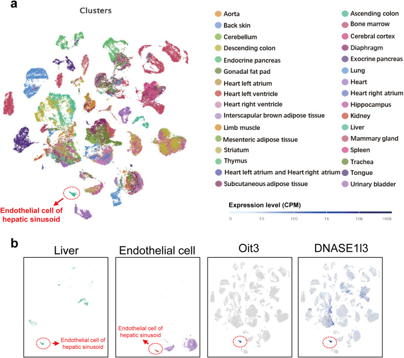
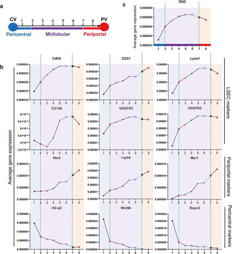
Liver sinusoidal endothelial cells (LSECs) play a pivotal role in maintaining liver homeostasis and influencing the pathological processes of various liver diseases. However, neither LSEC-specific hallmark genes nor a LSEC promoter-driven Cre mouse line has been introduced before, which largely restricts the study of liver diseases with vascular disorders. To explore LSEC-specific hallmark genes, we compared the top 50 marker genes between liver endothelial cells (ECs) and liver capillary ECs and identified 18 overlapping genes. After excluding globally expressed genes and those with low expression percentages, we narrowed our focus to two final candidates: Oit3 and Dnase1l3. Through single-cell RNA sequencing (scRNA-seq) and analysis of the NCBI database, we confirmed the extrahepatic expression of Dnase1l3. The paired-cell sequencing data further demonstrated that Oit3 was predominantly expressed in the midlobular liver ECs. Subsequently, we constructed inducible Oit3-CreERT2 transgenic mice, which were further crossed with ROSA26-tdTomato mice. Microscopy validated that the established Oit3-CreERT2-tdTomato mice exhibited significant fluorescence in the liver rather than in other organs. The staining analysis confirmed the colocalization of tdTomato and EC markers. Ex-vivo experiments further confirmed that isolated tdTomato+ cells exhibited well-differentiated fenestrae and highly expressed EC markers, confirming their identity as LSECs. Overall, Oit3 is a promising hallmark gene for tracing LSECs. The establishment of Oit3-CreERT2-tdTomato mice provides a valuable model for studying the complexities of LSECs in liver diseases.


Conflict of interest statement

The authors declare no competing interests.

Figures

Fig. 1
Fig. 1
Single-cell RNA sequencing reveals liver endothelial marker genes. a t-SNE plot of endothelial cells in different organs. b Venn diagram of the top 50 marker genes of liver-specific EC phenotypes and the top 50 marker genes of liver-specific capillary. c t-SNE plot of traditional markers used to label LSECs, including Cdh5, CD31, Lyve1, Vegfr2 and Vegfr3. d The list of the names and the expressing percentage of the 18 overlapped genes. e t-SNE plot of the 18 genes (EC Atlas; https://carmelietlab.sites.vib.be/en/softwaretools/scCycle)
Fig. 2
Fig. 2
The global gene expression of Oit3 and Dnase1l3. a The relative expression of Oit3 and Dnase1l3 in distinct organs of embryos and adults in mice, data were collected from NCBI database. b The expression level of Oit3 and Dnase1l3 in distinct organs in mice with different strains (www.ebi.ac.uk/gxa/home). c The relative expression level of Oit3 and Dnase1l3 in distinct organs of C57 mice (www.ebi.ac.uk/gxa/home)
Fig. 3
Fig. 3
Single-cell RNA-seq of global cells. a The clusters of 20 distinct organs and tissues from the previously established Tabula Muris of scRNA-seq analysis of individual mice. b The t-SNE plot of cells in liver and all endothelial cells is shown in the left two panels, while the right two panels show the expression of Oit3 and Dnase1l3 across various organs and tissues (GSE109774)
Fig. 4
Fig. 4
The LSEC-specific expression of Oit3. a The mRNA expression of Oit3 in liver, spleen, lung, brain, heart, testis, fat, intestine and kidney of mice, determined by qRT-PCR. β-actin was used as an internal control. b The mRNA expression of Oit3 in different kinds of liver cells, including hepatocytes, KCs, LSECs and HSCs. β-actin was used as an internal control. c The mRNA expression of Oit3 in hepatocytes, plasmacytoid dendritic cells, neutrophils, B cells, T cells, liver capsule macrophages, KCs and ECs, evaluated by the data of paired-cell sequencing of mice (GSE108561). d t-SNE plot revealed the spatial distribution of Oit3 in liver (GSE134037). e t-SNE plot revealed the spatial distribution of Oit3 in liver ECs (data from EC altas). Bars represent means ± SD, n = 3; *P < 0.05, **P < 0.01, ***P < 0.001
Fig. 5
Fig. 5
The spatial expression of endothelial markers. a A sketch mimicking the eight continuous regions from CV to PV. b The spatial expression of periportal, pericentral and EC marker genes in eight continuous regions (GSE108561). c The spatial expression of Oit3 in eight continuous regions of liver ECs (GSE108561)
Fig. 6
Fig. 6
The generation of Oit3-CreERT2-tdTomato mice. a Strategy of the generation of Oit3-CreERT2 knockin allele. b Schematic image of the genetic lineage tracing strategy. c Microscopy of tdTomato in livers of Oit3-CreERT2-tdTomato mice with or without tamoxifen injection. d Microscopy of tdTomato and DAPI in spleen, lung, brain, kidney, testis, intestine, heart and muscle of Oit3-CreERT2-tdTomato mice treated with tamoxifen. e Immunofluorescent staining of VE Cadherin, Lyve1 and DAPI in livers of Oit3-CreERT2-tdTomato mice treated with tamoxifen. f Immunofluorescent staining of Desmin, αSMA, F4/80, CD34, Albumin, CK19 and DAPI in livers of Oit3-CreERT2-tdTomato mice treated with tamoxifen. g FACS analysis of CD31+ percentage in isolated tdTomato+ cells. h SEM showed the well-differentiated fenestrae of cultured tdTomato+ cells. i The comparison of mRNA expression of CD31, VE-Cadherin, VEGFR2 and VEGFR3 between tdTomato+ and tdTomato- cells, determined by qRT-PCR. β-actin was used as an internal control. Bars represent means ± SD, n = 3; **P < 0.01, ***P < 0.001, ****P < 0.0001

References

    1. Gracia-Sancho J, Caparros E, Fernandez-Iglesias A, Frances R. Role of liver sinusoidal endothelial cells in liver diseases. Nat. Rev. Gastroenterol. Hepatol. 2021;18:411–431. - PubMed
    1. Furuta K, et al. Endotheliopathy in the metabolic syndrome: mechanisms and clinical implications. Pharm. Ther. 2023;244:108372. - PMC - PubMed
    1. Kumar S, Duan Q, Wu R, Harris EN, Su Q. Pathophysiological communication between hepatocytes and non-parenchymal cells in liver injury from NAFLD to liver fibrosis. Adv. Drug Deliv. Rev. 2021;176:113869. - PMC - PubMed
    1. McConnell MJ, Kostallari E, Ibrahim SH, Iwakiri Y. The evolving role of liver sinusoidal endothelial cells in liver health and disease. Hepatology. 2023;78:649–669. - PMC - PubMed
    1. Gage BK, et al. Generation of functional liver sinusoidal endothelial cells from human pluripotent stem-cell-derived venous angioblasts. Cell Stem Cell. 2020;27:254–269.e259. - PubMed

Substances