. 2023 Sep 11;13(1):14958.
doi: 10.1038/s41598-023-41026-3.

Systemic administration of clinical-grade multilineage-differentiating stress-enduring cells ameliorates hypoxic-ischemic brain injury in neonatal rats

Affiliations

Systemic administration of clinical-grade multilineage-differentiating stress-enduring cells ameliorates hypoxic-ischemic brain injury in neonatal rats

Kazuto Ueda et al. Sci Rep. .

Abstract

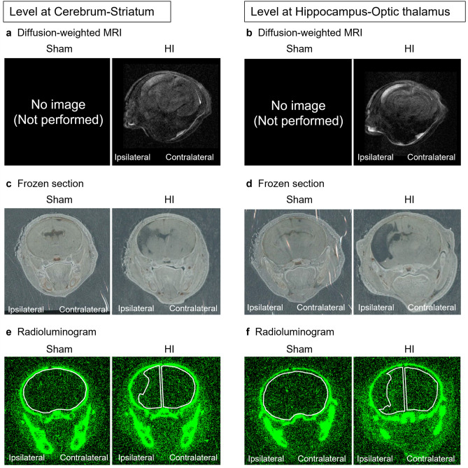
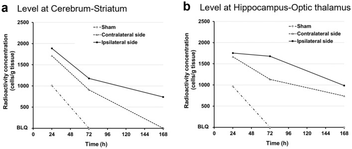
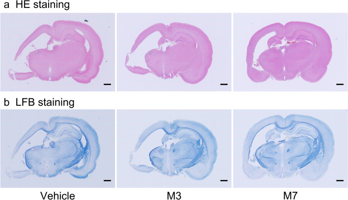
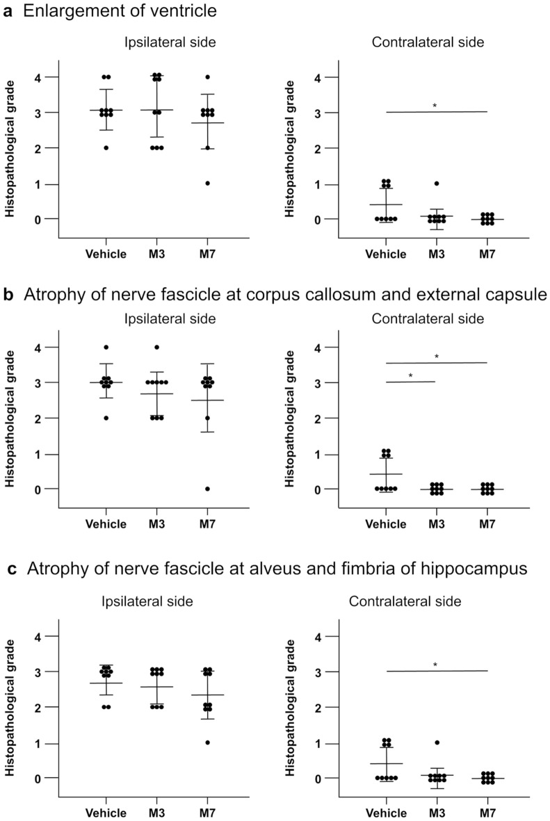
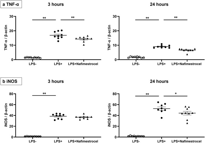
Multilineage-differentiating stress-enduring (Muse) cells are endogenous reparative pluripotent stem cells present in the bone marrow, peripheral blood, and organ connective tissues. We assessed the homing and therapeutic effects of systemically administered nafimestrocel, a clinical-grade human Muse cell-based product, without immunosuppressants in a neonatal hypoxic-ischemic (HI) rat model. HI injury was induced on postnatal day 7 (P7) and was confirmed by T2-weighted magnetic resonance imaging on P10. HI rats received a single dose nafimestrocel (1 × 10⁶ cells/body) or Hank's balanced salt solution (vehicle group) intravenously at either three days (on P10; M3 group) or seven days (on P14; M7 group) after HI insult. Radioisotope experiment demonstrated the homing of chromium-51-labeled nafimestrocel to the both cerebral hemispheres. The cylinder test (M3 and M7 groups) and open-field test (M7 group) showed significant amelioration of paralysis and hyperactivity at five weeks of age compared with those in the vehicle group. Nafimestrocel did not cause adverse events such as death or pathological changes in the lung at ten weeks in the both groups. Nafimestrocel attenuated the production of tumor necrosis factor-α and inducible nitric oxide synthase from activated cultured microglia in vitro. These results demonstrate the potential therapeutic benefits and safety of nafimestrocel.


Conflict of interest statement

YS, SS, TS, MM, MH and MD declare the following potential conflicts of interest with respect to the research, authorship, and/or publication of this article: YS and SS have a collaborative research agreement for perinatal disease and an investigator-initiated clinical trial agreement supplying CL2020 free of charge for HIE with Life Science Institute Inc. (LSII). SS has a contract for consulting with LSII. YS, SS, TS, MM, MH, and MD have a patent for the application of Muse cells for the treatment of perinatal brain damage and other indications. MD is party to a collaborative research agreement with LSII. MD has a patent for the application of Muse cells for the treatment of cerebral infarction. MD holds patents for Muse cells and the isolation method thereof, which are exclusively licensed to LSII. KU, AO, RM, SG, HM, YK, YY, KI, MT, KM and YT declare no competing interests.

Figures

Figure 1
Figure 1
Representative images of diffusion-weighted magnetic resonance imaging (MRI; a, b), histological frozen sections (c, d) and radioluminograms (e, f). Diffusion-weighted MRI was performed prior to treatment, and frozen sections and radioluminograms were prepared 72 h after a single intravenous administration of [51Cr]-nafimestrocel. Each region of interest (ROI) in the radioluminograms is indicated with a white outlined area.
Figure 2
Figure 2
Radioactivity concentration in the brain after a single intravenous administration of [51Cr]-nafimestrocel. (a) Level at cerebrum-striatum. (b) Level at hippocampus-optic thalamus. n = 1 at each time for the sham group (n = 3), the ipsilateral (left) side in the hypoxic–ischemic (HI) group (n = 3), and the contralateral side in the HI group (n = 3).
Figure 3
Figure 3
Body weight gain after birth throughout the observation period (n = 9 for the vehicle group, n = 9 for the M3 group, and n = 9 for the M7 group). Black dotted line, vehicle; green solid line, M3; red dashed line, M7. Data represent mean ± standard deviation.
Figure 4
Figure 4
(a) In the cylinder test, the average of the preference for the left (ipsilateral) forepaw was calculated on three consecutive days (postnatal day 36 (P36) to P38 (n = 9 for the vehicle group, n = 8 for the M3 group, and n = 9 for the M7 group). Rats in the M3 and M7 groups showed a significantly lower preference for the ipsilateral (left) forepaw than rats in the vehicle group. (b) In the open-field test, the distance traveled was evaluated on P42 (n = 9 for vehicle, n = 9 for M3, and n = 9 for M7). The distance traveled was significantly shorter in the M7 group than in the vehicle group. (c) In the water maze test, the average of the distance traveled on five consecutive days (P53–P57) was calculated (n = 9 for vehicle, n = 9 for M3, and n = 9 for M7). Data represent mean ± standard deviation. *p < 0.05 and **p < 0.01.
Figure 5
Figure 5
Representative images showing brain sections stained with hematoxylin–eosin (HE; a) and Luxol fast blue (LFB; b). (a) Enlargement of ventricles; vehicle group (grade 3); M3 group (grade 4); M7 group (grade 3). Bar = 1 mm. (b) Atrophy of nerve fascicles in the corpus callosum, external capsule, alveus of hippocampus, and fimbria of hippocampus; vehicle (grade 3); M3 (grade 3); M7 (grade 3). Bar = 1 mm.
Figure 6
Figure 6
Histopathological grade of enlargement of ventricles on ipsilateral (left) and contralateral sides (a), atrophy of nerve fascicles in the corpus callosum and external capsule on ipsilateral and contralateral sides (b), and atrophy of nerve fascicles in the alveus of hippocampus and fimbria of hippocampus on ipsilateral and contralateral sides (c); n = 9 for the vehicle group, n = 9 for the M3 group, and n = 9 for the M7 group. Data represent mean ± standard deviation. *p < 0.05 and **p < 0.01.
Figure 7
Figure 7
In vitro experiment using a coculture of microglia and nafimestrocel at 3 h (left) and 24 h after LPS administration. The production of TNF-α (a) and iNOS (b) was evaluated at 3 and 24 h after LPS administration. (a) The production of TNF-α was significantly lower in microglia cocultured with nafimestrocel at both 3 and 24 h after LPS administration. (b) The production of iNOS significantly decreased in microglia cocultured with nafimestrocel at 24 h after LPS administration. White circles (LPS −) correspond to microglia alone without LPS, black circles (LPS +) represent microglia alone with LPS, and black triangles (LPS + nafimestrocel) indicate microglia cocultured with nafimestrocel and LPS (n = 9 for LPS − , 9 for LPS + , 9 for LPS + nafimestrocel at 3 h, and n = 9 for LPS − , 8 for LPS + , 9 for LPS + nafimestrocel at 24 h). Data represent mean ± standard deviation. *p < 0.05 and **p < 0.01.

References

    1. Hayakawa M, et al. Incidence and prediction of outcome in hypoxic-ischemic encephalopathy in Japan. Pediatr. Int. 2014;56:215–221. doi: 10.1111/ped.12233. - DOI - PMC - PubMed
    1. Kurinczuk JJ, White-Koning M, Badawi N. Epidemiology of neonatal encephalopathy and hypoxic-ischaemic encephalopathy. Early Human Dev. 2010;86:329–338. doi: 10.1016/j.earlhumdev.2010.05.010. - DOI - PubMed
    1. Volpe JJ. Neonatal encephalopathy: An inadequate term for hypoxic-ischemic encephalopathy. Ann. Neurol. 2012;72:156–166. doi: 10.1002/ana.23647. - DOI - PubMed
    1. Shankaran S, et al. Whole-body hypothermia for neonates with hypoxic-ischemic encephalopathy. N. Engl. J. Med. 2005;353:1574–1584. doi: 10.1056/NEJMcps050929. - DOI - PubMed
    1. Edwards, A. D. et al. Neurological outcomes at 18 months of age after moderate hypothermia for perinatal hypoxic ischaemic encephalopathy: Synthesis and meta-analysis of trial data. BMJ (Clin. Res. ed.)340, c363. 10.1136/bmj.c363 (2010). - PMC - PubMed