. 2023 Sep 11;13(1):14988.
doi: 10.1038/s41598-023-41367-z.

Synthetic circuits based on split Cas9 to detect cellular events

Affiliations

Synthetic circuits based on split Cas9 to detect cellular events

Alicja Przybyszewska-Podstawka et al. Sci Rep. .

Abstract

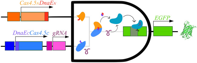
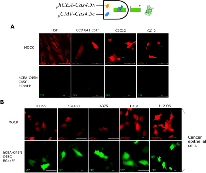
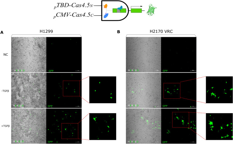
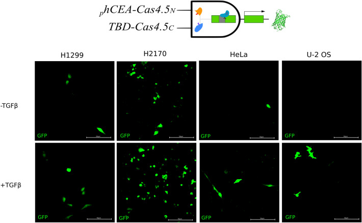
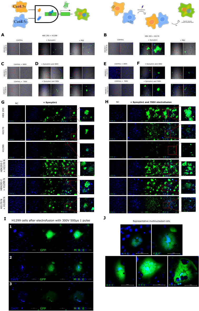
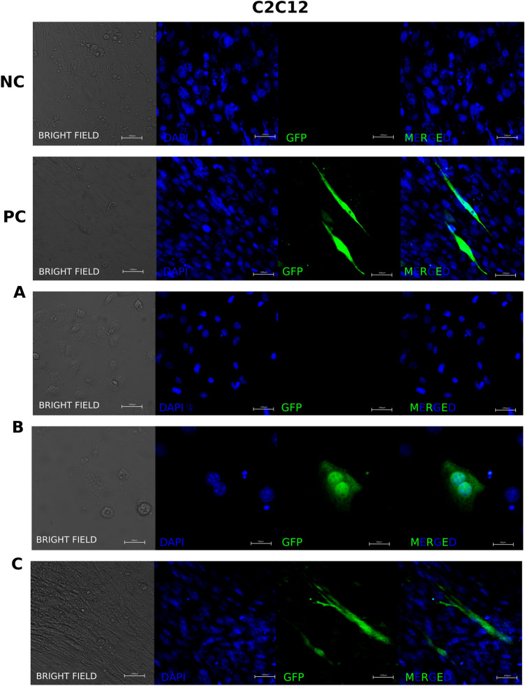
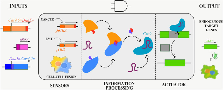
Synthetic biology involves the engineering of logic circuit gates that process different inputs to produce specific outputs, enabling the creation or control of biological functions. While CRISPR has become the tool of choice in molecular biology due to its RNA-guided targetability to other nucleic acids, it has not been frequently applied to logic gates beyond those controlling the guide RNA (gRNA). In this study, we present an adaptation of split Cas9 to generate logic gates capable of sensing biological events, leveraging a Cas9 reporter (EGxxFP) to detect occurrences such as cancer cell origin, epithelial to mesenchymal transition (EMT), and cell-cell fusion. First, we positioned the complementing halves of split Cas9 under different promoters-one specific to cancer cells of epithelial origin (phCEA) and the other a universal promoter. The use of self-assembling inteins facilitated the reconstitution of the Cas9 halves. Consequently, only cancer cells with an epithelial origin activated the reporter, exhibiting green fluorescence. Subsequently, we explored whether this system could detect biological processes such as epithelial to mesenchymal transition (EMT). To achieve this, we designed a logic gate where one half of Cas9 is expressed under the phCEA, while the other is activated by TWIST1. The results showed that cells undergoing EMT effectively activated the reporter. Next, we combined the two inputs (epithelial origin and EMT) to create a new logic gate, where only cancer epithelial cells undergoing EMT activated the reporter. Lastly, we applied the split-Cas9 logic gate as a sensor of cell-cell fusion, both in induced and naturally occurring scenarios. Each cell type expressed one half of split Cas9, and the induction of fusion resulted in the appearance of multinucleated syncytia and the fluorescent reporter. The simplicity of the split Cas9 system presented here allows for its integration into various cellular processes, not only as a sensor but also as an actuator.


Conflict of interest statement

The authors declare no competing interests.

Figures

Figure 1
Figure 1
Scheme of the principle of split-Cas9 as an AND logic gate.
Figure 2
Figure 2
Logic gate to identify only cancer cells of epithelial origin using halves of split Cas9 under a universal (pCMV) and a cell type-specific (phCEA) promoters. The expression of the green fluorescent protein reporter is observed only in cancer cells of epithelial origin. All cells were co-transfected with phCEA-Cas4.5N, pCMV-Cas4.5C/gRNA, the EGxxFP reporter and pCMV-mCherry (transfection control) constructs. Co-expression of the split Cas9 halves resulted in holo-enzyme formation via inteins, followed by Cas9-induced homologous recombination of EGFP. Only cancer cells of epithelial origin expressed both halves since the expression of Cas4.5N is restricted to these cells. Neither non-epithelial cell lines: human skin fibroblasts (HSF), murine myoblasts (C2C12), nor normal (non-cancerous) epithelial cells: CCD 841 CoTr and GC-2, showed any expression of the reporter (EGFP) (A), while all cancer cells with epithelial origin tested (H1299, SW480, A375, HeLa, and U-2 OS cells) expressed EGFP (B).
Figure 3
Figure 3
Logic gate for sensing cells that are undergoing EMT. Representative images of H1299 lung cancer cells (A) and H2170 VRCs cells (B), both partly mesenchymal. Both cell lines, when exposed to TGFβ (+ TGFβ) as an EMT enhancer, increased the number of EGFP expressing cells as a proof of undergoing EMT. Morphological changes were visible after activation of TGFβ showed features of cells of mesenchymal origin—more spindle-shaped, they had more protrusions facilitating migration in comparison to controls.
Figure 4
Figure 4
A two level AND logic gate, where epithelial cancer cells undergoing EMT are detected. Four epithelial cancer cell lines were transfected with split Cas9/gRNA system and the EGxxFP reporter. Then cells were stimulated to undergo EMT by the addition of TGFβ. While all these epithelial cancer cells were known to express the phCEA (Fig. 2), only cells with partial-EMT expressed the pTBD-Cas4,5C, without EMT stimulation (upper row, -TGFβ). But when EMT was induced (+ TGFβ), the number of positive cells increased dramatically in those partially mesenchymal (H1299 and H2170), and appeared in those more epithelial—HeLa and U-2 OS cells (lower row).
Figure 5
Figure 5
Logic gate to detect cell–cell fusion and schematic diagram of cell–cell electrofusion and hybrid cell formation. Images of admixed transiently transfected cells with complementing Cas9 halves HEK 293 + H1299 and HEK 293 + H2170 after fusion induction by overexpression of Syncytin-1, alone or in pair with electrofusion, or by PEG treatment. Control cells (NC) were transfected with only one half of Cas9 plus the reporter and fusogenic vectors (A,B). As we expected admixed cells HEK293 + H1299 expressing either one halve (as a control samples) and electrofused using 300V and 700V do not show any fusion events (C), compared to electrofusion of cells expressing both halves where we got results of a few cell fusion (D). After enhancement of fusion by electric pulses in pair with Syncytin1 overexpression, we visibly increased occurrence of fusion events (E). In control samples of admixed HEK293 + H2170 cells after electrofusion using 300 V and 700 V, also the fusion was undetectable (F), in comparison to Syncytin1-induced fusion cells plus electrofusion, where fusion cell were clearly visible (H). Images of admixed stable cell lines with complementing Cas9 halves (HEK293, H1299, H2170, HEK293 + H1299, HEK293 + H2170, and H1299 + H2170) after fusion induction by overexpression of Syncytin1, and Syncytin1 plus electrofusion. Control cells (NC) were transfected with only one half of Cas9 plus the reporter and fusogenic vectors. In relation to cells only expressed fusogenic Syncytin1 (G), the combination of Syncytin1 and electrical pulses (H) greatly enhanced the detection of syncytia. When fusing HEK293 with H1299 or H2170, the syncytia visibly formed contained more nuclei, than lung cancer cells in a mix within the same line. In summary, H1299 cells overexpressing Syncytin1 in pair with electrofusion at 700 V did not show significant fusion events (H). Higher efficiency was achieved by expression of Syncytin1 and lower voltage (300 V 500 µs one pulse) in stable expressing complementing Cas9 halves H1299 cells (I). Representative large multinucleated syncytia after electrofusion cells overexpressed Syncytin1 H1299 (using 300 V), H2170 cell lines (using 700 V), and mixed HEK 293 with H1299 cell line and HEK293 with H2170 cells (using 700 V) (J).
Figure 6
Figure 6
Applying split Cas9 logic gate to detect naturally occurring cell fusions. Murine C2C12 myoblast cells were transfected separately with either Cas9 half (Cas4.5C or Cas4.5N), gRNA and EGxxFP reporter, and then co-cultured together in differentiation medium. (A) C2C12 cells have a triangular shape while undifferentiated; (B) then they align with each other and begin to fuse—for better visualization of the two nuclei this image is show at higher magnification; (C) forming myotubules. NC—Negative control (cells with Split Cas9 halves, and EGxxFP reporter without gRNA); PC—(cells transfected with Cas9/gRNA and EGxxFP reporter). Additional images in Supplementary Fig. S1.
Figure 7
Figure 7
Engineering cells with CRISPR/Cas9 circuits.

References

    1. Gardner TS, Cantor CR, Collins JJ. Construction of a genetic toggle switch in Escherichia coli. Nature. 2000;403:339–342. - PubMed
    1. Elowitz MB, Leibier S. A synthetic oscillatory network of transcriptional regulators. Nature. 2000;403:335–338. - PubMed
    1. Bikard D, et al. Programmable repression and activation of bacterial gene expression using an engineered CRISPR-Cas system. Nucleic Acids Res. 2013;41:7429–7437. - PMC - PubMed
    1. Dong, C., Fontana, J., Patel, A., Carothers, J. M. & Zalatan, J. G. Synthetic CRISPR-Cas gene activators for transcriptional reprogramming in bacteria. Nat. Commun.9, (2018). - PMC - PubMed
    1. Kim H, Bojar D, Fussenegger M. A CRISPR/Cas9-based central processing unit to program complex logic computation in human cells. Proc. Natl. Acad. Sci. U.S.A. 2019;116:7214–7219. - PMC - PubMed

Publication types