Global proteomic identifies multiple cancer-related signaling pathways altered by a gut pathobiont associated with colorectal cancer
- PMID: 37696912
- PMCID: PMC10495336
- DOI: 10.1038/s41598-023-41951-3
Global proteomic identifies multiple cancer-related signaling pathways altered by a gut pathobiont associated with colorectal cancer
Erratum in
-
Author Correction: Global proteomic identifies multiple cancer-related signaling pathways altered by a gut pathobiont associated with colorectal cancer.Sci Rep. 2023 Oct 17;13(1):17676. doi: 10.1038/s41598-023-44834-9. Sci Rep. 2023. PMID: 37848491 Free PMC article. No abstract available.
Abstract
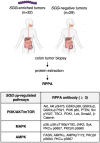
In this work, we investigated the oncogenic role of Streptococcus gallolyticus subsp. gallolyticus (SGG), a gut bacterium associated with colorectal cancer (CRC). We showed that SGG UCN34 accelerates colon tumor development in a chemically induced CRC murine model. Full proteome and phosphoproteome analysis of murine colons chronically colonized by SGG UCN34 revealed that 164 proteins and 725 phosphorylation sites were differentially regulated. Ingenuity Pathway Analysis (IPA) indicates a pro-tumoral shift specifically induced by SGG UCN34, as ~ 90% of proteins and phosphoproteins identified were associated with digestive cancer. Comprehensive analysis of the altered phosphoproteins using ROMA software revealed up-regulation of several cancer hallmark pathways such as MAPK, mTOR and integrin/ILK/actin, affecting epithelial and stromal colonic cells. Importantly, an independent analysis of protein arrays of human colon tumors colonized with SGG showed up-regulation of PI3K/Akt/mTOR and MAPK pathways, providing clinical relevance to our findings. To test SGG's capacity to induce pre-cancerous transformation of the murine colonic epithelium, we grew ex vivo organoids which revealed unusual structures with compact morphology. Taken together, our results demonstrate the oncogenic role of SGG UCN34 in a murine model of CRC associated with activation of multiple cancer-related signaling pathways.
© 2023. Springer Nature Limited.
Conflict of interest statement
The authors declare no competing interests.
Figures


Similar articles
-
Transcriptome profiling of human col\onic cells exposed to the gut pathobiont Streptococcus gallolyticus subsp. gallolyticus.PLoS One. 2023 Nov 30;18(11):e0294868. doi: 10.1371/journal.pone.0294868. eCollection 2023. PLoS One. 2023. PMID: 38033043 Free PMC article.
-
A pathogenicity locus of Streptococcus gallolyticus subspecies gallolyticus.Sci Rep. 2023 Apr 18;13(1):6291. doi: 10.1038/s41598-023-33178-z. Sci Rep. 2023. PMID: 37072463 Free PMC article.
-
A type VII secretion system of Streptococcus gallolyticus subsp. gallolyticus contributes to gut colonization and the development of colon tumors.PLoS Pathog. 2021 Jan 6;17(1):e1009182. doi: 10.1371/journal.ppat.1009182. eCollection 2021 Jan. PLoS Pathog. 2021. PMID: 33406160 Free PMC article.
-
Factors underlying the association between Streptococcus gallolyticus, subspecies gallolyticus infection and colorectal cancer: a mini review.Gut Microbiome (Camb). 2024 Nov 4;5:e9. doi: 10.1017/gmb.2024.11. eCollection 2024. Gut Microbiome (Camb). 2024. PMID: 39703539 Free PMC article. Review.
-
Significance of Streptococcus gallolyticus subsp. gallolyticus Association With Colorectal Cancer.Front Microbiol. 2018 Apr 3;9:614. doi: 10.3389/fmicb.2018.00614. eCollection 2018. Front Microbiol. 2018. PMID: 29666615 Free PMC article. Review.
Cited by
-
Transcriptome profiling of human col\onic cells exposed to the gut pathobiont Streptococcus gallolyticus subsp. gallolyticus.PLoS One. 2023 Nov 30;18(11):e0294868. doi: 10.1371/journal.pone.0294868. eCollection 2023. PLoS One. 2023. PMID: 38033043 Free PMC article.
-
Induction of intercrypt goblet cells upon bacterial infection: a promising therapeutic target to restore the mucosal barrier.Gut Microbes. 2024 Jan-Dec;16(1):2426609. doi: 10.1080/19490976.2024.2426609. Epub 2024 Nov 14. Gut Microbes. 2024. PMID: 39543081 Free PMC article.
-
Sciatica caused by spinal epidural abscess as the initial clinical presentation of colon cancer: a rare case report and review of literature.BMC Infect Dis. 2024 Mar 6;24(1):293. doi: 10.1186/s12879-024-09159-6. BMC Infect Dis. 2024. PMID: 38448866 Free PMC article. Review.
References
-
- Cancer today. http://gco.iarc.fr/today/home.
Publication types
MeSH terms
Substances
LinkOut - more resources
Full Text Sources
Molecular Biology Databases
Miscellaneous

