. 2023 Oct;30(10):1456-1467.
doi: 10.1038/s41594-023-01074-9. Epub 2023 Sep 11.

Dynamics of the DYNLL1-MRE11 complex regulate DNA end resection and recruitment of Shieldin to DSBs

Affiliations

Dynamics of the DYNLL1-MRE11 complex regulate DNA end resection and recruitment of Shieldin to DSBs

Michelle L Swift et al. Nat Struct Mol Biol. 2023 Oct.

Abstract

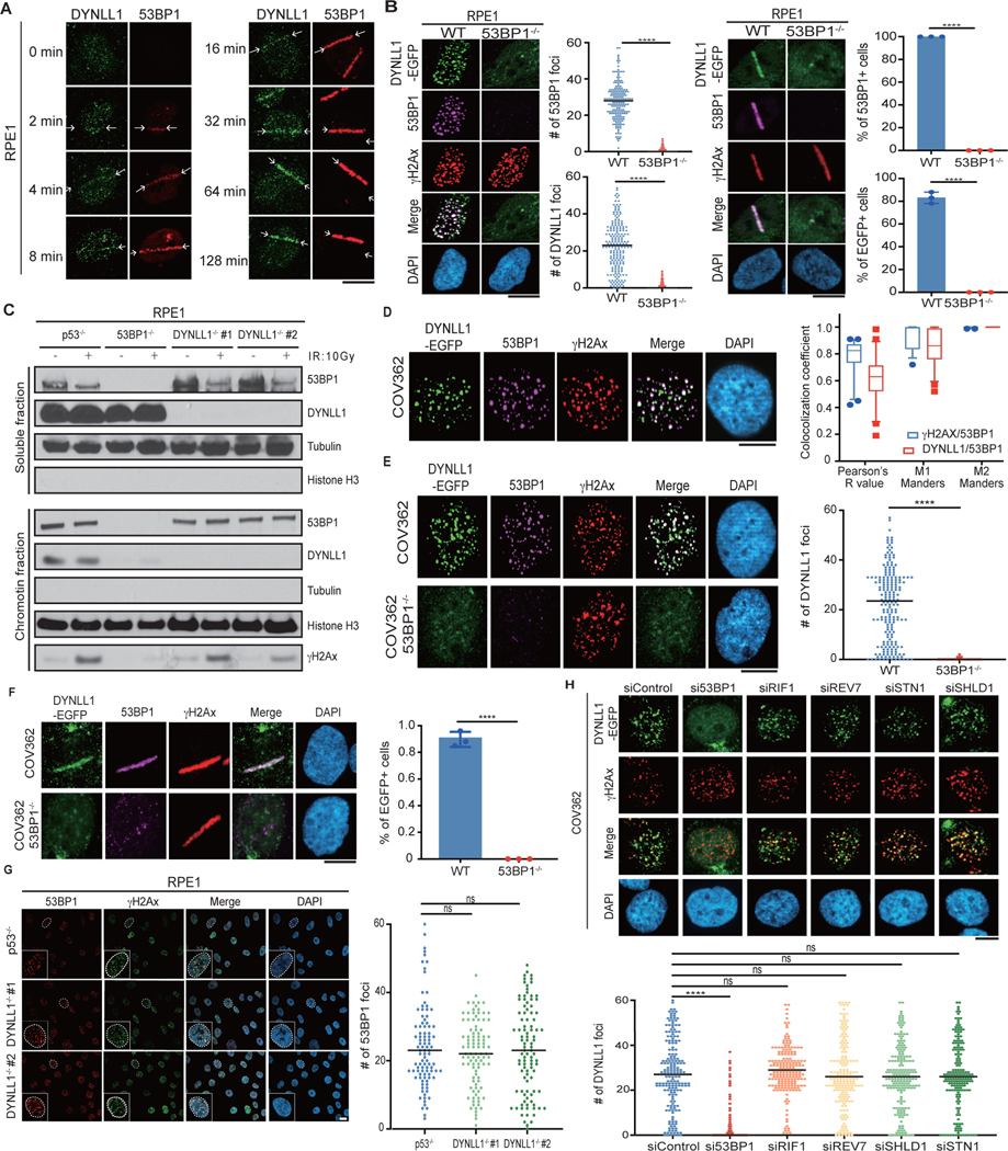
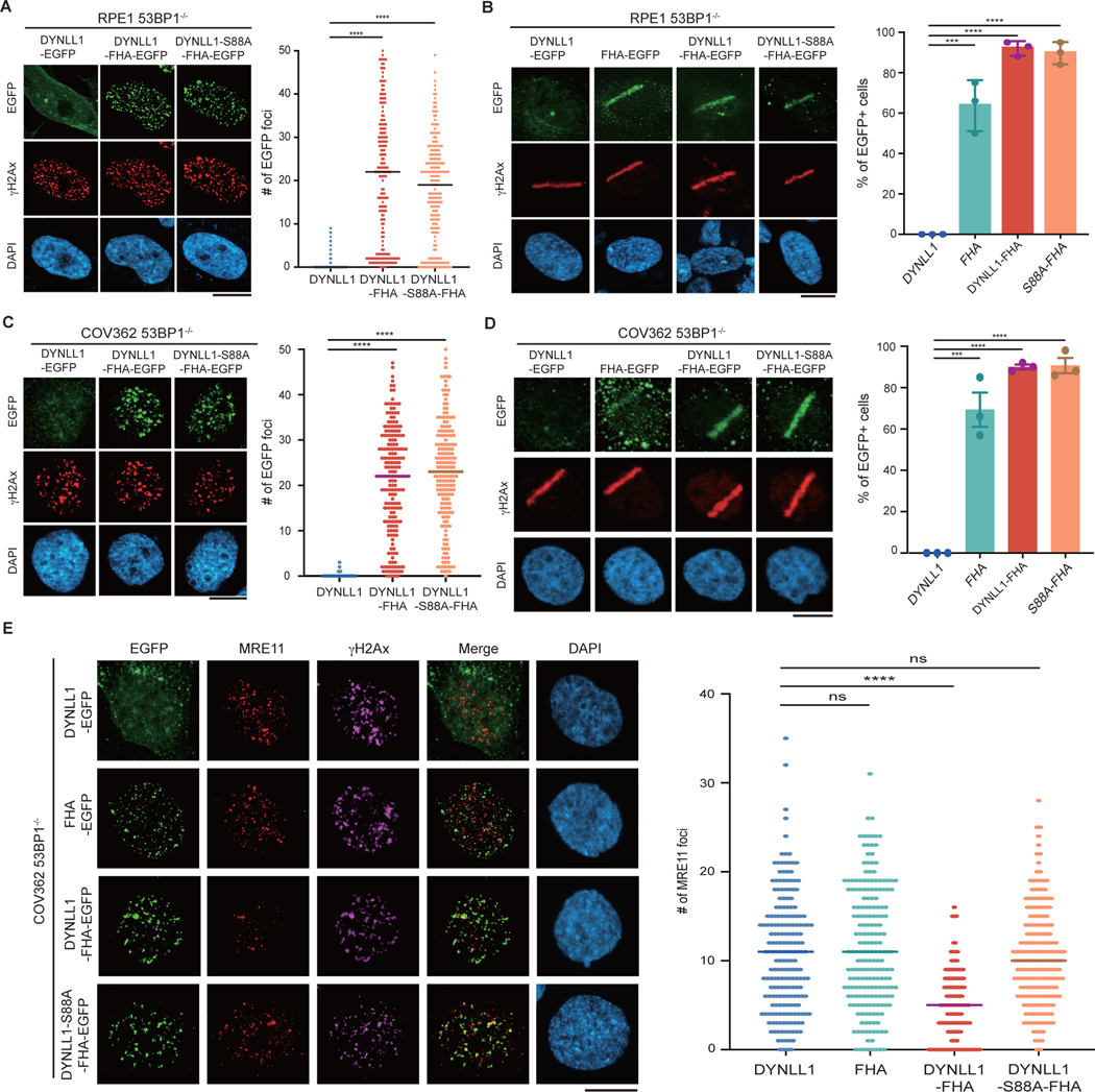
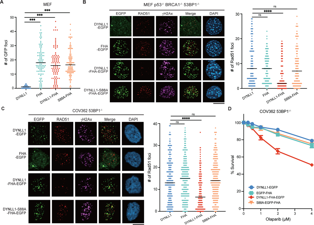
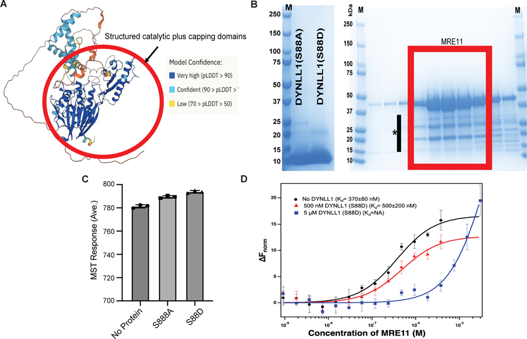
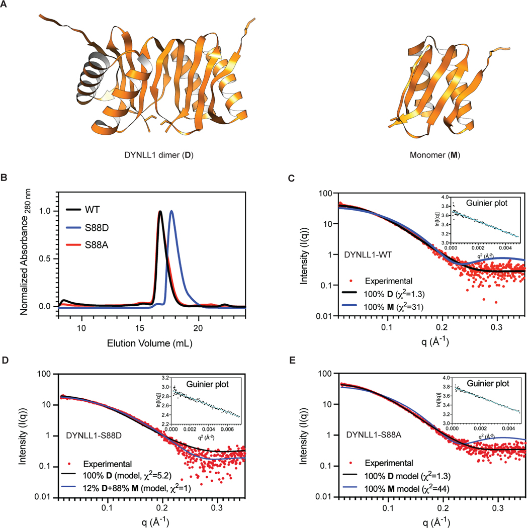
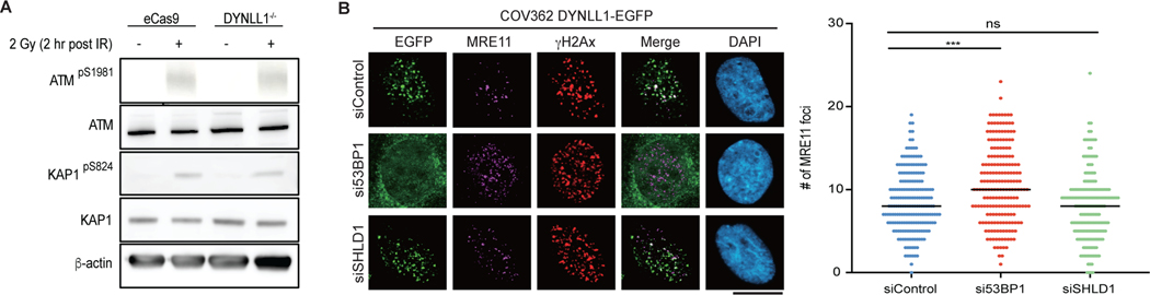
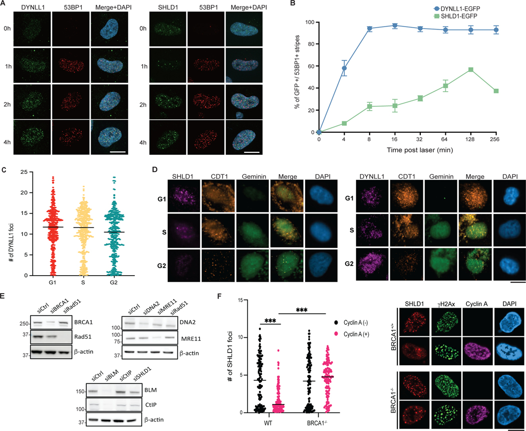
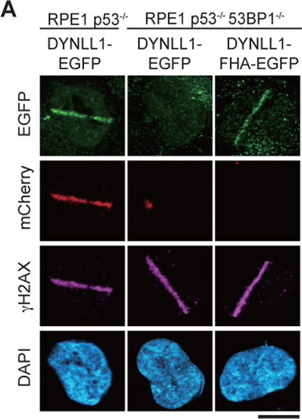
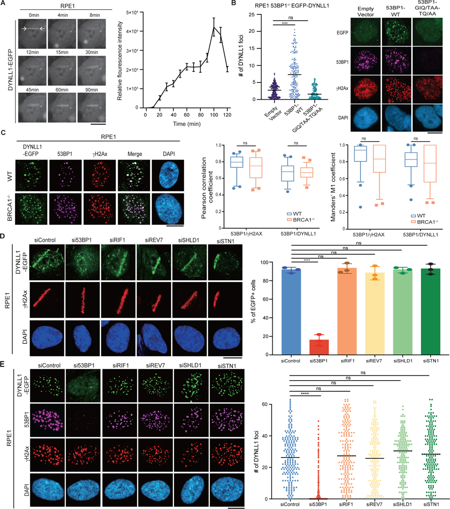
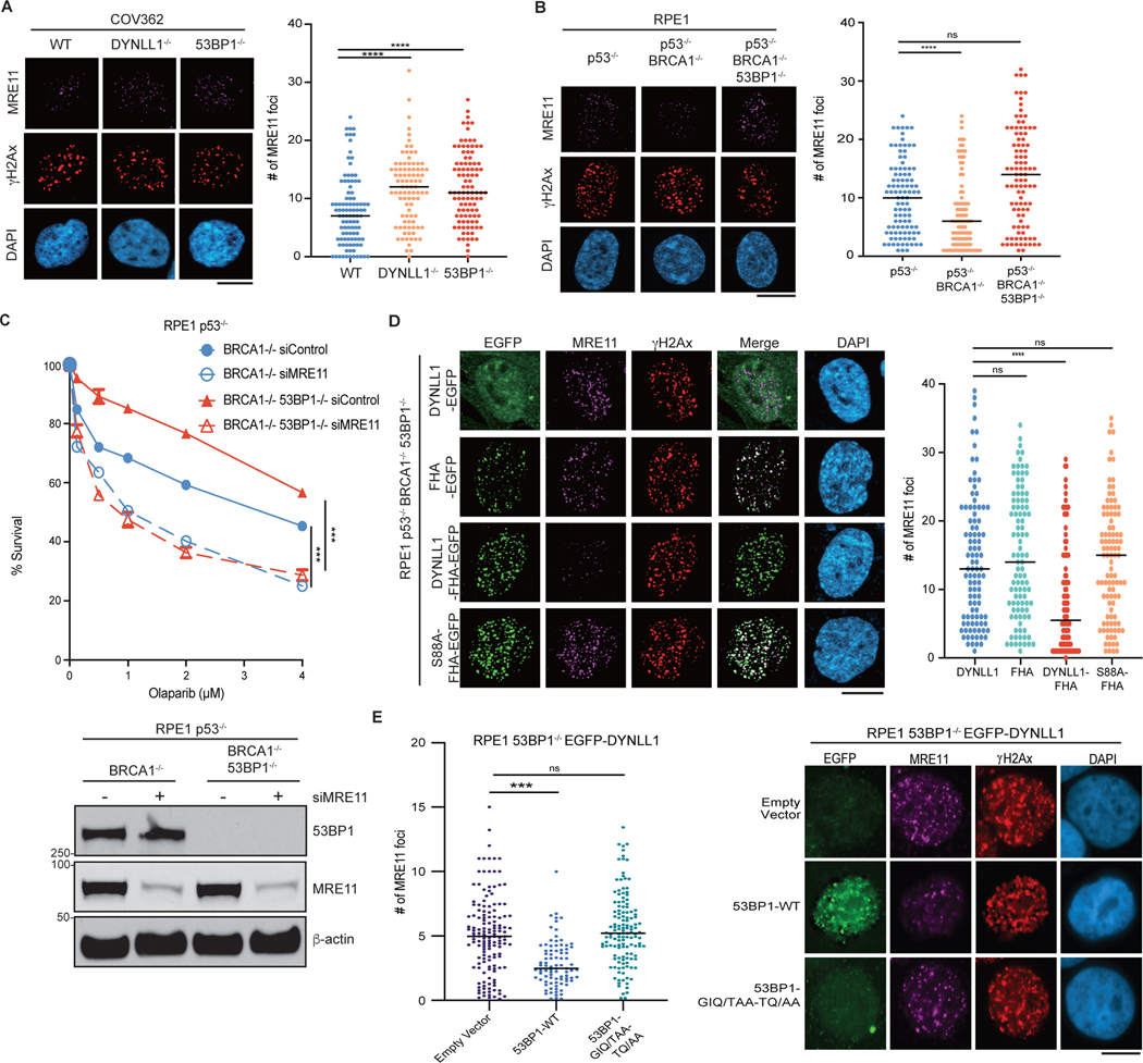
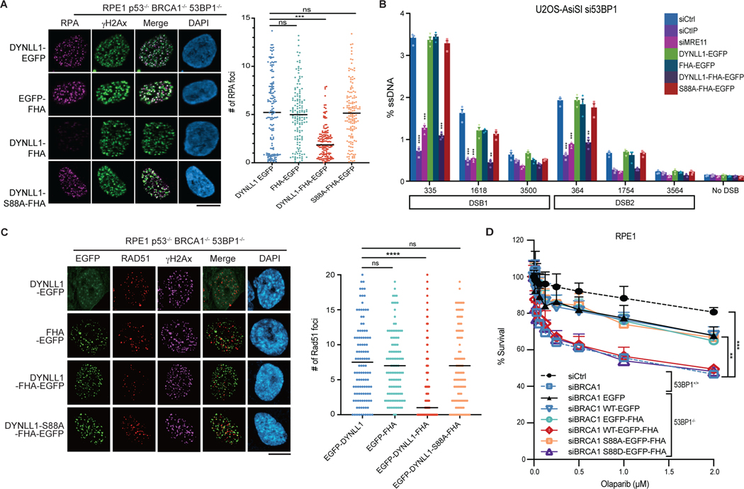
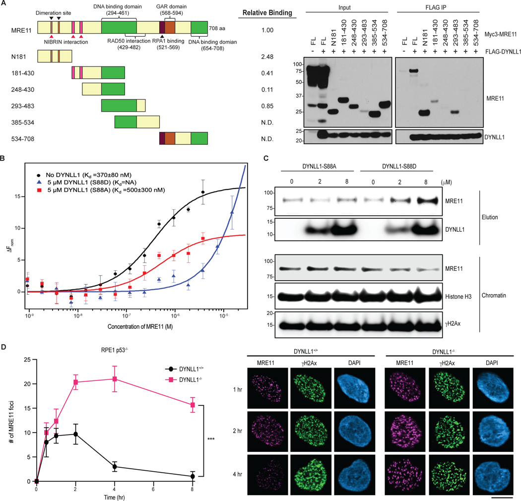
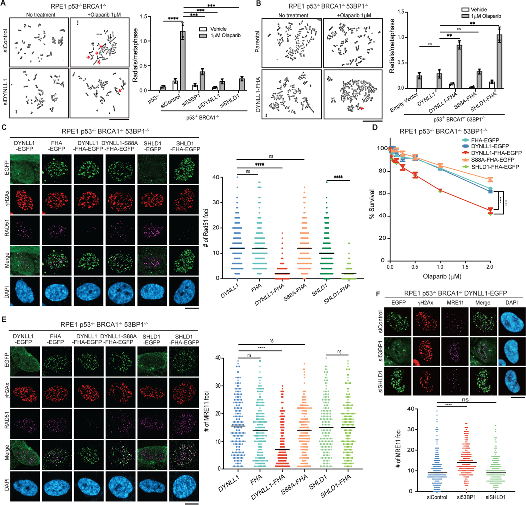
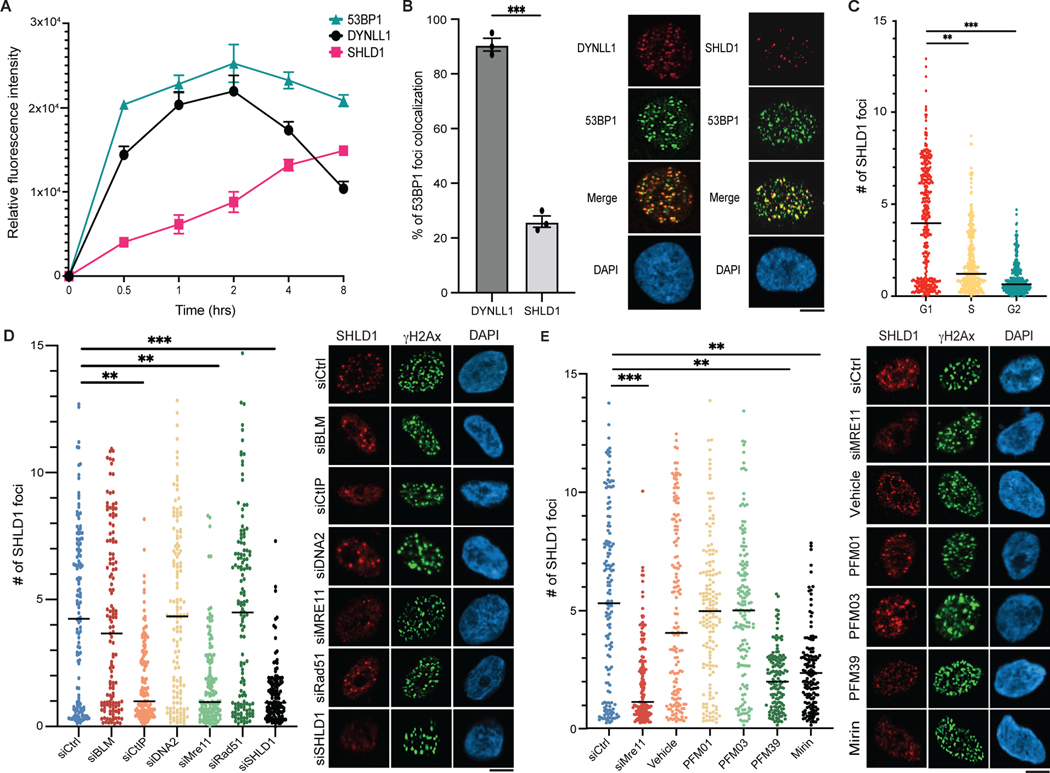
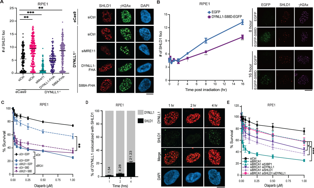
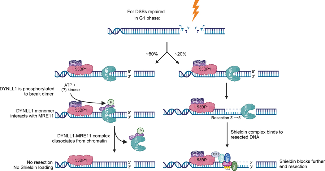
The extent and efficacy of DNA end resection at DNA double-strand breaks (DSB) determine the repair pathway choice. Here we describe how the 53BP1-associated protein DYNLL1 works in tandem with the Shieldin complex to protect DNA ends. DYNLL1 is recruited to DSBs by 53BP1, where it limits end resection by binding and disrupting the MRE11 dimer. The Shieldin complex is recruited to a fraction of 53BP1-positive DSBs hours after DYNLL1, predominantly in G1 cells. Shieldin localization to DSBs depends on MRE11 activity and is regulated by the interaction of DYNLL1 with MRE11. BRCA1-deficient cells rendered resistant to PARP inhibitors by the loss of Shieldin proteins can be resensitized by the constitutive association of DYNLL1 with MRE11. These results define the temporal and functional dynamics of the 53BP1-centric DNA end resection factors in cells.


Conflict of interest statement

A.D.D. reports consulting for AstraZeneca, Bayer AG, Blacksmith/ Lightstone Ventures, Bristol Myers Squibb, Cyteir Therapeutics, EMD Serono, Impact Therapeutics, PrimeFour Therapeutics, Pfizer, Tango Therapeutics and Zentalis Pharmaceuticals/Zeno Management; is an Advisory Board member for Cyteir and Impact Therapeutics; a stockholder in Cedilla Therapeutics, Cyteir, Impact Therapeutics and PrimeFour Therapeutics, and reports receiving commercial research grants from Bristol Myers Squibb, EMD Serono, Moderna and Tango Therapeutics. The remaining authors declare no competing interests.

Figures

Extended Data Fig. 1:
Extended Data Fig. 1:. 53BP1 is necessary for chromatin localization of DYNLL1. Related to Fig. 1.
(a) Representative immunofluorescence images of RPE1 cells subjected to laser microirradiation. Cells were fixed at indicated time points post laser microirradiation and processed for immunofluorescence with DYNLL1 and 53BP1 antibodies. (b) Representative images of RPE1 wild-type or 53BP1−/− cells 2 h after exposure to 2 Gy irradiation or laser microirradiation. Cells were fixed and processed for immunofluorescence using antibodies against 53BP1, GFP (DYNLL1), and γH2AX. (c) RPE1 cells depleted of p53, 53BP1, or DYNLL1 using CRISPR/Cas9 were exposed to 10 Gy irradiation. Protein was collected after 3 h. Localization of DYNLL1 to chromatin was evaluated by subcellular fractionation followed by immunoblotting for DYNLL1. (d–f) Representative images of COV362 cells (D) and COV362 wild-type or 53BP1−/− cells (e, f) exposed to 2 Gy irradiation (d, e) or laser microirradiation (F). 2 h post-recovery cells were fixed and processed for immunofluorescence using antibodies against 53BP1, GFP (DYNLL1), and γH2AX. Box plots show mean and center, quartiles (boxes), and range (whiskers)(d). (g) Representative immunofluorescence images of RPE1 cells depleted of DYNLL1 using CRISPR/Cas9 and exposed to 2 Gy irradiation. 1h post-irradiation, cells were fixed and processed for immunofluorescence using antibodies against 53BP1 and γH2AX. (a-g) n = 3 biologically independent experiments, counting ≥ 100 cells per experiment. Error bars represent the mean±s.e.m. P-values for foci quantification and “laser positive” cell analysis were calculated using two-sided unpaired t-tests. P values are indicated by nonsignificant (P >0.05), *(P < 0.05), **(P < 0.01), ***(P < 0.001), ****(P<0.0001). Black line in dot plots represent median. Scale bar = 20μm.
Extended Data Fig. 2:
Extended Data Fig. 2:. Force tethering DYNLL1 to chromatin inhibits MRE11 foci formation. Related to Fig. 2.
(a, b) RPE1 53BP1−/− cells were transfected with EGFP-tagged DYNLL1 or DYNLL1-FHA constructs. Cells were subjected to 2 Gy irradiation (A) or laser microirradiation (B). 2 h later cells were fixed and processed for immunofluorescence using antibodies against GFP (DYNLL1) and γH2AX. (c, d) COV362 53BP1−/− cells were transfected with a EGFP-tagged DYNLL1 and DYNLL1-FHA constructs. Cells were subjected to 2 Gy irradiation (c) or laser microirradiation (d). 2 h later cells were fixed and processed for immunofluorescence using antibodies against GFP (DYNLL1) and γH2AX. (e) COV362 53BP1−/− cells were transfected with EGFP-tagged DYNLL1 and DYNLL1- FHA constructs. Cells were exposed to 2 Gy irradiation. 2 h post-irradiation cells were fixed and processed for immunofluorescence using antibodies against MRE11, GFP (DYNLL1) and γH2AX. (A-E) n = 3 biologically independent experiments, counting ≥ 100 cells per experiment. Error bars represent the mean±s.e.m. P-values for foci quantification and “laser positive” cell analysis were calculated using two-sided unpaired t-tests. P values are indicated by nonsignificant (P >0.05), *(P < 0.05), **(P < 0.01), ***(P < 0.001), ****(P<0.0001). Black line in dot plots represent median. Scale bar = 20μm.
Extended Data Fig. 3:
Extended Data Fig. 3:. DYNLL1 chromatin binding suppresses 53BP1 loss- induced restoration of HR in BRCA1 deficient cells. Related to Fig. 3.
(a) MEFs expressing EGFP-tagged DYNLL1, and EGFP-tagged DYNLL1-FHA domains constructs were exposed to 2 Gy irradiation. 2 h after irradiation cells were fixed and processed for immunofluorescence using a GFP (DYNLL1) antibody. (b, c) MEF p53−/− BRCA1−/− 53BP1−/− cells (b) and COV362 53BP1−/− cells (c) were transfected with EGFP-tagged DYNLL, or EGFP-tagged DYNNL1-FHA constructs. Cells were exposed to 2 Gy irradiation. 2 h later, cells were fixed and processed for immunofluorescence using antibodies against GFP (DYNLL1), RAD51, and γH2AX. (d) COV362 53BP1−/− cells were transfected EGFP-tagged DYNLL, or EGFP-tagged DYNNL1-FHA constructs. Cells were treated with indicated concentrations of Olaparib for 6 days. Percent survival was determined via a cell viability assay. (a-d) n = 3 biologically independent experiments, counting ≥ 100 cells per experiment. Error bars represent the mean±s.e.m. P-values for foci quantification were calculated using two-sided unpaired t-tests. P-value measurements for cell survival curves were assessed by non-regression curve analysis. P values are indicated by nonsignificant (P >0.05), *(P < 0.05), **(P < 0.01), ***(P < 0.001),****(P<0.0001). Black line in dot plots represent median. Scale bar = 20μm.
Extended Data Fig. 4:
Extended Data Fig. 4:. DYNLL1 interferes with MRE11 dimerization. Related to Fig. 4.
(a) Predicted structure of full-length MRE11 created by AlphaFold Monomer V2 for Uniprot Accession number P49959. The structured catalytic domain of MRE11 is highlighted with a red circle. The model is color coded in terms of confidence in prediction and respective color schemes for the confidence is given in the figure. In general, disordered regions have less confidence in model prediction, thus indicating the unstructured regions of MRE11 beyond capping domain. (b) Coomassie-stained protein gels indicating the quality of the recombinant protein used in the current study. Left: DYNLL1 mutants after cleaving the His-tag with TEV protease. Right: MRE11 catalytic domain after the gel-filtration purification step. The red rectangle indicates the fractions that are combined. M indicates the protein standards and * indicates the MRE11 degradation bands. (c) Average MST response (n = 3) measured from labeled MRE11 in the MST buffer or MST buffer with 5 μM DYNLL1-S88A or DYNLL1-S88D mutant. (d) Change in the normalized fluorescence (ΔFnorm) as result of thermophoresis in the MST experiment plotted as a function of concentration of unlabeled MRE11. The resulting curves represents MRE11 dimerization in the absence of any DYNLL1 (black circles), in the presence of 500 nM DYNLL1-S88D (red triangles) or in the presence of 5 μM DYNLL1-S88D (blue squares). The Kd values are measured by fitting the curves with Kd model in the analysis software. (B-D) The data points represent average of three independent measurements and error bars represents standard deviation.
Extended Data Fig. 5:
Extended Data Fig. 5:. Solution structures of DYNLL1-WT and mutants.
(a) AlphaFold2 predicted models of DYNLL1 dimer (left) and monomer (right). (b) Size exclusion chromatography elution profiles for DYNLL1-WT, S88A and S88D mutants. For clarity, initial 8 mL (pre-void volume with no peaks) were omitted from the chromatograms. (c–e) FoXS fitting of experimental X-ray scattering data (red dots) to theoretical SAXS profiles (solid lines) derived from structural models of DYNLL1 monomer (M) and dimer (D) or mixture of monomer and dimer state. The goodness of the fit is evaluated by χ2. Guinier plots from the measured scattering intensity (I(q)) as a function of scattering vector (q) in the low q region shown as insets for WT and mutant proteins.
Extended Data Fig. 6:
Extended Data Fig. 6:. Depletion of the Shieldin complex does not affect MRE11 recruitment. Related to Fig. 5.
(a) Protein expression from lysates collected from RPE1 wild-type or DYNLL1−/− cells 2 h after 2 Gy irradiation. (b) COV362 cells overexpressing EGFP-DYNLL1 and transfected with siRNA targeting either 53BP1, or SHLD1 were exposed to 2 Gy irradiation. Cells were fixed 2 h post exposure and processed for immunofluorescence using antibodies against GFP (DYNLL1), γH2AX, and MRE11. (a, b) n = 3 biologically independent experiments, counting ≥ 100 cells per experiment. P-values for foci quantification were calculated using two-sided unpaired t-tests. P values are indicated by nonsignificant (P >0.05), *(P < 0.05), **(P < 0.01), ***(P < 0.001), ****(P<0.0001). Black line in dot plots represent median. Scale bar = 20μm.
Extended Data Fig. 7:
Extended Data Fig. 7:. Shieldin is recruited to DSBs later than DYNLL1 and in G1 phase only. Related to Fig. 6.
(a) Representative images from Fig. 6a. (b) Quantification of GFP positive stripes for cells expressing DYNLL1-EGFP or SHLD1-EGFP after exposure to laser microirradiation and fixed at indicated time points. (c) Quantification of number of DYNLL1 foci for RPE1 cells transduced with lentivirus comprised of the Fucci system reporter assay. Cells were exposed to 10 Gy irradiation, fixed 6 h later, and processed using antibodies against Geminin, Cdt1, and DYNLL1. (d) Representative images for Fig. 6c and Extended Data Fig. 6c. (e) Representative western blots showing knockdown of indicated proteins for Fig. 6d. (f) RPE1 BRCA1−/− were subjected to 10 Gy IR and 4 h later were fixed and processed for immunofluorescence using antibodies against SHLD1, Cyclin A, and γH2AX. (a-f) n = 3 biologically independent experiments, counting ≥ 100 cells per experiment. Error bars represent the mean±s.e.m. P-values for foci quantification and ‘laser positive’ cell analysis was calculated using two-sided unpaired t-tests. P values are indicated by nonsignificant (P >0.05), *(P < 0.05), **(P < 0.01), ***(P < 0.001), ****(P<0.0001). Black line in dot plots represent median. Scale bar = 20μm.
Extended Data Fig. 8:
Extended Data Fig. 8:. Shieldin functions downstream of DYNLL1, but is not dependent on DYNLL1 for its localization to chromatin. Related to Fig. 7.
(a) RPE1 p53−/− and RPE1 p53−/− 53BP1−/− cells were transfected with EGFP-tagged DYNLL1 or EGFP-tagged DYNLL1-FHA. Cells were then transfected with mCherry-SHLD1 and subjected to laser microirradiation. 2 h after laser microirradiation cells were fixed and processed for immunofluorescence using antibodies against GFP (DYNLL1), mCherry (SHLD1), and γH2AX. n = 3 biologically independent experiments. Scale bar = 20μm.
Fig. 1 |
Fig. 1 |. DYNLL1 recruitment to DSBs is dependent on 53BP1 but independent of other 53BP1-associated factors.
a, Representative live cell images of RPE1 cells expressing DYNLL1-EGFP after laser microirradiation. b, RPE1 53BP1−/− cells coexpressing EGFP-DYNLL1 and 53BP1–DYNLL1-binding mutants were subjected to 2 Gy of irradiation and 2 h later were processed for immunofluorescence. c, Representative immunofluorescent images of wild-type (WT) RPE1 or BRCA1−/− cells 2 h after exposure to 2 Gy of irradiation. Box plots show the mean, upper and lower quartiles (boxes) and range (whiskers). d,e, Representative images of RPE1 cells coexpressing EGFP-tagged DYNLL1 and short interfering RNA (siRNA) constructs and subjected to laser microirradiation (d) or 2 Gy of irradiation (e) and fixed 1 h later. siControl denotes siRNA against non-targeting control. In ae, n = 3 biologically independent experiments, counting at least 100 cells per experiment. Error bars represent mean ± s.e.m. P values were determined by Mann–Whitney test (b, c, e) or two-sided unpaired t-tests (d). ****P < 0.0001. NS, not significant (P < 0.05). Black lines in dot plots represent the median. DAPI, 4,6-diamidino-2-phenylindole. Scale bars, 20 μm.
Fig. 2:
Fig. 2:. DYNLL1 regulates MRE11 activity independent of 53BP1.
a,b, COV362 cells depleted of DYNLL1 or 53BP1 (a) and RPE1 cells depleted of p53, BRCA1 and/or 53BP1 (b) using CRISPR–Cas9 were exposed to 2 Gy of irradiation for 2 h. Cells were fixed and immunostained using antibodies against MRE11 and γH2AX. c, RPE1 p53−/− BRCA1−/− and p53−/− BRCA1−/− 53BP1−/− cells were depleted of MRE11 using siRNA (siMRE11), and treated with indicated concentrations of olaparib for 6 days. Survival was determined via a cell viability assay (top), and a representative western blot shows successful knockdown of MRE11 using siRNA (bottom). d, Immunofluorescence of RPE1 p53−/− BRCA1−/− 53BP1−/− cells expressing EGFP-tagged DYNLL1 constructs exposed to 2 Gy of irradiation for 2 h, and stained using antibodies against MRE11, GFP (DYNLL1) and γH2AX. e, Immunofluorescence of RPE1 53BP1−/− cells coexpressing EGFP-DYNLL1 and 53BP1–DYNLL1-binding mutants exposed to 2 Gy of irradiation for 2 h, and stained using antibodies against MRE11, 53BP1 and γH2AX. In ae, n = 3 biologically independent experiments, counting at least 100 cells per experiment. P values determined by two-sided unpaired t-tests (a,b,d,e) or nonregression curve analysis (c). ***P < 0.001, ****P < 0.0001. Black lines in dot plots represent medians. Scale bars, 20 μm.
Fig. 3:
Fig. 3:. Functional effect of DYNLL1 at DSBs in 53BP1-deficient cells.
a, Immunofluorescence of RPE1 p53−/− BRCA1−/− 53BP1−/− cells expressing EGFP-tagged DYNLL1 or FHA–DYNNL1 constructs exposed to 2 Gy of irradiation for 4 h, and stained with, antibodies against GFP (DYNLL1), RPA32 and γH2AX. b, U2OS AsiSI cells cotransfected with siRNA against 53BP1 and EGFP-tagged DYNLL1 or FHA–DYNLL1 constructs were treated with 300 nM 4-OHT for 4 h, and ssDNA formation was determined at various sites downstream of the break via PCR quantification. siCtrl denotes non-targeting control siRNA; siCtIP denotes siRNA against CtIP (also known as RBBP8). c, Immunofluorescence of RPE1 p53−/− BRCA1−/− 53BP1−/− cells expressing EGFP-tagged DYNLL1 or FHA–DYNNL1 constructs exposed to 2 Gy of irradiation for 4 h. Cells were stained with antibodies against GFP (DYNLL1), RAD51 and γH2AX. d, RPE1 p53−/− 53BP1−/− cells were cotransfected with siRNA against BRCA1 (siBRCA1) and EGFP-tagged DYNLL1 or FHA–DYNLL1 constructs, and treated with indicated concentrations of olaparib for 6 days. Survival was determined via a cell viability assay. In ad, n = 3 biologically independent experiments, counting at least 100 cells per experiment. Error bars represent mean ± s.e.m. P values were calculated using two-sided unpaired t-tests (a,c) or nonregression curve analysis (d). **P < 0.01, ***P < 0.001, ****P < 0.0001. Black lines in dot plots represent medians. Scale bars, 20 μm.
Fig. 4:
Fig. 4:. DYNLL1 disrupts MRE11 dimerization to impair its retention on chromatin.
a, Left: schematic of MRE11 truncation mutants. Right: relative binding of MRE11 mutants and DYNLL1. Immunoprecipitation (IP) from cells coexpressing Flag–DYNLL1 and Myc3–MRE11 truncation mutants. ND, not detected. b, Change in the normalized fluorescence (ΔFnorm) as a result of thermophoresis in the MST experiment plotted as a function of the concentration of unlabeled MRE11. The resulting curves represent MRE11 dimerization in the absence of any DYNLL1 (black circles), or in the presence of 5 μM DYNLL1-S88D (blue triangles) or 5 μM DYNLL1-S88A (red squares). The dissociation constant (Kd) values are measured by fitting the curves with the Kd model in the analysis software. Data are mean and s.d. of three independent measurements. c, Western blot analysis of purified DYNLL1-S88D and DYNLL1-S88A proteins (Extended Data Fig. 4b) incubated with pre-extracted chromatin from HEK293T cells after 5 Gy of irradiation. Incubation of recombinant DYNLL1-S88D but not DYNLL1-S88A protein resulted in increased MRE11 elution from damaged chromatin. d, Immunofluorescence of DYNLL1+/+ and DYNLL1−/− cells exposed to 2 Gy of irradiation and fixed at various time points after irradiation. ad, n = 3 biologically independent experiments, counting at least 100 cells per experiment. Error bars represent s.d. (b) or mean ± s.e.m. (d). P values determined by two-sided unpaired t-tests. ***P < 0.001. Scale bar, 20 μm.
Fig. 5:
Fig. 5:. Functional comparison of DYNLL1 and the Shieldin complex.
a, RPE1 p53−/− BRCA1−/− cells depleted of 53BP1, DYNLL1 or SHLD1 were treated with olaparib. Representative images of metaphase spreads (left) and quantification of the number of redials per cell (right) are shown. b, RPE1 p53−/− BRCA1−/− 53BP1−/− cells expressing DYNLL1–FHA or SHLD1–FHA constructs were treated with olaparib. Representative images of metaphase spreads (left) and quantification of the number of radials per cell (right) are shown. c, Immunofluorescence of cells expressing EGFP-tagged DYNLL1, FHA–DYNLL1 or SHLD1–FHA constructs exposed to 2 Gy of irradiation for 4 h, and stained using antibodies against RAD51, GFP (DYNLL1) and γH2AX. d, RPE1 p53−/− BRCA1−/− 53BP1−/− cells expressing EGFP-tagged DYNLL1, FHA–DYNLL1 or SHLD1–FHA constructs were treated with indicated concentrations of olaparib for 6 days, and cell survival was determined via a cell viability assay. e, Immunofluorescence of cells expressing EGFP-tagged DYNLL1, FHA–DYNLL1, or SHLD1–FHA constructs exposed to 2 Gy of irradiation for 2 h, and stained using antibodies against MRE11, GFP (DYNLL1) and γH2AX. f, Immunofluorescence of RPE1 p53−/− BRCA1−/− 53BP1−/− cells coexpressing EGFP- tagged DYNLL1 and indicated siRNAs exposed to 2 Gy of irradiation for 2 h, and stained using antibodies against GFP, γH2AX and MRE11. n = 2 (a,b) or n = 3 (cf) biologically independent experiments, counting at least 100 cells per experiment. Error bars represent mean ± s.e.m. P values were determined by two-sided unpaired t-tests (ac,e,f) or nonregression curve analysis (d). **P < 0.01, ***P < 0.001, ****P < 0.0001. Black lines in dot plots represent medians. Scale bars, 20 μm.
Fig. 6:
Fig. 6:. Kinetics and dependencies of Shieldin complex recruitment to DSBs.
a, RPE1 cells were pretreated with EdU, exposed to 10 Gy of irradiation, and fixed at indicated time points. Cells were processed for immunofluorescence using antibodies against 53BP1, DYNLL1, SHLD1 and γH2AX. Relative fluorescence intensity is normalized to γH2AX. EdU-negative cells were quantified. b, Immuofluorescence of RPE1 cells pretreated with EdU, exposed to 10 Gy of irradiation for 4 h, and stained using antibodies against DYNLL1 and 53BP1, or SHLD1 and 53BP1. Colocalization of DYNLL1 or SHLD1 foci with 53BP1 foci in EdU-negative cells is quantified. c, RPE1 cells were transduced with lentivirus composed of the Fucci system reporter assay. Cells were exposed to 10 Gy of irradiation, fixed 6 h later and processed using antibodies against geminin, CDT1 and SHLD1. d,e, Cells were transfected with the indicated siRNA (d) or treated with MRE11 endo- and exonuclease inhibitors (e). Cells were pretreated with EdU for 30 min, then exposed to 10 Gy of irradiation and fixed 6 h later to be processed for immunofluorescence. EdU-negative cells were quantified. ae, n = 3 biologically independent experiments, counting at least 100 cells per experiment. Error bars represent mean ± s.e.m. P values were determined using two-sided unpaired t-tests. **P < 0.01, ***P < 0.001, ****P < 0.0001. Black lines in dot plots represent medians. Scale bars, 20 μm.
Fig. 7:
Fig. 7:. DYNLL1 is required for Shieldin loading to DSBs.
a, Wild-type or DYNLL1−/− RPE1 cells expressing DYNLL1–FHA constructs were pretreated with EdU for 30 min, exposed to 10 Gy of irradiation, and fixed 6 h later for immunofluorescence. EdU-negative cells were quantified. b, RPE1 cells expressing EGFP-tagged wild-type DYNLL1 or its phosphomimetic, S88D, were exposed to 10 Gy of irradiation and fixed at the indicated time points. Cells were stained using antibodies against GFP (DYNLL1), γH2AX and SHLD1. c, BRCA1-depleted RPE1 cells coexpressing DYNLL1 constructs and siRNA against SHLD1 (siSHLD1) were treated with indicated concentrations of olaparib. Cell viability assays were performed 6 days after treatment. d, RPE1 cells expressing EGFP-DYNLL1 were pretreated with EdU then exposed to 10 Gy of irradiation for 4 h. Cells were fixed and processed using antibodies against GFP (DYNLL1), SHLD1 and 53BP1. Colocalization between DYNLL1 and SHLD1 is quantified in EdU-negative cells. e, RPE1 cells depleted of BRCA1, 53BP1, DYNLL1 and/or SHLD1 were treated with indicated concentrations of olaparib. Cell viability assays were performed 6 days after treatment. For all experiments, n = 3 biologically independent experiments, counting at least 100 cells per experiment. Error bars represent mean ± s.e.m. P values determined using two-sided unpaired t-tests (a,b,d) or nonregression curve analysis (c,e). *P < 0.05, **P < 0.01, ***P < 0.001, ****P < 0.0001. Black lines in dot plots represent medians. Scale bars 20 μm.
Fig 8:
Fig 8:. Phosphorylated DYNLL1 negatively regulates end resection and Shieldin complex recruitment at DSBs.
The model shows that in about 80% of DSB repair that does not require end resection, phosphorylated DYNLL1-S88 binds to and removes MRE11 from chromatin to inhibit resection. However, in a subset of DSBs, 53BP1 bound DYNLL1 remains unphosphorylated allowing MRE11 activity. MRE11 exonuclease activity creates ssDNA permitting Shieldin complex binding and inhibition of long-range end resection.

Update of

References

    1. Zhao W, Wiese C, Kwon Y, Hromas R. & Sung P. The BRCA Tumor Suppressor Network in Chromosome Damage Repair by Homologous Recombination. Annu Rev Biochem 88, 221–245 (2019). - PMC - PubMed
    1. Zhao B, Rothenberg E, Ramsden DA & Lieber MR The molecular basis and disease relevance of non-homologous DNA end joining. Nat Rev Mol Cell Biol 21, 765–781 (2020). - PMC - PubMed
    1. Hustedt N. & Durocher D. The control of DNA repair by the cell cycle. Nat Cell Biol 19, 1–9 (2016). - PubMed
    1. Gnugge R. & Symington LS DNA end resection during homologous recombination. Curr Opin Genet Dev 71, 99–105 (2021). - PMC - PubMed
    1. Bunting SF et al. 53BP1 inhibits homologous recombination in Brca1-deficient cells by blocking resection of DNA breaks. Cell 141, 243–54 (2010). - PMC - PubMed

Publication types

MeSH terms