. 2023 Sep 11;13(1):14951.
doi: 10.1038/s41598-023-42339-z.

Continuous anesthesia for 60 days in an isosmotic environment does not impair limb or cardiac regeneration in the axolotl

Affiliations

Continuous anesthesia for 60 days in an isosmotic environment does not impair limb or cardiac regeneration in the axolotl

Sofie Amalie Andersson et al. Sci Rep. .

Abstract

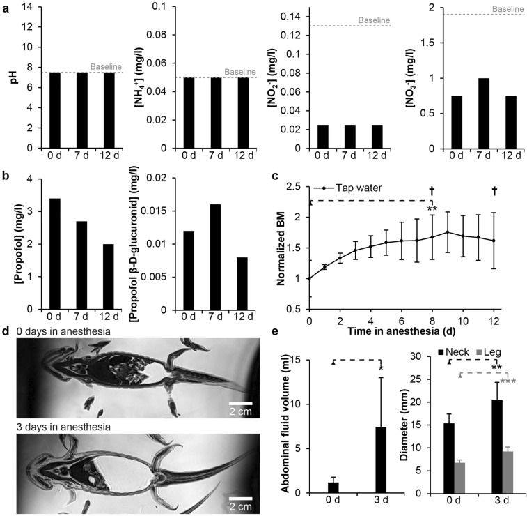
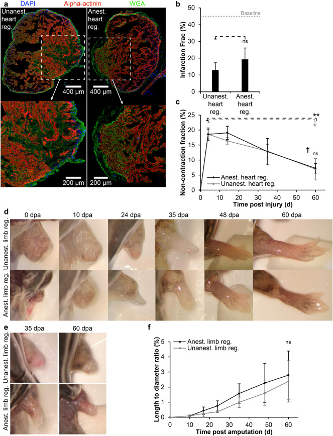
Longitudinal animal experiments in the field of regenerative biology often require repeated use of short-term anesthesia (minutes to a few hours). Regain of consciousness limits the level of acceptable invasiveness of procedures, and it makes it difficult to untangle behavioral changes caused by injury to physiological processes involved in the regenerative response. Therefore, a method to keep a regenerative research animal in a comatose state under continuous anesthesia during regenerative experiments often spanning months, would be ethically and experimentally desirable. Here we report on a method using propofol based anesthesia in an isosmotic environment that allows for continuous anesthesia of regenerating axolotls for 60 days with a 75% survival rate, thus spanning the majority of a full regenerative cycle following limb amputation or cryoinjury to the heart. No differences were detected in the axolotl's ability to regenerate amputated limbs and cardiac cryo-injury while anesthetized, however some regenerative failures in the limb were observed in both anesthetized and unanesthetized control groups, most likely caused by prolonged fasting. Sixty days of anesthesia may be approaching a level were kidney function is affected, but the 75% surviving anesthetized animals recovered well after anesthesia and showed a full behavioral recovery within 17 days.


Conflict of interest statement

The authors declare no competing interests.

Figures

Figure 1
Figure 1
Waste production, propofol consumption and long-term anesthesia in conventional tap water housing medium. (a) pH, ammonium ion, nitrite ion and nitrate ion concentration over time in 10 l housing medium containing six axolotls during 12 days of continuous anesthesia. (b) Propofol and propofol β-d-glucuronid concentration over time in housing medium. (c) Normalized body mass (BM) over time. Body mass was significantly increased (**p < 0.01, based on paired t-test; n = 6) until an animal died at day 8 and again at day 12 (cross symbol) and anesthesia was ended. (d) Representative virtual coronal magnetic resonance imaging slices of the same axolotl at the beginning of anesthesia and after 3 days showing gradual built up of abdominal fluid. (e) Significant increase in abdominal fluid (*p < 0.05, based on paired t-test; n = 6) and diameter at the level of the neck (**p < 0.01, based on paired t-test; n = 6) and hind legs (***p < 0.001, based on paired t-test; n = 6) following 3 days in propofol anesthesia in tap water housing medium.
Figure 2
Figure 2
The effect of regulating osmolarity in housing medium and of fasting on body mass over time and cardiac regeneration. (a) Normalized body mass (BM) over the course of 3 days of propofol anesthetized axolotls (three animals per group) housed in hyposmotic tap water, hyposmotic 100% Holtfreter’s solution or isosmotic axolotl adjusted Ringer’s solution. (b) Normalized body mass of conscious axolotls with a cardiac cryoinfarction housed in either tap water or Ringer’s solution with or without feeding for 60 days. Axolotls housed in Ringer’s solution with feeding significantly increased their body mass over the course of the experiment (*p < 0.05, based on paired t-test; n = 4), whereas fasted animals housed in both tap water and Ringer’s solution significantly decreased their body mass over time (***p < 0.001, based on paired t-test; n = 4 and *p < 0.05, based on paired t-test; n = 4, respectively). (c) The non-contraction fraction of the ventricle in axolotls with a cardiac cryoinfarction housed in either tap water or Ringer’s solution with or without feeding for 60 days. All groups showed functional regeneration of the heart expressed as a decrease in the non-contraction fraction (Tap water, fasting: *p < 0.05, Ringer’s solution, fed: **p < 0.01, Ringer’s solution fasted: **p < 0.01, respectively, all based on paired t-tests; n = 4) and there was no significant difference in non-contraction fraction between groups (ns not significant, based on one-way ANOVA; n = 4 pr. group).
Figure 3
Figure 3
Survival rate, propofol infusion rate and body mass over time in continuously anesthetized axolotls in isosmotic housing medium. (a) Survival rate over time in anesthetized control axolotls (Anest. control) without injury, anesthetized and unanesthetized axolotls performing limb regeneration (Anest. limb reg. and Unanest. limb reg.), anesthetized and unanesthetized axolotls performing heart regeneration (Anest. heart reg. and Unanest. heart reg.), and combined for all anesthetized axolotls. (b) Propofol infusion rate over the course of 60 day. After the loss of two anesthetized control animals at day 9, infusion was briefly stopped to allow the animals to metabolize some of the built up propofol in the tissue while still being under complete anesthesia, before propofol infusion was resumed. (c) Normalized body mass over time of all groups. The loss of two anesthetized control animals at day 9 and one anesthetized heart regeneration animal at day 56 are indicated by cross symbols.
Figure 4
Figure 4
Heart and limb regeneration during continuous propofol anesthesia. (a) Representative cryosections of the cardiac infarction zone in an unanesthetized (left) and an anesthetized axolotl (right) after 60 days of regeneration. The injury zone is still visible via wheat-germ agglutinin staining in both hearts at this time point but it is highly infiltrated by cardiomyocytes made visible by alpha-actinin staining. White boxes are magnified ×2 in images at the bottom. (b) There was no significant difference in infarction fraction measured by quantitative histology between hearts at 60 days post injury in the unanesthetized (Unanest. heart reg) and the anesthetized group (Anest. heart reg) (ns, not significant, based on unpaired t-test, n = 3 for each group). (c) The non-contraction fraction of the ventricle in unanesthetized and anesthetized axolotls with a cardiac cryoinfarction over the course of 60 days. Both groups showed a significant decrease in non-contraction fraction from day 4 to day 60 post injury (*p < 0.05 and **p < 0.01, respectively, based on paired t-tests; n = 3 for each group) and there was no significant difference between unanesthetized and anesthetized animals at day 60 (ns not significant, based on unpaired t-test; n = 3 for each group). (d,e) Representative photos of amputated right front limb in anesthetized and unanesthetized axolotls at 0, 10, 24, 35, 48 and 60 days post amputation (dpa). Most axolotls showed complete limb regeneration except from one animal from each of the anesthetized and unanesthetized groups showing a regression of the initially regenerating limb (e). (f) Length to diameter ratio of regenerating limbs over time. There was no significant difference of limb size at day 60 between anesthetized and unanesthetized axolotls (unpaired t-test, n = 4 and 3, p = 0.77).
Figure 5
Figure 5
Oxygen consumption, skin toxicity and behavioral effects of continuous propofol anesthesia. (a) Oxygen consumption rate over time in anesthetized control axolotls (Anest. control) without injury, anesthetized and unanesthetized axolotls performing limb regeneration (Anest. limb reg. and Unanest. limb reg.), anesthetized and unanesthetized axolotls performing heart regeneration (Anest. heart reg. and Unanest. heart reg.). Oxygen consumption rate was significantly different from unanesthetized baseline at some time points in the anesthetized limb regeneration group and in both the anesthetized and the unanesthetized heart regeneration groups (see text for details). (b) plasma creatinine level in the different treatment groups over time. Although not significantly regulated, there was a tendency of an increase in creatinine level over time. (c) Small skin lesions were visible on the ventral surface of some anesthetized axolotls (black arrows). White arrow points to healing skin at the heart injury zone. (d) Lactate dehydrogenase (LDH) release from skin samples showed increased toxicity of propofol at high concentrations although not pronounced within the used therapeutic zone. (e) Percent time of axolotls being active at baseline and again at 7 days of recovery and 17 days of recovery following 60 days of anesthesia. Data is shown for both previously anesthetized control animals as well as these animals in combination with previously anesthetized limb regeneration animals (All). Long term propofol anesthesia significantly affected activity levels 7 days after recovery (p = 0.014), whereas at 17 days post recovery it was normalized to pre anesthesia level (p = 0.35).

Similar articles

References

    1. Laflamme MA, Murry CE. Heart regeneration. Nature. 2011;473:326–335. doi: 10.1038/nature10147. - DOI - PMC - PubMed
    1. Pfefferli C, Jaźwińska A. The art of fin regeneration in zebrafish. Regen (Oxf., Engl.) 2015;2:72–83. - PMC - PubMed
    1. Roy S, Gatien S. Regeneration in axolotls: A model to aim for! Exp. Gerontol. 2008;43:968–973. doi: 10.1016/j.exger.2008.09.003. - DOI - PubMed
    1. Poss KD, Wilson LG, Keating MT. Heart regeneration in zebrafish. Science. 2002;298:2188–2190. doi: 10.1126/science.1077857. - DOI - PubMed
    1. Maden M, Manwell LA, Ormerod BK. Proliferation zones in the axolotl brain and regeneration of the telencephalon. Neural Dev. 2013;8:25. doi: 10.1186/1749-8104-8-1. - DOI - PMC - PubMed

Publication types