. 2023 Sep 12;18(1):60.
doi: 10.1186/s13024-023-00647-y.

Tracking reactive astrogliosis in autosomal dominant and sporadic Alzheimer's disease with multi-modal PET and plasma GFAP

Affiliations

Tracking reactive astrogliosis in autosomal dominant and sporadic Alzheimer's disease with multi-modal PET and plasma GFAP

Konstantinos Chiotis et al. Mol Neurodegener. .

Abstract

Background: Plasma assays for the detection of Alzheimer's disease neuropathological changes are receiving ever increasing interest. The concentration of plasma glial fibrillary acidic protein (GFAP) has been suggested as a potential marker of astrocytes or recently, amyloid-β burden, although this hypothesis remains unproven. We compared plasma GFAP levels with the astrocyte tracer 11C-Deuterium-L-Deprenyl (11C-DED) in a multi-modal PET design in participants with sporadic and Autosomal Dominant Alzheimer's disease.

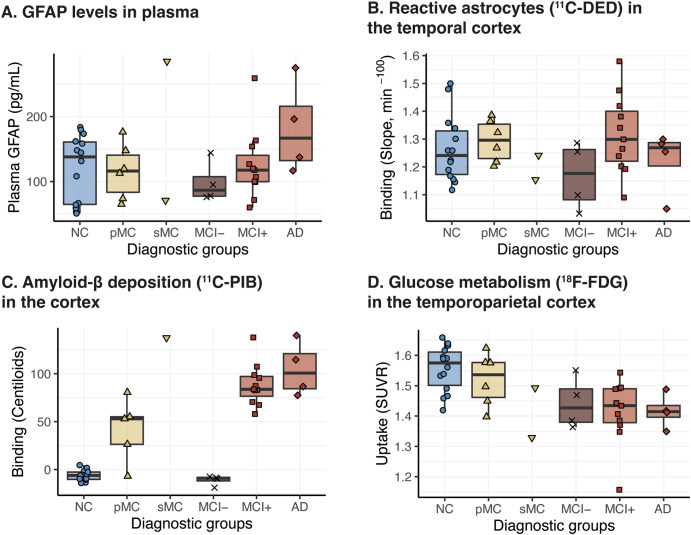
Methods: Twenty-four individuals from families with known Autosomal Dominant Alzheimer's Disease mutations (mutation carriers = 10; non-carriers = 14) and fifteen patients with sporadic Alzheimer's disease were included. The individuals underwent PET imaging with 11C-DED, 11C-PIB and 18F-FDG, as markers of reactive astrogliosis, amyloid-β deposition, and glucose metabolism, respectively, and plasma sampling for measuring GFAP concentrations. Twenty-one participants from the Autosomal Dominant Alzheimer's Disease group underwent follow-up plasma sampling and ten of these participants underwent follow-up PET imaging.

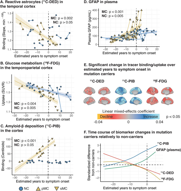
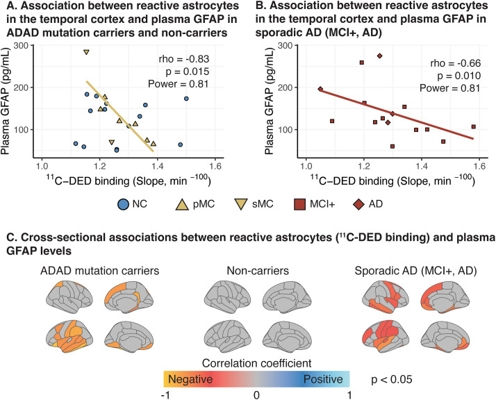
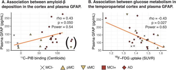
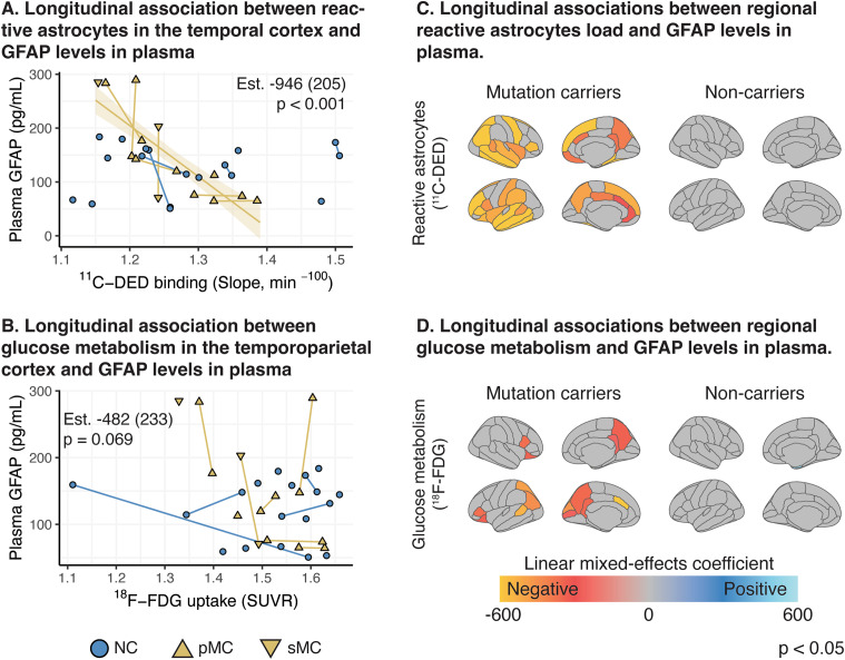
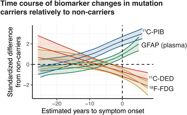
Results: In mutation carriers, plasma GFAP levels and 11C-PIB binding increased, while 11C-DED binding and 18F-FDG uptake significantly decreased across the estimated years to symptom onset. Cross-sectionally, plasma GFAP demonstrated a negative correlation with 11C-DED binding in both mutation carriers and patients with sporadic disease. Plasma GFAP indicated cross-sectionally a significant positive correlation with 11C-PIB binding and a significant negative correlation with 18F-FDG in the whole sample. The longitudinal levels of 11C-DED binding showed a significant negative correlation with longitudinal plasma GFAP concentrations over the follow-up interval.

Conclusions: Plasma GFAP concentration and astrocyte 11C-DED brain binding levels followed divergent trajectories and may reflect different underlying processes. The strong negative association between plasma GFAP and 11C-DED binding in Autosomal Dominant and sporadic Alzheimer's disease brains may indicate that if both are markers of reactive astrogliosis, they may detect different states or subtypes of astrogliosis. Increased 11C-DED brain binding seems to be an earlier phenomenon in Alzheimer's disease progression than increased plasma GFAP concentration.

Keywords: Alzheimer’s disease; Astrocytes; Astrogliosis; Autosomal Dominant Alzheimer’s disease; Deprenyl; Plasma GFAP.


Conflict of interest statement

KC, CJ, and ERV have no competing interests. AN has received a consulting fee from AVVA Pharmaceuticals, H Lundbeck A/S, Hoffman La Roche, honorarium for lecture Hoffman La Roche, Roche and hold a patent WO 2022/255915. Patent No. PCT/SE2022/050413 PET Tracers. HZ has served at scientific advisory boards and/or as a consultant for Abbvie, Acumen, Alector, Alzinova, ALZPath, Annexon, Apellis, Artery Therapeutics, AZTherapies, CogRx, Denali, Eisai, Nervgen, Novo Nordisk, Optoceutics, Passage Bio, Pinteon Therapeutics, Prothena, Red Abbey Labs, reMYND, Roche, Samumed, Siemens Healthineers, Triplet Therapeutics, and Wave, has given lectures in symposia sponsored by Cellectricon, Fujirebio, Alzecure, Biogen, and Roche, and is a co-founder of Brain Biomarker Solutions in Gothenburg AB (BBS), which is a part of the GU Ventures Incubator Program (outside submitted work).

Figures

Fig. 1
Fig. 1
Longitudinal trajectories of biomarker changes based on mixed-effects modelling in ADAD mutation carriers. (A-D) Regression plots illustrating the association between estimated time to expected symptom onset and biomarker level in ADAD mutation carriers (linear mixed-effects models). (E) Surface maps depicting the ROIs with a significant change in the PET binding/uptake values over the estimated time to expected symptom onset in ADAD mutation carriers. (F) Regression plots comparing the longitudinal non-linear trajectories of the different biomarkers in ADAD mutation carriers across the estimated time to expected symptom onset (generalized additive mixed-effects models); the biomarker values are presented as standardised difference from non-carriers (NC). For the generalized additive mixed model (F) we illustrate the 11C-DED, 11C-PIB and 18F-FDG binding/uptake values in the composite temporal, global cortical and temporoparietal ROIs, respectively. The p value of the models is shown (A-D). pMC: presymptomatic mutation carriers; sMC: symptomatic mutation carriers. In Fig. 1A, B, D, an ADAD mutation carrier is denoted by a normal triangle, even though the individual surpassed the baseline point (0) on the estimated years to symptom onset axis. This discrepancy is due to the fact that the participant did not exhibit any signs of cognitive impairment at that time, and this inconsistency falls within the expected error between the estimated age of symptom onset and the actual age of symptom onset at the individual level. Subsequently, during the follow-up assessment, this same participant showed clear objective evidence of cognitive impairment, as indicated by the use of an inverted triangle. For the analyses pertaining to 11C-PIB binding, APParc mutation carriers were excluded due to the known mutation-specific relative sparsity of fibrillar amyloid-β that causes exceptionally low 11C-PIB binding levels
Fig. 2
Fig. 2
Boxplots of the biomarker levels in the different diagnostic groups. Inferential statistics were not used for group comparisons due to the small size of the individual groups. For the analyses pertaining to 11C-PIB binding, APParc mutation carriers were excluded due to the known mutation-specific relative sparsity of fibrillar amyloid-β that causes exceptionally low 11C-PIB binding levels. NC: non-carriers; pMC: presymptomatic mutation carriers; sMC: symptomatic mutation carriers; MCI-: MCI with a negative amyloid-β PET scan; MCI+: MCI with a positive amyloid-β PET scan; AD: Alzheimer’s disease dementia
Fig. 3
Fig. 3
Cross-sectional correlation analyses between 11C-DED PET binding and plasma GFAP levels. (A-B) Scatterplots evaluating the relationship between 11C-DED PET binding and plasma GFAP levels in carriers of ADAD mutations and patients with sporadic Alzheimer’s disease for composite temporal ROIs, and (C) surface maps illustrating in colour the cortical ROIs with a significant cross-sectional association between 11C-DED PET binding and plasma GFAP. The Spearman’s correlation coefficient (rho), the p and power values are shown. NC: non-carriers; pMC: presymptomatic mutation carriers; sMC: symptomatic mutation carriers; MCI+: MCI with a positive amyloid-β PET scan; AD: Alzheimer’s disease dementia
Fig. 4
Fig. 4
Cross-sectional correlation analyses evaluating the relationship between 11C-PIB, 18F-FDG PET tracers’ binding/uptake and plasma GFAP. The analyses were performed for composite cortical ROIs (A, B) in the whole sample, not considering the non-carrier group. For the correlation analyses between 11C-PIB binding and plasma GFAP levels, the individuals with extremely high 11C-PIB binding (marked in parentheses in the figures) were excluded given the known non-linear association between the biomarkers in high amyloid-β levels [48]. For the analyses pertaining to 11C-PIB binding, APParc mutation carriers were excluded due to the known mutation-specific relative sparsity of fibrillar amyloid-β that causes exceptionally low 11C-PIB binding levels. The Spearman’s correlation coefficient (rho), the p and power values are shown. NC: non-carriers; pMC: presymptomatic mutation carriers; sMC: symptomatic mutation carriers; MCI-: MCI with a negative amyloid-β PET scan; MCI+: MCI with a positive amyloid-β PET scan; AD: Alzheimer’s disease dementia
Fig. 5
Fig. 5
Longitudinal linear mixed-effects model analyses evaluating the relationship between PET tracers’ binding/uptake and plasma GFAP. (A, B) Scatterplots for composite cortical ROIs and (C, D) surface maps where cortical ROIs with a significant longitudinal association between PET tracers’ binding/uptake and plasma GFAP are depicted in colour. The estimate (Est.), the standard error (parentheses) and p value of the models are shown. NC: non-carriers; pMC: presymptomatic mutation carriers; sMC: symptomatic mutation carriers
Fig. 6
Fig. 6
Longitudinal trajectories of biomarker changes based on generalized additive mixed-effects modelling in ADAD mutation carriers. Regression plots comparing the longitudinal nonlinear trajectories of the different biomarkers in ADAD mutation carriers across the estimated time to expected symptom onset; the biomarker values are presented as standardised difference from non-carriers (NC) and the shaded areas depict the 95% confidence interval for each regression curve. The 11C-DED, 11C-PIB and 18F-FDG binding/uptake were quantified in the composite temporal, global cortical and temporoparietal ROIs, respectively. pMC: presymptomatic mutation carriers; sMC: symptomatic mutation carriers

Similar articles

Cited by

References

    1. Price BR, Johnson LA, Norris CM. Reactive astrocytes: the nexus of pathological and clinical hallmarks of Alzheimer’s disease. Ageing Res Rev. 2021;68:101335. doi: 10.1016/j.arr.2021.101335. - DOI - PMC - PubMed
    1. Kumar A, Fontana IC, Nordberg A. Reactive astrogliosis: a friend or foe in the pathogenesis of Alzheimer’s disease. J Neurochem. 2022;jnc.15565. - PubMed
    1. Arranz AM, De Strooper B. The role of astroglia in Alzheimer’s disease: pathophysiology and clinical implications. Lancet Neurol. 2019;18(4):406–14. doi: 10.1016/S1474-4422(18)30490-3. - DOI - PubMed
    1. Sofroniew MV. Astrocyte reactivity: subtypes, States, and functions in CNS innate immunity. Trends Immunol. 2020;41(9):758–70. doi: 10.1016/j.it.2020.07.004. - DOI - PMC - PubMed
    1. Escartin C, Galea E, Lakatos A, O’Callaghan JP, Petzold GC, Serrano-Pozo A, et al. Reactive astrocyte nomenclature, definitions, and future directions. Nat Neurosci. 2021;24(3):312–25. doi: 10.1038/s41593-020-00783-4. - DOI - PMC - PubMed

Publication types

Substances