. 2023 Sep 11;21(1):230.
doi: 10.1186/s12964-023-01254-6.

Sex hormone-binding globulin (SHBG) mitigates ER stress and improves viability and insulin sensitivity in adipose-derived mesenchymal stem cells (ASC) of equine metabolic syndrome (EMS)-affected horses

Affiliations

Sex hormone-binding globulin (SHBG) mitigates ER stress and improves viability and insulin sensitivity in adipose-derived mesenchymal stem cells (ASC) of equine metabolic syndrome (EMS)-affected horses

Nabila Bourebaba et al. Cell Commun Signal. .

Abstract

Background: Equine metabolic syndrome (EMS), which encompasses insulin resistance, low-grade inflammation and predisposition to laminitis is a critical endocrine disorder among the most prevalent conditions affecting horses from different breeds. According to the most recent research, low human sex hormone-binding globulin (SHBG) serum levels correlate with an increased risk of obesity, insulin resistance and diabetes, and may contribute to overall metabolic dysregulations. This study aimed to test whether exogenous SHBG could protect EMS affected adipose-derived stromal stem cells (EqASCEMS) from apoptosis, oxidative stress, ER stress and thus improve insulin sensitivity.

Methods: EqASCEMS wells were treated with two different concentrations (50 and 100 nM) of exogenous SHBG, whose biocompatibility was tested after 24, 48 and 72 h of incubation. Several parameters including cell viability, apoptosis, cell cycle, reactive oxygen species levels, ER stress, Pi3K/MAPK activation and insulin transducers expression were analysed.

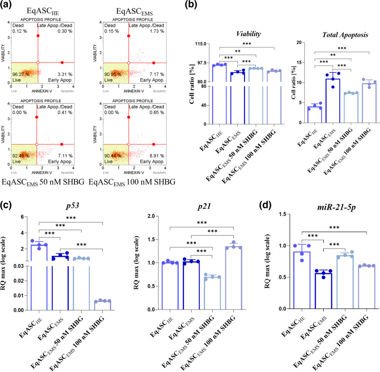
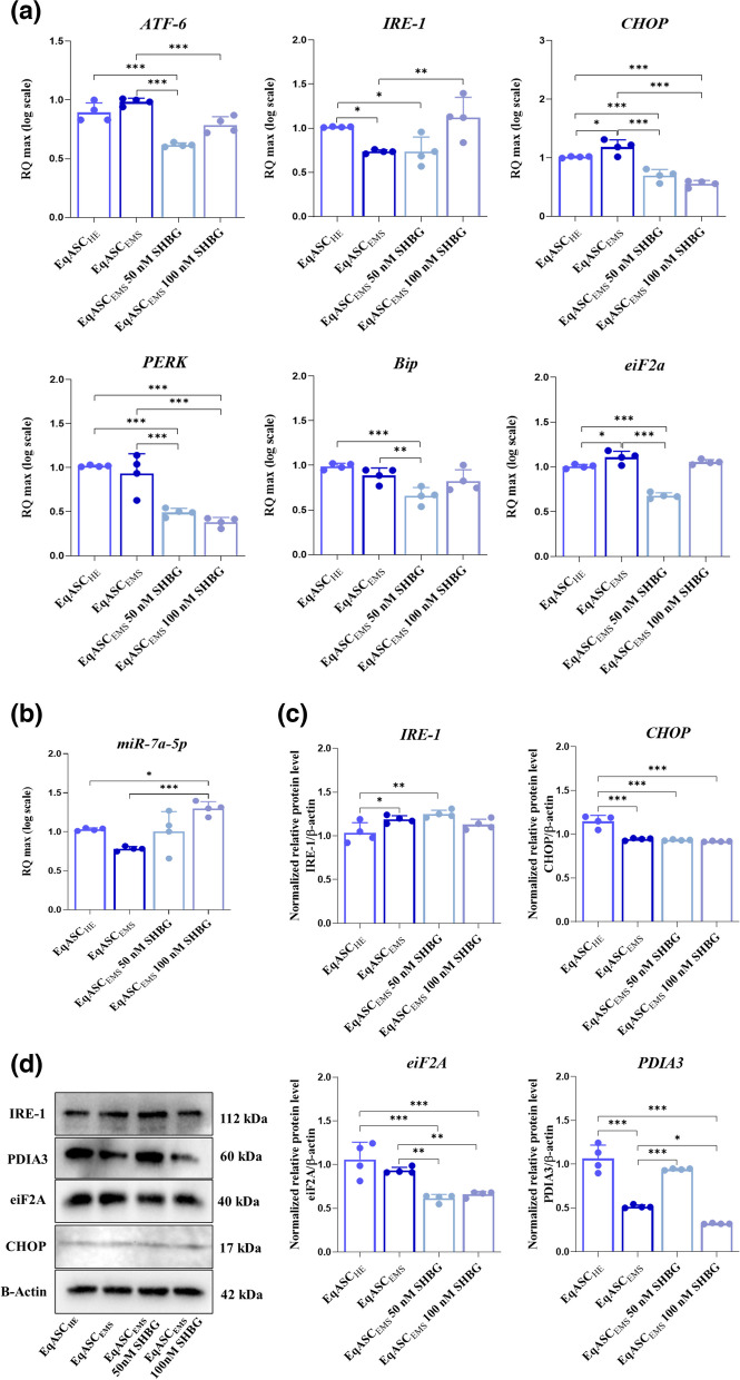
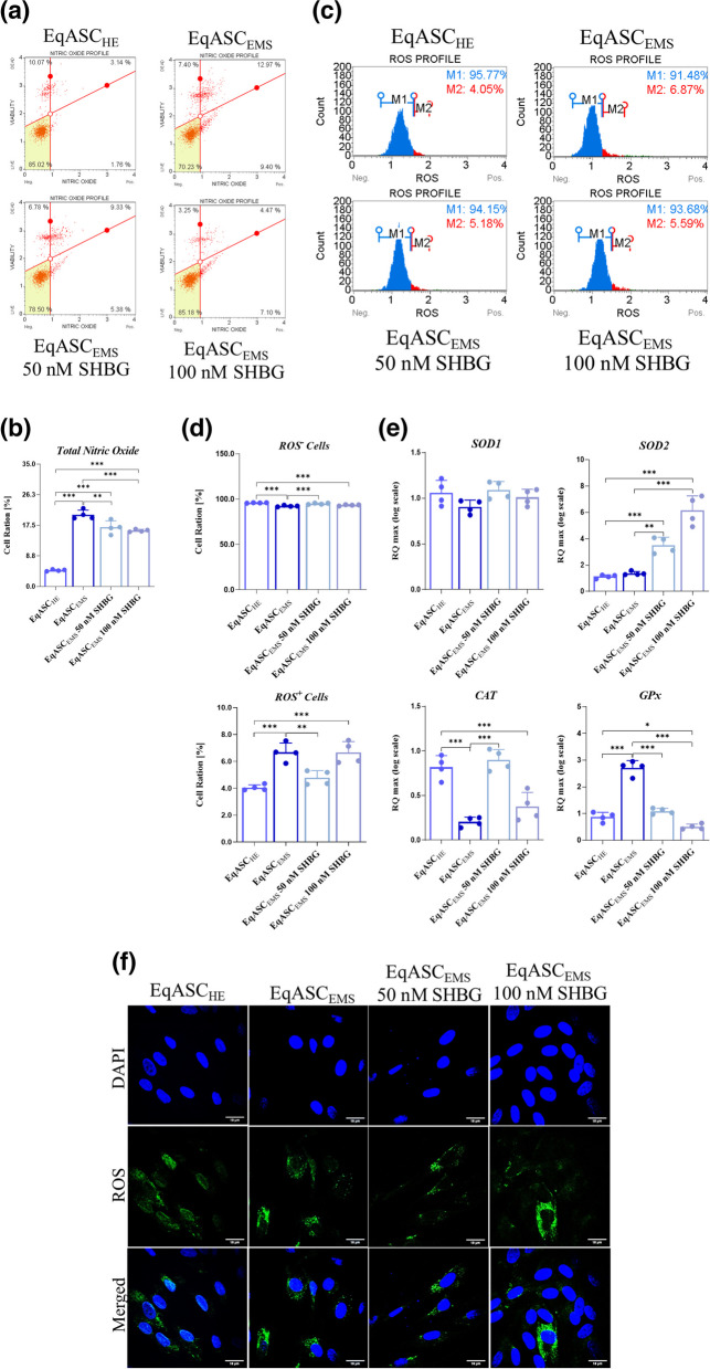
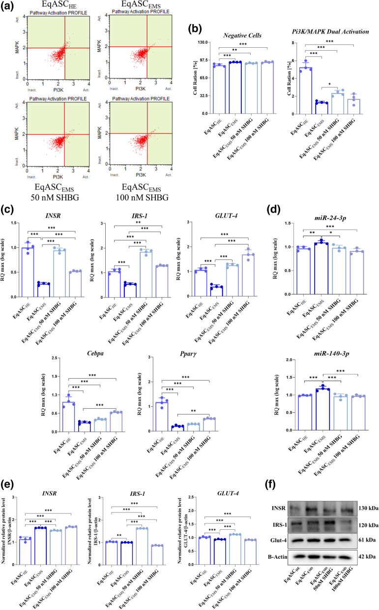
Results: Obtained data demonstrated that exogenous SHBG treatment significantly promoted ASCs cells proliferation, cell cycle and survival with reduced expression of p53 and p21 pro-apoptotic mediators. Furthermore, SHBG alleviated the oxidative stress caused by EMS and reduced the overaccumulation of intracellular ROS, by reducing ROS + cell percentage and regulating gene expression of endogenous antioxidant enzymes (Sod 1, Cat, GPx), SHBG treatment exhibited antioxidant activity by modulating total nitric oxide (NO) levels in EMS cells as well. SHBG treatment dampened the activation of ER stress sensors and effectors in EqASCEMS cells via the upregulation of MiR-7a-5p, the decrease in the expression levels of ATF-6, CHOP and eiF2A and the restoration of PDIA3 chaperone protein levels. As a consequence, SHBG application substantially improved insulin sensitivity through the modulation of Pi3K/Akt/Glut4 insulin signalling cascades.

Conclusion: Our results suggest that the SHBG is endowed with crucial beneficial effects on ASCs metabolic activities and could serve as a valuable therapeutic target for the development of efficient EMS treatment protocols. Video Abstract.

Keywords: ASC; Apoptosis; EMS; ER Stress; Insulin resistance; PDIA3; SHBG.


Conflict of interest statement

Not applicable.

Figures

Fig. 1
Fig. 1
Effect on cell proliferation and viability of sex hormone binding globulin (SHBG) on EqASCEMS. a Histograms represent the average absorbance at 490 nm of MTS tetrazolium. b Histograms represent the average absorbance at 490 nm and 680 nM of lactate dehydrogenase (LDH). Representative data from three independent experiments are shown ± SD (n = 4). An asterisk (*) indicates a comparison of treated group to untreated healthy cells. * p < 0.05, ** p < 0.01, *** p < 0.001. EqASCHE: healthy equine stem cells; EqASCEMS: EMS equine stem cells; EqASCEMS 50 nM SHBG: equine EMS stem cells treated with 50 nM of SHBG for 24 h; EqASCEMS 100 nM SHBG: equine EMS stem cells treated with 100 nM of SHBG for 24 h
Fig. 2
Fig. 2
The influence of SHBG on the regulation of cells apoptosis in the EqASCEMS. a Representative dot-plots for Annexin V & Dead Cell assay. b Bar-charts depicting the quantitative analysis of live, early, and late apoptosis and cell death. c Relative expression quantitation of main apoptosis-associated markers levels (p53 and p21). d Relative expression quantitation of apoptosis-associated miR-21-5p level. Representative data from three independent experiments are shown ± SD (n = 4). An asterisk (*) indicates a comparison of treated group to untreated healthy cells. * p < 0.05, ** p < 0.01, *** p < 0.001. EqASCHE: healthy equine stem cells; EqASCEMS: EMS equine stem cells; EqASCEMS 50 nM SHBG: equine EMS stem cells treated with 50 nM of SHBG for 24 h; EqASCEMS 100 nM SHBG: equine EMS stem cells treated with 100 nM of SHBG for 24 h
Fig. 3
Fig. 3
The effect of the SHBG on EqASCEMS’ cell cycle. a Representative dot-plots for cell cycle analysis of DNA content by the Muse™ Cell Cycle assay. b Bar-charts depicting the quantitative analysis of G0/G1, S and G2/M phases. Representative data from three independent experiments are shown ± SD (n = 4). An asterisk (*) indicates a comparison of treated group to untreated healthy cells. * p < 0.05, ** p < 0.01, *** p < 0.001. EqASCHE: healthy equine stem cells; EqASCEMS: EMS equine stem cells; EqASCEMS 50 nM SHBG: equine EMS stem cells treated with 50 nM of SHBG for 24 h; EqASCEMS 100 nM SHBG: equine EMS stem cells treated with 100 nM of SHBG for 24 h
Fig. 4
Fig. 4
Preventive effect of SHBG in EqASCEMS cells against endoplasmic reticulum stress. a Relative gene expression of ATF6, IRE1, CHOP, PERK, Bip and eiF2A ER stress transcripts. b Relative expression quantitation of ER stress-associated miR-7a-5p level. c Quantitative representation of IRE1, CHOP, eiF2A and PDIA3 proteins. d Representative immunoblots for each assayed protein detected by chemiluminescence. Representative data from three independent experiments are shown ± SD (n = 4). An asterisk (*) indicates a comparison of treated group to untreated healthy cells. * p < 0.05, ** p < 0.01, *** p < 0.001. EqASCHE: healthy equine stem cells; EqASCEMS: EMS equine stem cells; EqASCEMS 50 nM SHBG: equine EMS stem cells treated with 50 nM of SHBG for 24 h; EqASCEMS 100 nM SHBG: equine EMS stem cells treated with 100 nM of SHBG for 24 h
Fig. 5
Fig. 5
Sex hormone binding globulin (SHBG) moderates oxidative stress in EqASCEMS. a Dot-Plots for intracellular nitric oxide production detected by Muse Nitric Oxide Kit. b Average percentages of total nitric oxide production cells in each experimental group. c Dot-Plots for intracellular ROS production detected by Muse™ Oxidative Stress Kit. d Average percentages of total ROS+ and ROS cells in each experimental group. e Relative gene expression of Sod1, Sod2, CAT and GPx antioxidant enzymes transcripts. f Representative photomicrographs of CM-H2DCFDA staining assay obtained by confocal epi-fluorescent microscopy; Bar size 18 μm; magnification × 60. Representative data from three independent experiments are shown ± SD (n = 4). An asterisk (*) indicates a comparison of treated group to untreated healthy cells. * p < 0.05, ** p < 0.01, *** p < 0.001. EqASCHE: healthy equine stem cells; EqASCEMS: EMS equine stem cells; EqASCEMS 50 nM SHBG: equine EMS stem cells treated with 50 nM of SHBG for 24 h; EqASCEMS 100 nM SHBG: equine EMS stem cells treated with 100 nM of SHBG for 24 h
Fig. 6
Fig. 6
Reversing effect of sex hormone binding globulin on defective insulin signalling in EqASCEMS. a Dot-Plots for the dual Pi3K/MAPK pathway activation. b Average percentages of negative and positive Pi3K/MAPK pathway activation in each experimental group. c Main genes (INSR, IRS-1, Glut-4) expression levels involved in insulin signalling execution. d Relative expression quantitation of insulin signalling pathway miR-24-3p and miR-140-3p levels. e Quantitative analysis of insulin signalling-related proteins expression using western blot. f Representative immunoblots for protein insulin signalling detected by chemiluminescence. Representative data from three independent experiments are shown ± SD (n = 4). An asterisk (*) indicates a comparison of treated group to untreated healthy cells. * p < 0.05, ** p < 0.01, *** p < 0.001. EqASCHE: healthy equine stem cells; EqASCEMS: EMS equine stem cells; EqASCEMS 50 nM SHBG: equine EMS stem cells treated with 50 nM of SHBG for 24 h; EqASCEMS 100 nM SHBG: equine EMS stem cells treated with 100 nM of SHBG for 24 h

References

    1. Morgan R, Keen J, McGowan C. Equine metabolic syndrome. Veterinary Record. 2015;177(7):173–179. doi: 10.1136/vr.103226. - DOI - PMC - PubMed
    1. Marycz K, Michalak I, Kornicka K. Advanced nutritional and stem cells approaches to prevent equine metabolic syndrome. Res Vet Sci. 2018;118:115–125. doi: 10.1016/j.rvsc.2018.01.015. - DOI - PubMed
    1. Bourebaba L, Marycz K. Pathophysiological Implication of Fetuin-A Glycoprotein in the Development of Metabolic Disorders: A Concise Review. JCM. 2019;8(12):2033. doi: 10.3390/jcm8122033. - DOI - PMC - PubMed
    1. Marycz K, Kornicka K, Basinska K, Czyrek A. Equine Metabolic Syndrome Affects Viability, Senescence, and Stress Factors of Equine Adipose-Derived Mesenchymal Stromal Stem Cells: New Insight into EqASCs Isolated from EMS Horses in the Context of Their Aging. Oxid Med Cell Longev. 2016;2016:1–17. doi: 10.1155/2016/4710326. - DOI - PMC - PubMed
    1. Foss-Freitas MC, Ferraz RC, Monteiro LZ, et al. Endoplasmic reticulum stress activation in adipose tissue induces metabolic syndrome in individuals with familial partial lipodystrophy of the Dunnigan type. Diabetol Metab Syndr. 2018;10(1):6. doi: 10.1186/s13098-017-0301-6. - DOI - PMC - PubMed

Publication types