. 2023 Oct 27;51(19):10484-10505.
doi: 10.1093/nar/gkad733.

RNF8 ubiquitylation of XRN2 facilitates R-loop resolution and restrains genomic instability in BRCA1 mutant cells

Affiliations

RNF8 ubiquitylation of XRN2 facilitates R-loop resolution and restrains genomic instability in BRCA1 mutant cells

Rehna Krishnan et al. Nucleic Acids Res. .

Abstract

Breast cancer linked with BRCA1/2 mutations commonly recur and resist current therapies, including PARP inhibitors. Given the lack of effective targeted therapies for BRCA1-mutant cancers, we sought to identify novel targets to selectively kill these cancers. Here, we report that loss of RNF8 significantly protects Brca1-mutant mice against mammary tumorigenesis. RNF8 deficiency in human BRCA1-mutant breast cancer cells was found to promote R-loop accumulation and replication fork instability, leading to increased DNA damage, senescence, and synthetic lethality. Mechanistically, RNF8 interacts with XRN2, which is crucial for transcription termination and R-loop resolution. We report that RNF8 ubiquitylates XRN2 to facilitate its recruitment to R-loop-prone genomic loci and that RNF8 deficiency in BRCA1-mutant breast cancer cells decreases XRN2 occupancy at R-loop-prone sites, thereby promoting R-loop accumulation, transcription-replication collisions, excessive genomic instability, and cancer cell death. Collectively, our work identifies a synthetic lethal interaction between RNF8 and BRCA1, which is mediated by a pathological accumulation of R-loops.


Figures

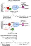
Graphical Abstract
Graphical Abstract
Figure 1.
Figure 1.
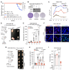
Increased expression of RNF8 in HRD breast cancer and its deficiency markedly protects mouse models of Brca1 mutation from mammary tumorigenesis. (A) Graph showing mammary tumor-free survival of mice with the cohorts of WT (n = 40), Rnf8–/– (n = 36), Wap-Cre Brca1−/− (n = 35), and Rnf8–/–;Wap-Cre Brca1−/− (n = 31). (B) Western blot (WB) analysis of the indicated MDA-MB-436 cells (upper panel). MDA-MB-436 cells transduced with Tet-On shRNF8 and shScr were treated with Dox for 7 days (BRCA1Mut: BRCA1 mutant). β-Actin was used as a loading control. The indicated Dox-induced MDA-MB-436 cells (100 000) were seeded per 6 cm dish, grown for 14 days, and stained with crystal violet (bottom panel). (C) Relative growth of primary cultures of the indicated PDX cells expressing mutated (IB-1) and WT (BRC#141) BRCA1 (n = 3). The chart depicts relative light units (RLU) detected for each primary xenograft, measured daily for 10 days. (D) A schematic of the experimental setting used to examine the effect of RNF8 deficiency on in vivo growth of xenografts of BRCA1-mutant TNBC cells and representative images of the indicated MDA-MB-436 mammary tumors resected 3 weeks post-injection (n = 5 per condition; bar = 1 cm). (E) Graph depicting changes in tumor volume over 21 days as in (D). (F) Tumors in (D) were sectioned and immunostained with RNF8, Ki67, and cleaved CASP3 antibodies. Representative images are shown (bar = 12.5 μm). (G) A schematic of the experimental setting used to examine in vivo effects of RNF8 depletion on established BRCA1-mutant tumors. MDA-MB-436 cancer cells were injected into NSG mice, and tumors were allowed to grow for two weeks before giving all cohorts of mice Dox in drinking water for another 3 weeks. Representative images 5 weeks post transplantation are shown for the indicated MDA-MB-436 mammary tumors resected from NSG mice (n > 4 per condition; bar = 1 cm). (H, I) Tumor volumes and mass from mice in (G) were monitored for 5 weeks. Graphs are depicted as mean ± SEM unless otherwise indicated. Data were analyzed using the log-rank test (A), two-tailed, paired Student's t-test (C), two-way ANOVA with Sidak's multiple comparisons test (E, H), and one-way ANOVA with Tukey's multiple comparisons test (I). ns: not significant. WT: wildtype. Mut: mutant.
Figure 2.
Figure 2.
RNF8 depletion increases genomic instability and senescence in BRCA1/2-deficient cells. (A) Western blot analysis of the indicated MDA-MB-231 cells. β-Actin was used as a loading control. (B) The indicated MDA-MB-231 cells were stained with anti-γH2AX and DAPI, and cells with more than 15 γH2AX foci were quantified. Scale bar, 25 μm; n = 3. (C) The indicated MDA-MB-436 cells (untreated (UT), 6 or 24 h post-IR 8Gy) were stained with anti-γH2AX and DAPI, and cells with more than 10 γH2AX foci were scored. Scale bar, 25 μm; n = 3. (D) The indicated Kuramochi cells were stained with anti-γH2AX, and cells with more than 10 γH2AX foci were scored. Scale bar, 25 μm; n = 3. (E) Quantification of γH2AX staining of the indicated Mammary epithelial cells (n = 3). (F) Box plots depicting the efficiency of DSB repair pathways (homologous recombination (HR), non-homologous DNA end joining (NHEJ), and alternative end-joining (a-EJ) pathways) in mediating the repair of I-SceI–induced DSBs in indicated cells (n = 3). (G) Cell cycle distribution analysis using live Hoechst staining of the indicated cells (n = 3). (H) Representative images of SA-β-gal staining of the indicated mammary epithelial cells. Scale bar, 100 μm; n = 3. Graphs are depicted as mean ± SEM unless otherwise indicated. Data were analyzed using a one-way ANOVA with Tukey's multiple comparisons test (B, C, E), unpaired Student's t test (D, G) and unpaired t-test with Welch's correction (F). ns: not significant. WT: wildtype. Mut: mutant.
Figure 3.
Figure 3.
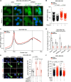
RNF8 deficiency in BRCA1 mutant breast cancer cells potentiates R-loop accumulation leading to increased genomic instability. (A, B) Representative images and quantification of R-loop levels as detected by GFP-tagged dRHN1 in indicated cells after mock or RNaseH treatment. Bar = 25 μm. (C) Metagene plot of the distribution of S9.6 signals (IP-Input) along all the expressed protein coding genes (TPM > 0; 12 080 genes) and flanking regions (±5 kb) in control (blue) and shRNF8 (purple) MDA-MB-436 cells. (D) ChIP-qPCR analysis of RNF8 recruitment to the R-loop positive (AFAP1, ZNF425 and HNRNPUL2) and R-loop negative (PRKCSH and TAF4) loci (n = 3). (E) Representative images of RNaseH1 and γH2AX staining of the indicated MDA-MB-436 cells transiently transfected with RNaseH1. Scale bar, 10 μm. γH2AX foci were counted only in RNaseH1 overexpressing cells (n = 3). More than 200 nuclei were scored per condition. (F) Quantification of nuclear γH2AX foci in the indicated cells post-treatment with flavopiridol (Flavo) and actinomycin D (ActD) or vehicle control (n = 3). Scale bar = 25 μm; n = 3. Graphs are depicted as mean ± SEM. Data were analyzed using a Mann–Whitney U test (B, F), a two-tailed unpaired student's t-test (D), and a one-way ANOVA with Tukey's multiple comparisons test (E). Mut: mutant.
Figure 4.
Figure 4.
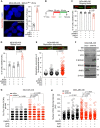
RNF8 deficiency promotes replication stress in BRCA1-mutant breast cancer cells. (A) Representative images and quantification of PLA foci indicating the interaction between RNA Pol II and PCNA (scale bar = 25 μm; n = 3). (B) Schematic of the experimental design used for DNA fibre analysis. shRNF8 and shScr MDA-MB-436 cells were treated with Dox for 4 days and sequentially pulse-labelled with two thymidine analogues CldU and IdU for 20 min each before being harvested for DNA fibre analysis. (C. D) The percent of ongoing and stalled replication forks in the indicated cells is plotted and representative images are shown. (E) The replication fork asymmetry in the indicated cells is plotted and representative images are shown. For the replication fork structures (C. D: ongoing and stalled forks) more than 1250 CldU-labelled fibres in total were counted for the whole experiment. For the replication fork asymmetry (E) over 50 bi-directional first label origins in total were counted for the whole experiment. All the fibre data (C–E) are representative of three independent experimental replicates. (F) Western blot showing the expression of the indicated proteins in RNF8-depleted MDA-MB-436 cells and their shScr controls. (G) Quantification of PCNA-RNAPII CTD PLA foci per nucleus in the indicated MDA-MB-436 cells treated for 1hr with DMSO, APH (500 nM) or ActD (0.5 μM). (H) Quantification of nuclear γH2AX foci in shRNF8 and shScr MDA-MB-436 cells after 1 h treatment with APH, ActD, and DMSO as indicated (n = 3). Graphs are depicted as mean ± SEM. Data were analysed using two-tailed unpaired Student's t-test (A, C, D), Mann–Whitney U test (E), and one-way ANOVA with Tukey's multiple comparisons test (G, H). Mut: mutant.
Figure 5.
Figure 5.
RNF8 interacts with XRN2, mediating its ubiquitylation and recruitment to R-loop-prone loci. (A) Interaction of endogenous RNF8 and XRN2 in MDA-MB-436 cells was examined by IP/WB as indicated. (B) Schematic of XRN2 full length wildtype (WT), its deletion mutants, and IP/WB showing XRN2 domains interacting with RNF8. (C) Representative WB showing the expression of XRN2 protein in the indicated MDA-MB-436 and MDA-MB-231 cells. (D, E) Representative WB of the indicated MDA-MB-436 and MDA-MB-231 cells subjected to IP with anti-XRN2 antibody followed by immunoblotting using anti-Ubiquitin (Ub) antibodies. WB analysis confirmed the IP of XRN2 and indicated the level of RNF8 and β-Actin in WCL. (F) HEK293T cells were transfected with indicated Flag-tagged mouse Rnf8, RNF8C406S and empty vector (EV) as indicated. WCL were subjected to IP with anti-XRN2 antibody and membrane was probed with anti-ubiquitin antibody. In vivo ubiquitylation of XRN2 in indicated cells is shown. (G) In vitro ubiquitylation of recombinant XRN2 in the presence of recombinant RNF8, UBE1 (E1), UBE2 (E2) and Ub proteins. (H) ChIP-qPCR analysis of shRNF8 and shScr MD-MB-436 cells for XRN2 recruitment to R-loop positive (AFAP1, ZNF425 and HNRNPUL2) and negative (PRKCSH and TAF4) loci in indicated cells (n = 4). Graphs are depicted as mean ± SEM. The data was analyzed using a two-tailed unpaired Student's t-test (G). ns: not significant.
Figure 6.
Figure 6.
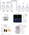
Reconstitution of RNF8-depleted cells with wildtype RNF8 rescues XRN2 recruitment to R-loops. (A) Anti-XRN2 ChIP-qPCR was performed using MDA-MB-436 cells to examine the effect of RNF8-WT and its C406S mutant on the recruitment of XRN2 to the indicated R-loop–prone loci (n = 3). (B) Representative WB validating XRN2 knockdown in MDA-MB-436 cells. (C) Representative images and quantification of γH2AX foci in shXRN2 and shScr MDA-MB-436 cells (n = 3). Scale bar, 25 μm. (D) Quantification of R-loop levels as detected by GFP-tagged dRHN1 in indicated cells after mock or RNaseH treatment. (n = 3; Bar = 25 μm). (E) Representative images and quantification of the colony-forming ability of shXRN2 and shScr MDA-MB-436 cells (n = 3). Graphs are depicted as mean ± SEM. The data was analyzed using the 2-way ANOVA with Tukey's multiple-comparison test (A), two-tailed unpaired Student's t-test (C), Mann–Whitney U test (D), unpaired t-test with Welch's correction (E). ns: not significant. Prom: promoter.
Figure 7.
Figure 7.
Schematic of RNF8 and XRN2 mediated R-loop regulation to control BRCA1-mutant cancers. The proposed model depicts a new role for RNF8 in mediating the ubiquitylation of XRN2, facilitating its recruitment to DNA–RNA hybrids. In BRCA1 mutant cancer cells, RNF8-dependent recruitment of XRN2 to R-loops allows it to resolve the DNA–RNA hybrids, limiting genomic instability. RNF8 deficiency in BRCA1-mutant breast cancer cells induces R-loop accumulation and transcription-replication collisions, leading to increased DNA damage, further exacerbating genomic instability, senescence, and growth defects in these cancer cells. RNF8 deficiency also increases the sensitivity to PARP inhibitors (PARPi) and irradiation of BRCA1-mutant cells and protects against BRCA1-mutated breast cancer.

References

    1. Kuchenbaecker K.B., Hopper J.L., Barnes D.R., Phillips K.A., Mooij T.M., Roos-Blom M.J., Jervis S., van Leeuwen F.E., Milne R.L., Andrieu N.et al. .. Risks of breast, ovarian, and contralateral breast cancer for BRCA1 and BRCA2 mutation carriers. JAMA. 2017; 317:2402–2416. - PubMed
    1. Ashworth A., Lord C.J.. Synthetic lethal therapies for cancer: what's next after PARP inhibitors?. Nat. Rev. Clin. Oncol. 2018; 15:564–576. - PubMed
    1. Chen C.C., Feng W., Lim P.X., Kass E.M., Jasin M.. Homology-directed repair and the role of BRCA1, BRCA2, and related proteins in genome integrity and cancer. Annu Rev Cancer Biol. 2018; 2:313–336. - PMC - PubMed
    1. Krais J.J., Johnson N.. BRCA1 Mutations in cancer: coordinating deficiencies in homologous recombination with tumorigenesis. Cancer Res. 2020; 80:4601–4609. - PMC - PubMed
    1. Hill S.J., Rolland T., Adelmant G., Xia X., Owen M.S., Dricot A., Zack T.I., Sahni N., Jacob Y., Hao T.et al. .. Systematic screening reveals a role for BRCA1 in the response to transcription-associated DNA damage. Genes Dev. 2014; 28:1957–1975. - PMC - PubMed

Publication types