. 2023 Sep 20;145(37):20539-20550.
doi: 10.1021/jacs.3c06703. Epub 2023 Sep 11.

Hormone Analogues with Unique Signaling Profiles from Replacement of α-Residue Triads with β/γ Diads

Affiliations

Hormone Analogues with Unique Signaling Profiles from Replacement of α-Residue Triads with β/γ Diads

Ruslan Gibadullin et al. J Am Chem Soc. .

Abstract

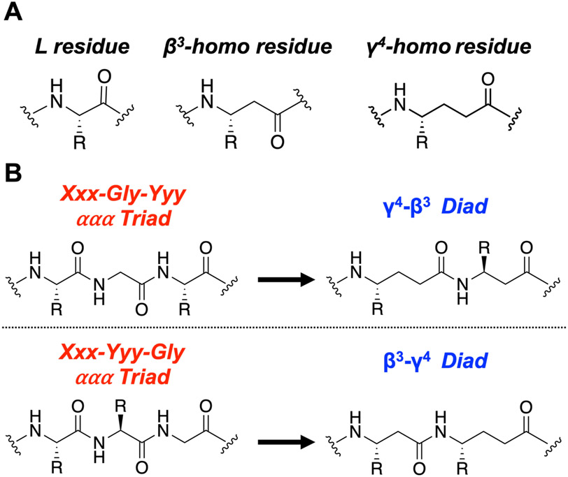
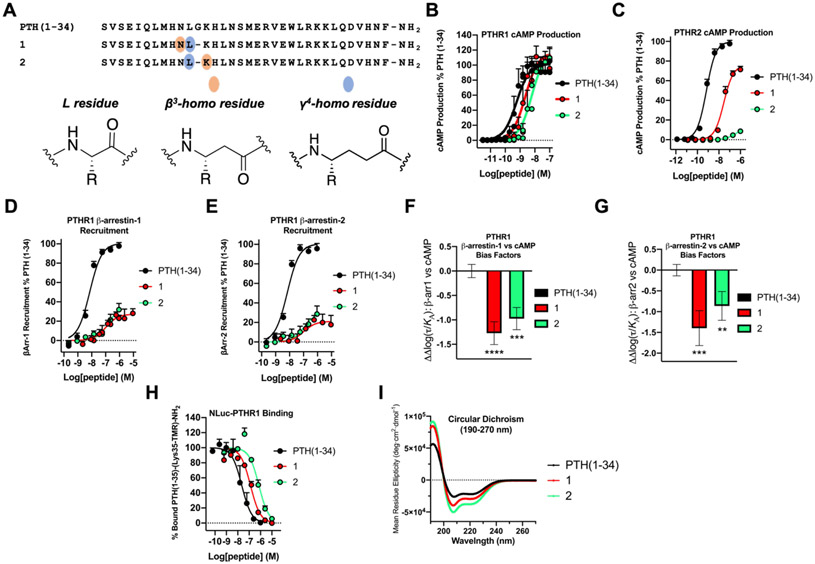
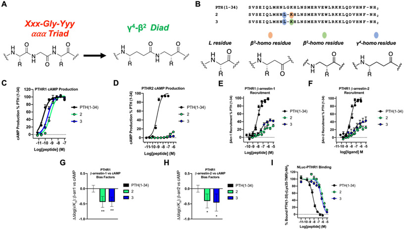
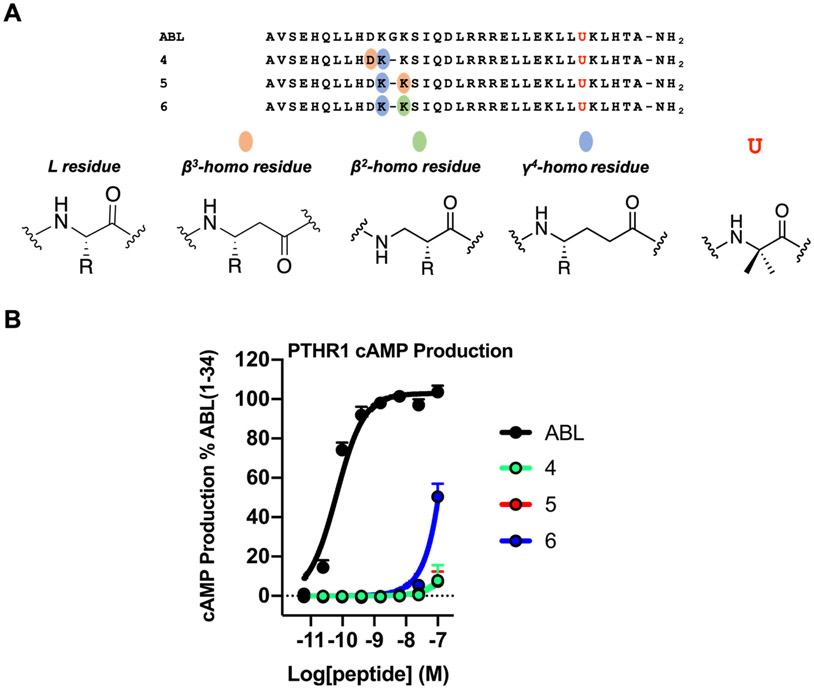
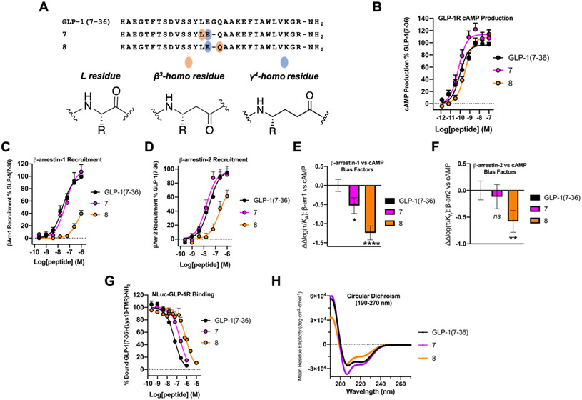
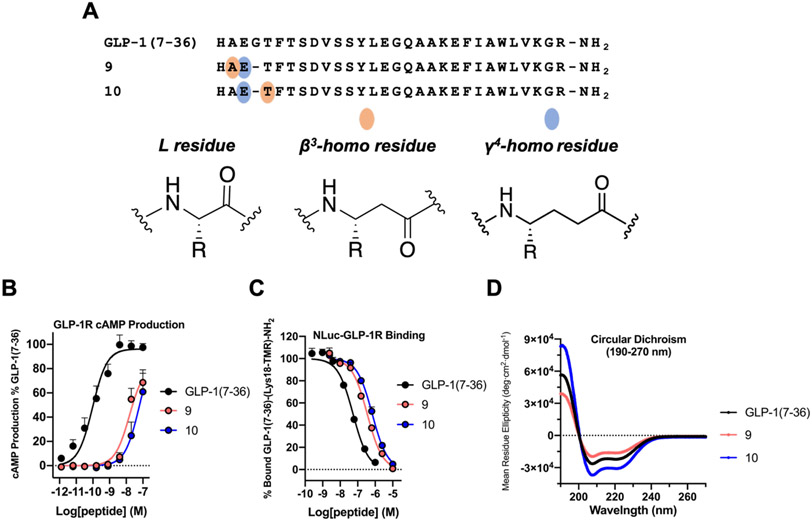
We have applied an underexplored backbone modification strategy to generate new analogues of peptides that activate two clinically important class B1 G protein-coupled receptors (GPCRs). Most peptide modification strategies involve changing side chains or, less commonly, changing the configuration at side chain-bearing carbons (i.e., l residues replaced by d residues). In contrast, backbone modifications alter the number of backbone atoms and the identities of backbone atoms relative to a poly-α-amino acid backbone. Starting from the peptide agonists PTH(1-34) (the first 34 residues of the parathyroid hormone, used clinically as the drug teriparatide) and glucagon-like peptide-1 (7-36) (GLP-1(7-36)), we replaced native α-residue triads with a diad composed of a β-amino acid residue and a γ-amino acid residue. The β/γ diad retains the number of backbone atoms in the ααα triad. Because the β and γ residue each bear a single side chain, we implemented ααα→βγ replacements at sites that contained a Gly residue (i.e., at α-residue triads that presented only two side chains). All seven of the α/β/γ-peptides derived from PTH(1-34) or GLP-1(7-36) bind to the cognate receptor (the PTHR1 or the GLP-1R), but they vary considerably in their activity profiles. Outcomes include functional mimicry of the all-α agonist, receptor-selective agonist activity, biased agonism, or strong binding with weak activation, which could lead to antagonist development. Collectively, these findings demonstrate that ααα→βγ replacements, which are easily implemented via solid-phase synthesis, can generate peptide hormone analogues that display unique and potentially useful signaling behavior.


Figures

Figure 1.
Figure 1.
The ααα→βγ backbone replacement approach utilized in this work. (A) Generic structures of L-amino acid residues and β3- and γ4-homoamino acid residues. (B) Overview of two backbone replacements starting with either the Xxx-Gly-Yyy or Xxx-Yyy-Gly ααα triad; each of these replacements retains original side chains and the total number of backbone atoms found in the original ααα triad. Xxx or Yyy = any chiral residue; Gly = achiral Gly residue.
Figure 2.
Figure 2.
PTH(1-34) α/β/γ-analogues display PTHR1 selectivity and bias toward cAMP production and away from β-arrestin recruitment. (A) Sequences of PTH(1-34) and α/β/γ-analogues 1 and 2. (B) PTHR1 activation, via cAMP production, in HEK293 cells stably expressing the PTHR1 and the cAMP-sensing protein GloSensor. Data points represent the average of 4 independent experiments. (C) PTHR2 activation, via cAMP production, in HEK293 cells stably expressing PTHR2 and GloSensor. Data is derived from ≥4 independent experiments. (D,E) PTHR1 β-arrestin-1 and −2 recruitment dose-responses. Assays were performed in CHO-FlpIn cells stably transfected with the PTHR1-RLuc8 and β-arrestin-1-Venus or β-arrestin-2-Venus. Data represent the mean of ≥4 (β-arrestin-1) or ≥3 (β-arrestin-2) independent experiments. All cAMP, β-arrestin-1 and β-arrestin-2 data was normalized against PTH(1-34) and all uncertainties are expressed as SEM. (F,G) PTHR1 β-arrestin-1 or −2 vs. cAMP bias factors were generated from dose-response curves in Figures 2B, D, and E. Negative values indicate bias towards cAMP production. Bias factor uncertainties are expressed in SD. ** = p ≤ 0.01; *** = p ≤ 0.001; **** = p ≤ 0.0001; by one-way ANOVA followed by Dunnett test. (H) Competition binding measurements by bioluminescence resonance energy transfer (BRET) in HEK293 cells expressing the NLuc-PTHR1. The peptide tracer was a tetramethylrhodamine (TMR)-labeled derivative of PTH(1-34), PTH(1-35)-(Lys35-TMR)-NH2. Data (as IC50 values) represent the average of 3 independent experiments. (I) Circular dichroism data measured in 50 mM PBS buffer, pH 7.3, 20% 2,2,2-trifluoroethanol (TFE).
Figure 3.
Figure 3.
PTH(1-34) γ43 (peptide 2) and γ42 (peptide 3) analogues display comparable affinity and signaling outcomes at PTHR1. (A) Backbone replacement of Xxx-Gly-Yyy ααα triad with a γ42 diad. For comparison, replacement of Xxx-Gly-Yyy ααα triad with a γ43 diad is shown in Figure 1B. (B) Sequences of PTH(1-34) and α/β/γ-analogues 2 and 3. (C) PTHR1 activation, via cAMP production, in HEK293 cells stably expressing the PTHR1 and the cAMP-sensing protein GloSensor. Data points represent the average of 5 independent experiments. (D) PTHR2 activation, via cAMP production, in HEK293 cells stably expressing PTHR2 and GloSensor. Data is derived from 3 independent experiments. (E,F) PTHR1 β-arrestin-1 and −2 recruitment dose-responses. Assays were performed in CHO-FlpIn cells stably transfected with the PTHR1-RLuc8 and β-arrestin-1-Venus or β-arrestin-2-Venus. Data represent the mean of ≥5 (β-arrestin-1) or ≥4 (β-arrestin-2) independent experiments. All cAMP, β-arrestin-1 and β-arrestin-2 data was normalized against PTH(1-34) and all uncertainties are expressed as SEM. (G,H) PTHR1 β-arrestin-1 or −2 vs. cAMP bias factors were generated from dose-response curves in Figures 3C, E, and F. Negative values indicate bias towards cAMP production. Bias factor uncertainties are expressed in SD. * = p ≤ 0.05; ** = p ≤ 0.01; by one-way ANOVA followed by Dunnett test. (I) Competition binding measurements by bioluminescence resonance energy transfer (BRET) in HEK293 cells expressing the NLuc-PTHR1. The peptide tracer was a tetramethylrhodamine (TMR)-labeled derivative of PTH(1-34), PTH(1-35)-(Lys35-TMR)-NH2. Data (as IC50 values) represent the average of 3 independent experiments.
Figure 4.
Figure 4.
ABL α/β/γ-analogues 4-6 are weak activators of PTHR1. (A) Sequences of ABL and α/β/γ-analogues 4, 5 and 6. Letter (U) denotes 2-aminoisobutyric acid. (B) PTHR1 activation, via cAMP production, in HEK293 cells stably expressing the PTHR1 and the cAMP-sensing protein GloSensor. Data points represent the average of ≥3 independent experiments. Data was normalized against ABL and all error bars uncertainties are expressed as SEM. Note: data points for ABL α/β/γ-analogues 4 and 5 overlap.
Figure 5.
Figure 5.
Evaluation of ααα→βγ replacements at Gly22 of GLP-1R agonist, GLP-1(7-36). (A) Sequences of GLP-1(7-36) and α/β/γ-analogues 7 and 8. (B) GLP-1R activation, measured via stimulation of intracellular cAMP, in HEK293 cells transiently expressing the GLP-1R and stably expressing the GloSensor protein. Data points represent the average of ≥3 independent experiments. (C,D) β-arrestin-1 and −2 recruitment dose-response curves for GLP-1(7-36), 7 and 8. Assays were performed in HEK293 cells transiently transfected with GLP-1R-RLuc8 and GFP2-tagged β-arrestin-1 or β-arrestin-2. Data represent the mean of ≥2 (β-arrestin-1) and ≥3 (β-arrestin-2) independent experiments. All cAMP, β-arrestin-1 and β-arrestin-2 data was normalized against GLP-1(7-36) and all uncertainties are expressed as SEM. (E,F) GLP-1R β-arrestin-1 or −2 vs. cAMP bias factors generated from curves in Figures 5B, C, and D. Negative values indicate bias towards cAMP production. Bias factor uncertainties are expressed in SD. ns= non-significant; * = p ≤ 0.05; ** = p ≤ 0.01; **** = p ≤ 0.0001; by one-way ANOVA followed by Dunnett test. (G) Competition NLuc-GLP-1R affinity measurements in HEK293 cells. GLP-1(7-36)-(Lys18-TMR)-NH2 was used as a labeled peptide tracer. IC50 values represent the average of ≥3 independent experiments. (H) Circular dichroism data for GLP-1(7-36), 7 and 8 measured in 50 mM PBS buffer, pH 7.3, 20% 2,2,2-trifluoroethanol (TFE).
Figure 6.
Figure 6.
ααα→βγ replacements at the N-terminus (Gly10) of GLP-1(7-36) are not well tolerated. (A) Sequences of GLP-1(7-36) and α/β/γ-analogues 9 and 10. (B) GLP-1R activation, measured via cAMP production, in HEK293 cells transiently expressing the GLP-1R and stably expressing the GloSensor protein. Data points represent the average of ≥3 independent experiments. The cAMP data was normalized against GLP-1(7-36) and all uncertainties are expressed as SEM. (C) Competition affinity measurements in HEK293 cells transiently transfected with the NLuc-GLP-1R. GLP-1(7-36)-(Lys18-TMR)-NH2 was used as a labeled peptide tracer. IC50 values represent the average of ≥3 independent experiments. (D) Circular dichroism data for GLP-1(7-36), 9 and 10 measured in 50 mM PBS buffer, pH 7.3, 20% 2,2,2-trifluoroethanol (TFE).

Similar articles

References

    1. Cheloha RW; Gellman SH; Vilardaga JP; Gardella TJ PTH Receptor-1 Signalling—Mechanistic Insights and Therapeutic Prospects. Nature Reviews Endocrinology 2015, 11 (12), 712–724. 10.1038/nrendo.2015.139. - DOI - PMC - PubMed
    1. Davenport AP; Scully CCG; de Graaf C; Brown AJH; Maguire JJ Advances in Therapeutic Peptides Targeting G Protein-Coupled Receptors. Nat Rev Drug Discov 2020, 19 (6), 389–413. 10.1038/s41573-020-0062-z. - DOI - PubMed
    1. Suzuki R; Brown GA; Christopher JA; Scully CCG; Congreve M Recent Developments in Therapeutic Peptides for the Glucagon-like Peptide 1 and 2 Receptors. J Med Chem 2020, 63 (3), 905–927. 10.1021/ACS.JMEDCHEM.9B00835. - DOI - PubMed
    1. Wootten D; Christopoulos A; Marti-Solano M; Babu MM; Sexton PM Mechanisms of Signalling and Biased Agonism in G Protein-Coupled Receptors. Nature Reviews Molecular Cell Biology 2018, 19 (10), 638–653. 10.1038/s41580-018-0049-3. - DOI - PubMed
    1. Gurevich VV; Gurevich EV GPCR Signaling Regulation: The Role of GRKs and Arrestins. Front Pharmacol 2019, 10 (125). 10.3389/FPHAR.2019.00125. - DOI - PMC - PubMed

Publication types

LinkOut - more resources