. 2023 Nov 21;64(6):880-892.
doi: 10.1093/jrr/rrad065.

Therapeutic mechanism of Liangxue-Guyuan-Yishen decoction on intestinal stem cells and tight junction proteins in gastrointestinal acute radiation syndrome rats

Affiliations

Therapeutic mechanism of Liangxue-Guyuan-Yishen decoction on intestinal stem cells and tight junction proteins in gastrointestinal acute radiation syndrome rats

Ziqiao Yan et al. J Radiat Res. .

Abstract

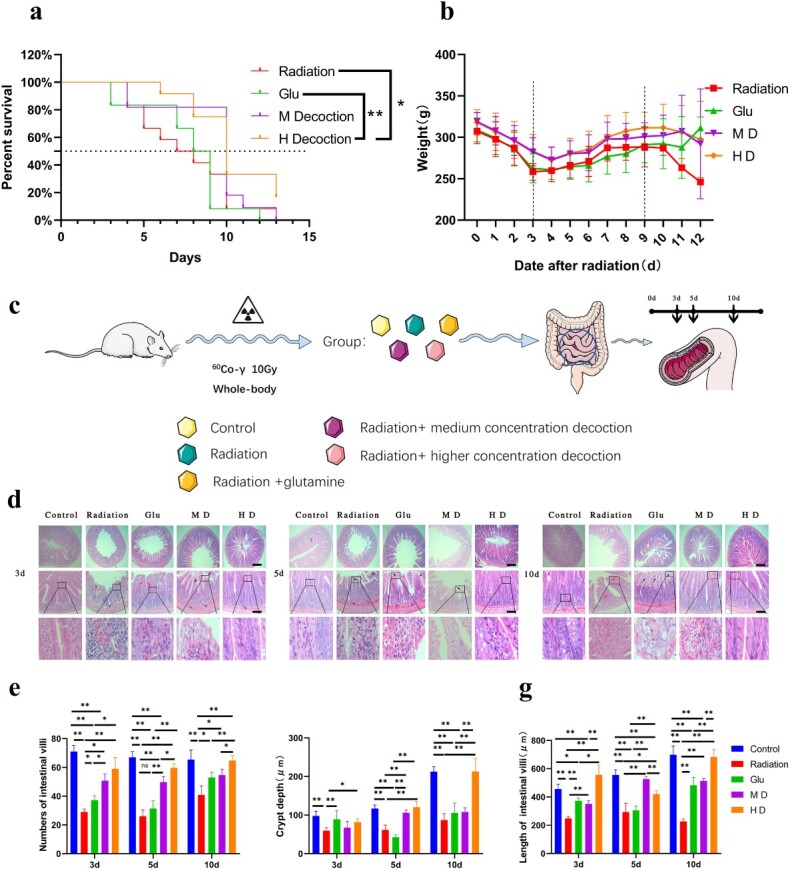
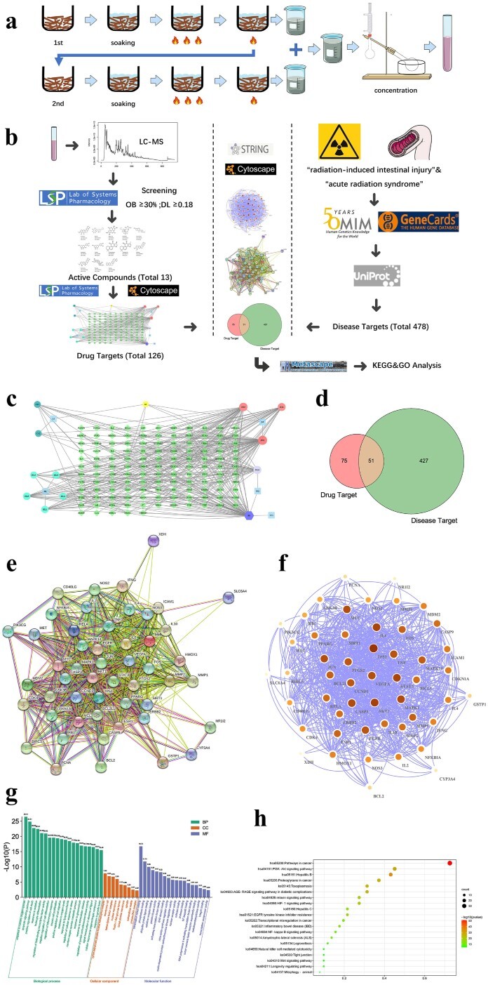
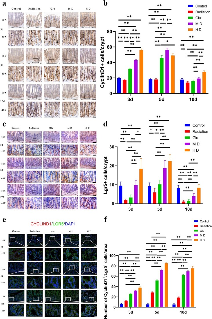
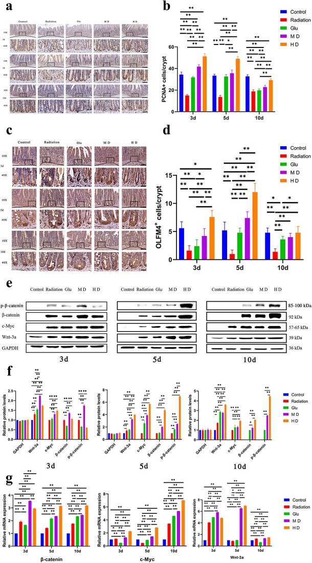
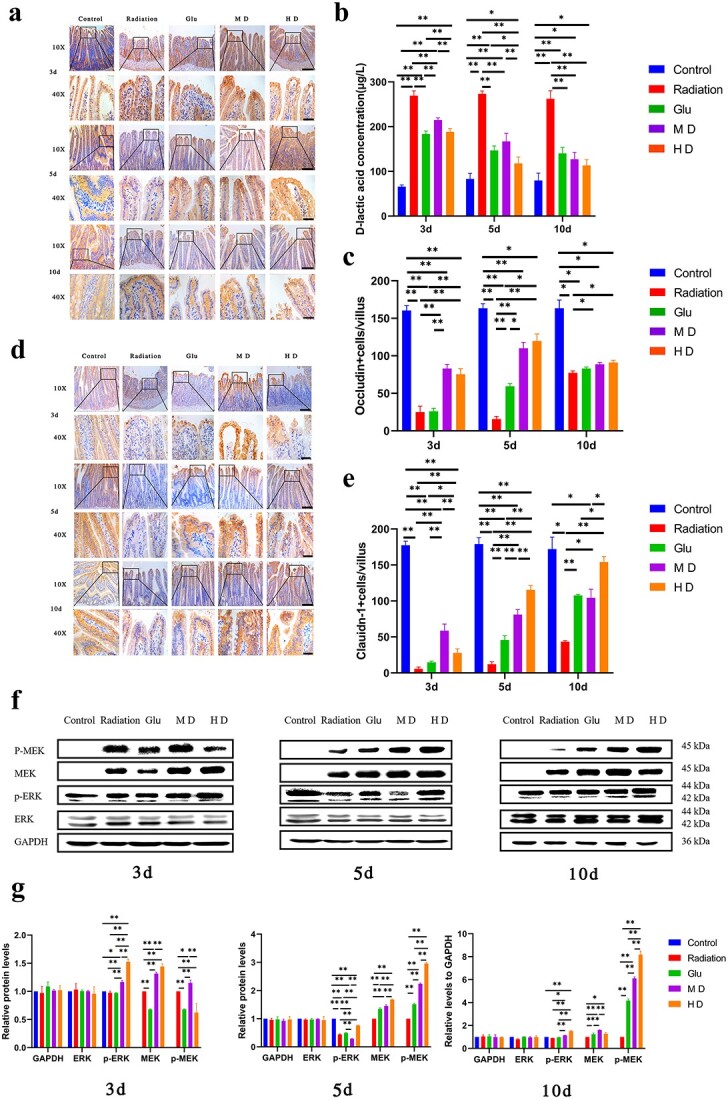
On the basis of the previous research, the Traditional Chinese Medicine theory was used to improve the drug composition for gastrointestinal acute radiation syndrome (GI-ARS). The purpose of this study was to study the therapeutic mechanism of Liangxue-Guyuan-Yishen decoction (LGYD) on GI-ARS and to provide a new scheme for the treatment of radiation injury. Here, we investigated the effects of LGYD on intestinal stem cells (ISCs) in a GI-ARS rat model. Rat health and survival and the protective efficacy of LGYD on the intestines were analyzed. The active principles in LGYD were detected using liquid chromatography-mass spectrometry (LC-MS). ISC proliferation, intestinal epithelial tight junction (TJ) protein expression and regulatory pathways were explored using immunohistochemistry, western blotting (WB) and reverse transcription quantitative polymerase chain reaction (RT-qPCR), respectively. Involvement of the WNT and MEK/ERK pathways in intestinal recovery was screened using network pharmacology analysis and validated by WB and RT-qPCR. LGYD administration significantly improved health and survival in GI-ARS rats. Pathological analysis showed that LGYD ameliorated radiation-induced intestinal injury and significantly promoted LGR5+ stem cell regeneration in the intestinal crypts, upregulated TJ protein, and accelerated crypt reconstruction in the irradiated rats. LC-MS revealed ≥13 constituents that might contribute to LGYD's protective effects. Collectively, LGYD can promote crypt cell proliferation and ISCs after radiation damage, the above effect may be related to WNT and MEK/ERK pathway.

Keywords: Chinese herbal medicine; gastrointestinal acute radiation syndrome; intestinal stem cell; tight junction protein.


Conflict of interest statement

All authors disclosed no relevant relationships.

Figures

Fig. 1
Fig. 1
LXGYD can improve the survival status and intestinal injury state of rats after total body irradiation with a lethal dose. (a) Survival curves of rats in different treatment groups after radiation. (b) Weight change of rats in different treatment groups after radiation. (c) Rats were randomly divided into Control, Radiation, Glu, MD and HD groups. Excluding the Control, all groups were subjected to single whole-body irradiation. From Day 1 postirradiation, the Control and Radiation groups were administered normal saline. The Glu group was administered a Glu suspension and the MD and HD groups were administered 2.73 and 5.46 g/ml LXGYD, respectively. Five rats were randomly selected on Days 3, 5 and 10 postirradiation and 5 cm of small intestine, and bilateral femur was dissected and interpreted. (d) HE staining revealed that the epithelial cells of the intestinal villi of rats in the Radiation and Glu groups were enlarged and had a pale cytoplasm on Days 3, 5 and 10 postirradiation (d). Fibrous tissue proliferation was observed in the submucosa with widened gaps. After radiation, fibrous tissue proliferated in the glandular interstitium. However, in the group treated with TCM, pathological damage of the small intestine was not obvious, the epithelial cells of the intestinal villi were edematous with a light cytoplasm (d) and a relatively obvious inflammatory cell infiltration was observed in Radiation group and Glu group. A few intestinal villi exhibited microvascular hyperplasia and dilation of the lamina propria. Quantification of villus number (e), length (f) and crypt depth (g). Bars represent 200 and 100 μm, respectively, *P < 0.05, **P < 0.01.
Fig. 2
Fig. 2
Drug preparation and network pharmacological study. (a) The drug was prepared, adjusted to the appropriate concentration, and stored at 4°C. (b) Network pharmacological procedure. (c) Target interaction relationship of core active components. Among them, light blue is the main drug (CS: red peony root, HL: coptis, DS: salvia miltiorrhiza, GG: pueraria root, HQ: Astragalus membranaceus), dark green is the active ingredient of CS (CS1: Ellagic acid, CS2: Paeoniflorin), lighter blue is the effective ingredient of HL (HL1: Berberine, HL2: Berberrubine, HL3, Palmatine, HL4, Coptisine, HL5: Berlambine), red is the DS effective components (DS1: Luteolin, DS2: Cryptotanshinone, DS3: Tanshinone IIA), lilac is the active component of HQ (HQ1: Isorhamnetin), purple is the common component of HQ and GG (B1: Formononetin) and yellow is the common component of CS and DS (A1: Baicalin). (d) The intersecting gene-disease target and core active component gene target. (e) The interaction network of the intersecting genes. Colored nodes: query proteins and first shell of interactors; white nodes: second shell of interactors; empty nodes: proteins of unknown 3D structure; filled nodes: some 3D structure is known or predicted; edges: known interactions (light blue: from curated databases; purple: experimentally determined), predicted interactions (green: gene neighborhood; red: gene fusions; blue: gene co-occurrence) and others (yellow: text mining; black: co-expression; lavender: protein homology). (f) Cytoscape map of the intersecting genes; node color and size represent how close the connections are. GO enrichment analysis (g) and KEGG enrichment analysis (h) of the intersecting genes.
Fig. 3
Fig. 3
Immunohistochemical and immunofluorescence (IF) staining of Lgr5 and CyclinD1. (a, b) Immunohistochemical staining of CyclinD1 in cells at the bottom of the crypt. (c, d) Immunohistochemical staining of Lgr5-positive cells at the bottom of the crypt. (e, f) IF staining of Lgr5- and CyclinD1-positive cells in the crypt (Cyclind1 in red)/Lgr5 in green)/DAPI in blue). Bars represent 100 (10×) and 20 μm (40×), respectively; *P < 0.05, **P < 0.01.
Fig. 4
Fig. 4
Immunohistochemical staining of PCNA and OLFM4 and WB and RT-qPCR to measure relative expression levels of β-catenin, p-β-catenin, C-MYC and WNT-3A. (a, b) Immunohistochemical staining of PCNA in cells at the bottom of the crypt. (c, d) Immunohistochemical staining of OLMF4-positive cells at the bottom of the crypt. Bars represent 100 (10×) and 20 μm (40×), respectively; *P < 0.05, **P < 0.01. (e, f) WB to measure the expression of proteins involved in the Wnt pathway. (g) RT-qPCR to measure the expression of genes involved in the Wnt pathway.
Fig. 5
Fig. 5
Immunohistochemical staining of occludin and claudin-1, D-lactic acid concentration and WB to measure the expression of proteins involved in the MEK/ERK pathway. (a, c, d, e) Immunohistochemical staining of occludin and claudin-1. Bars in (a) and (d) represent 100 (10×) and 20 μm (40×), respectively; *P < 0.05, **P < 0.01. (b) Rat D-Lactate ELISA Kit was used to measure D-lactic acid concentrations in the peripheral blood of rats. (f, g) Expression of key proteins involved in the MEK/ERK pathway, ERK, p-ERK, MEK and p-MEK, was measured using WB, and semi-quantitative analysis was performed.

Similar articles

Cited by

References

    1. Chapel A, Francois S, Douay L et al. New insights for pelvic radiation disease treatment: multipotent stromal cell is a promise mainstay treatment for the restoration of abdominopelvic severe chronic damages induced by radiotherapy. World J Stem Cells 2013;5:106–11. - PMC - PubMed
    1. Qiu W, Leibowitz B, Zhang L, Yu J. Growth factors protect intestinal stem cells from radiation-induced apoptosis by suppressing PUMA through the PI3K/AKT/p53 axis. Oncogene 2010;29:1622–32. - PMC - PubMed
    1. Wei L, Leibowitz BJ, Wang X et al. Inhibition of CDK4/6 protects against radiation-induced intestinal injury in mice. J Clin Invest 2016;126:4076–87. - PMC - PubMed
    1. Bhanja P, Norris A, Gupta-Saraf P et al. BCN057 induces intestinal stem cell repair and mitigates radiation-induced intestinal injury. Stem Cell Res Ther 2018;9:26. - PMC - PubMed
    1. Maisin JR. Bacq and Alexander award lecture chemical radioprotection: past, present and future prospects. Int J Radiat Biol 1998;73:443–50. - PubMed

MeSH terms

Substances