

The emerging roles of deep crypt secretory cells in colonic physiology
- PMID: 37697924
- PMCID: PMC10887841
- DOI: 10.1152/ajpgi.00093.2023
The emerging roles of deep crypt secretory cells in colonic physiology
Abstract
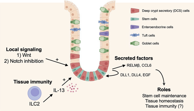
Deep crypt secretory (DCS) cells are a population of epithelial cells located at the colonic crypt base that share some similarities to Paneth and goblet cells. They were initially defined as c-Kit expressing cells, though subsequent work showed that they are more specifically marked by Reg4 in the murine colon. The best-understood function of DCS cells at present is supporting the stem cell niche by generating Notch and EGF ligands. However, as these cells also express immunoregulatory (e.g., Ccl6) and host defense (e.g., Retnlb) genes, it is likely they have additional functions in maintaining colonic health outside of maintenance of the stem niche. Recent advances in single-cell transcriptomic profiling hint at additional epithelial and immune roles that may exist for these cells and have aided in elucidating their developmental lineage. This review highlights the emerging evidence supporting a crucial role for DCS cells in intestinal physiology, the current understanding of how these cells are regulated, and their potential role(s) in colonic disease.
Keywords: IBD; colon; epithelium; secretory cells.
Conflict of interest statement
No conflicts of interest, financial or otherwise, are declared by the authors.
Figures
Similar articles
-
Reg4+ deep crypt secretory cells function as epithelial niche for Lgr5+ stem cells in colon.Proc Natl Acad Sci U S A. 2016 Sep 13;113(37):E5399-407. doi: 10.1073/pnas.1607327113. Epub 2016 Aug 29. Proc Natl Acad Sci U S A. 2016. PMID: 27573849 Free PMC article.
-
Deep Crypt Secretory Cell Differentiation in the Colonic Epithelium Is Regulated by Sprouty2 and Interleukin 13.Cell Mol Gastroenterol Hepatol. 2023;15(4):971-984. doi: 10.1016/j.jcmgh.2022.11.004. Epub 2022 Nov 19. Cell Mol Gastroenterol Hepatol. 2023. PMID: 36414210 Free PMC article.
-
Identification of a cKit(+) colonic crypt base secretory cell that supports Lgr5(+) stem cells in mice.Gastroenterology. 2012 May;142(5):1195-1205.e6. doi: 10.1053/j.gastro.2012.02.006. Epub 2012 Feb 11. Gastroenterology. 2012. PMID: 22333952 Free PMC article.
-
Paneth cells and their multiple functions.Cell Biol Int. 2022 May;46(5):701-710. doi: 10.1002/cbin.11764. Epub 2022 Jan 23. Cell Biol Int. 2022. PMID: 35032139 Review.
-
Development, validation and implementation of an in vitro model for the study of metabolic and immune function in normal and inflamed human colonic epithelium.Dan Med J. 2015 Jan;62(1):B4973. Dan Med J. 2015. PMID: 25557335 Review.
Cited by
-
Enterocloster clostridioformis protects against Salmonella pathogenesis and modulates epithelial and mucosal immune function.Microbiome. 2025 Feb 28;13(1):61. doi: 10.1186/s40168-025-02050-9. Microbiome. 2025. PMID: 40022210 Free PMC article.
-
Paneth Cells: Dispensable yet Irreplaceable for the Intestinal Stem Cell Niche.Cell Mol Gastroenterol Hepatol. 2025;19(4):101443. doi: 10.1016/j.jcmgh.2024.101443. Epub 2024 Dec 19. Cell Mol Gastroenterol Hepatol. 2025. PMID: 39708920 Free PMC article. Review.
-
Secretagogin Downregulation Impairs Nerve Cell Migration in Hirschsprung Disease via Inhibition of the LEF-1/NCAM1 Axis.Mol Cell Proteomics. 2025 Jul 11;24(8):101032. doi: 10.1016/j.mcpro.2025.101032. Online ahead of print. Mol Cell Proteomics. 2025. PMID: 40653016 Free PMC article.
-
A Review of Epithelial Ion Transporters and Their Roles in Equine Infectious Colitis.Vet Sci. 2024 Oct 7;11(10):480. doi: 10.3390/vetsci11100480. Vet Sci. 2024. PMID: 39453072 Free PMC article. Review.
-
BEST4+ cells in the intestinal epithelium.Am J Physiol Cell Physiol. 2024 May 1;326(5):C1345-C1352. doi: 10.1152/ajpcell.00042.2024. Epub 2024 Apr 1. Am J Physiol Cell Physiol. 2024. PMID: 38557358 Free PMC article. Review.
References
-
- Tanaka M, Saito H, Kusumi T, Fukuda S, Shimoyama T, Sasaki Y, Suto K, Munakata A, Kudo H. Spatial distribution and histogenesis of colorectal Paneth cell metaplasia in idiopathic inflammatory bowel disease. J Gastroenterol Hepatol 16: 1353–1359, 2001. doi:10.1046/j.1440-1746.2001.02629.x. - DOI - PubMed
Publication types
MeSH terms
Grants and funding
LinkOut - more resources
Full Text Sources