. 2023 Sep 29;133(8):704-719.
doi: 10.1161/CIRCRESAHA.123.322740. Epub 2023 Sep 12.

Altered Smooth Muscle Cell Histone Acetylome by the SPHK2/S1P Axis Promotes Pulmonary Hypertension

Affiliations

Altered Smooth Muscle Cell Histone Acetylome by the SPHK2/S1P Axis Promotes Pulmonary Hypertension

A Dushani C U Ranasinghe et al. Circ Res. .

Abstract

Background: Epigenetic regulation of vascular remodeling in pulmonary hypertension (PH) is poorly understood. Transcription regulating, histone acetylation code alters chromatin accessibility to promote transcriptional activation. Our goal was to identify upstream mechanisms that disrupt epigenetic equilibrium in PH.

Methods: Human pulmonary artery smooth muscle cells (PASMCs), human idiopathic pulmonary arterial hypertension (iPAH):human PASMCs, iPAH lung tissue, failed donor lung tissue, human pulmonary microvascular endothelial cells, iPAH:PASMC and non-iPAH:PASMC RNA-seq databases, NanoString nCounter, and cleavage under targets and release using nuclease were utilized to investigate histone acetylation, hyperacetylation targets, protein and gene expression, sphingolipid activation, cell proliferation, and gene target identification. SPHK2 (sphingosine kinase 2) knockout was compared with control C57BL/6NJ mice after 3 weeks of hypoxia and assessed for indices of PH.

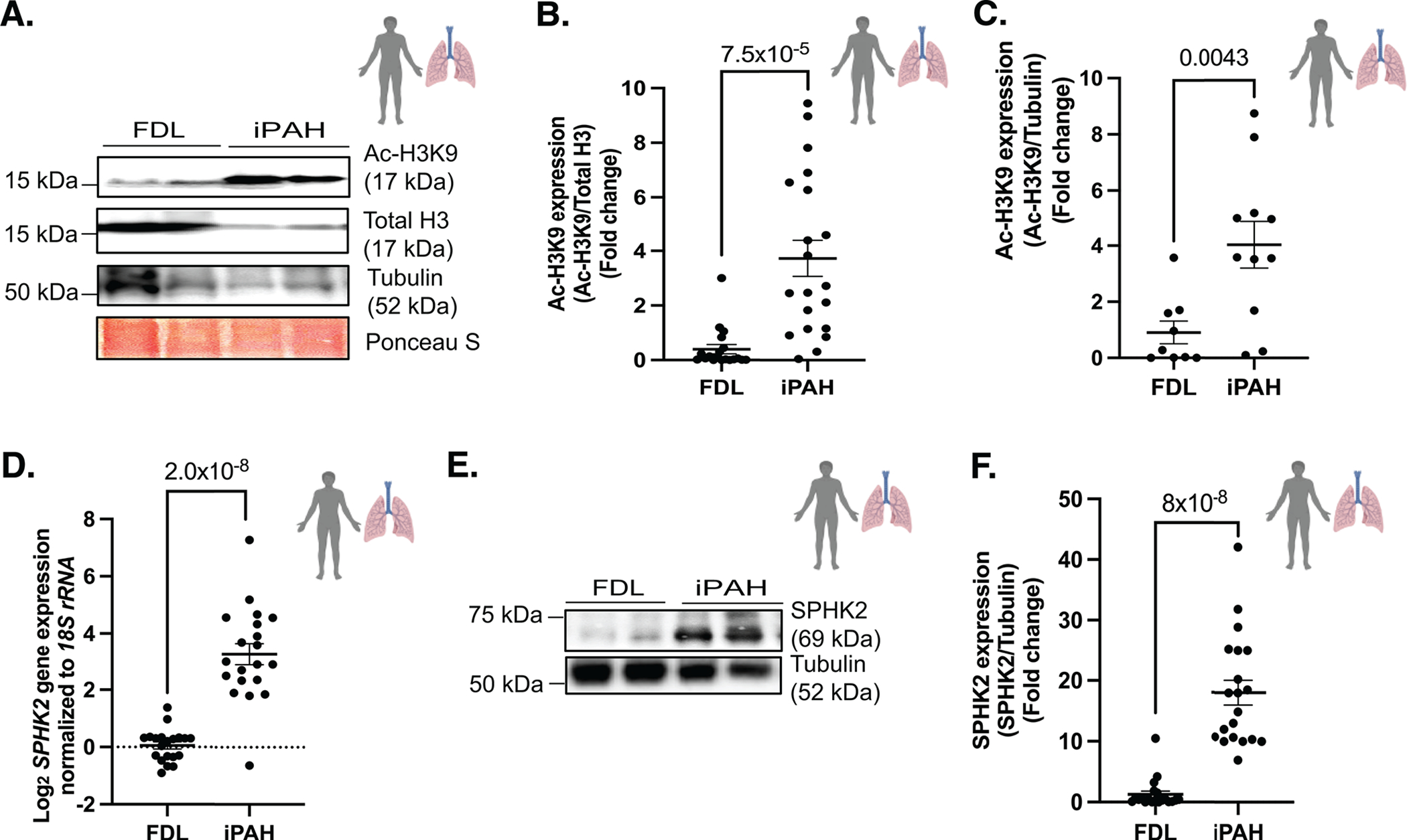
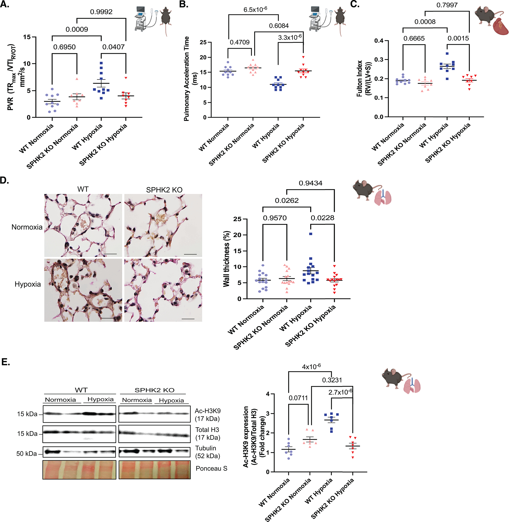
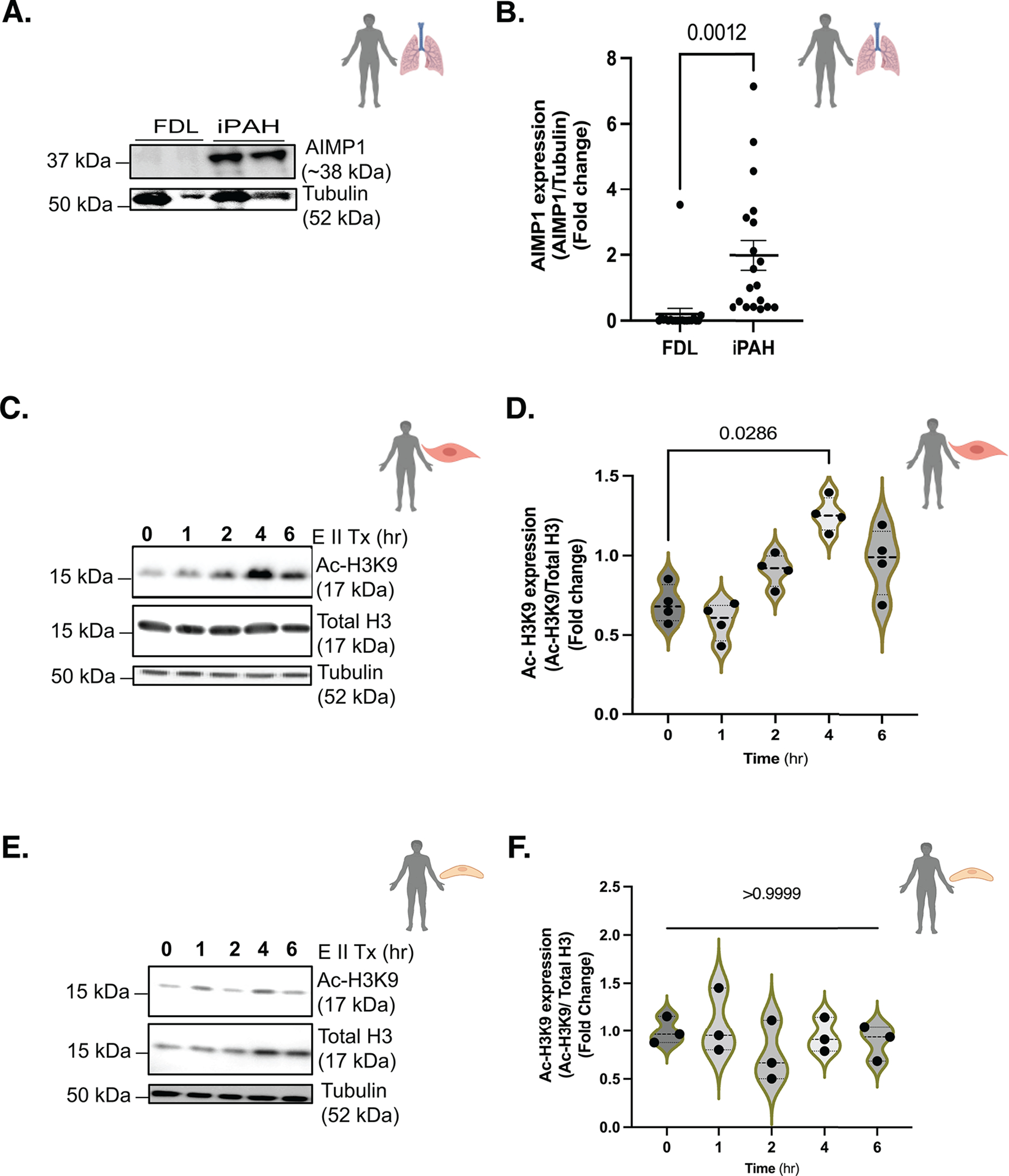
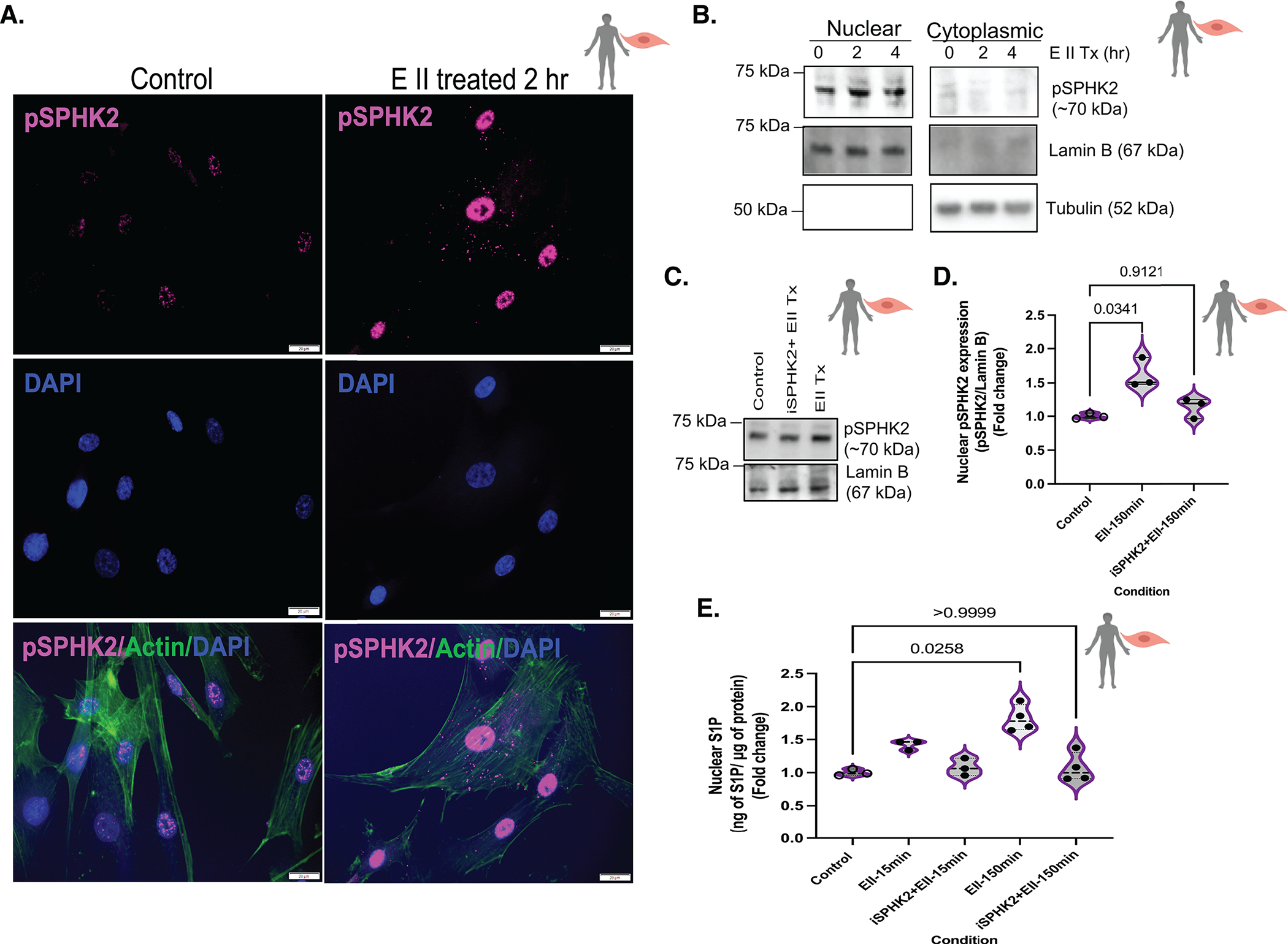
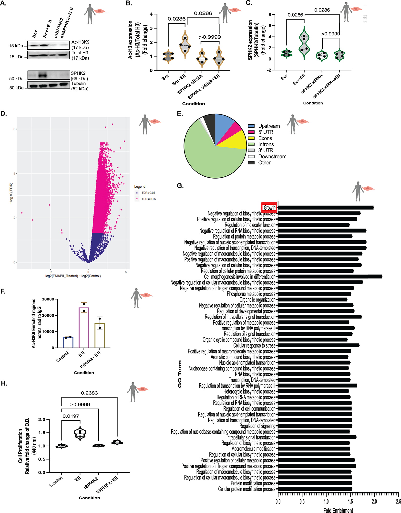
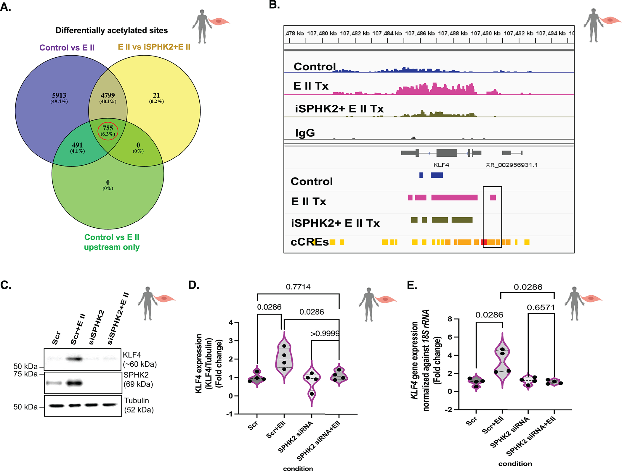
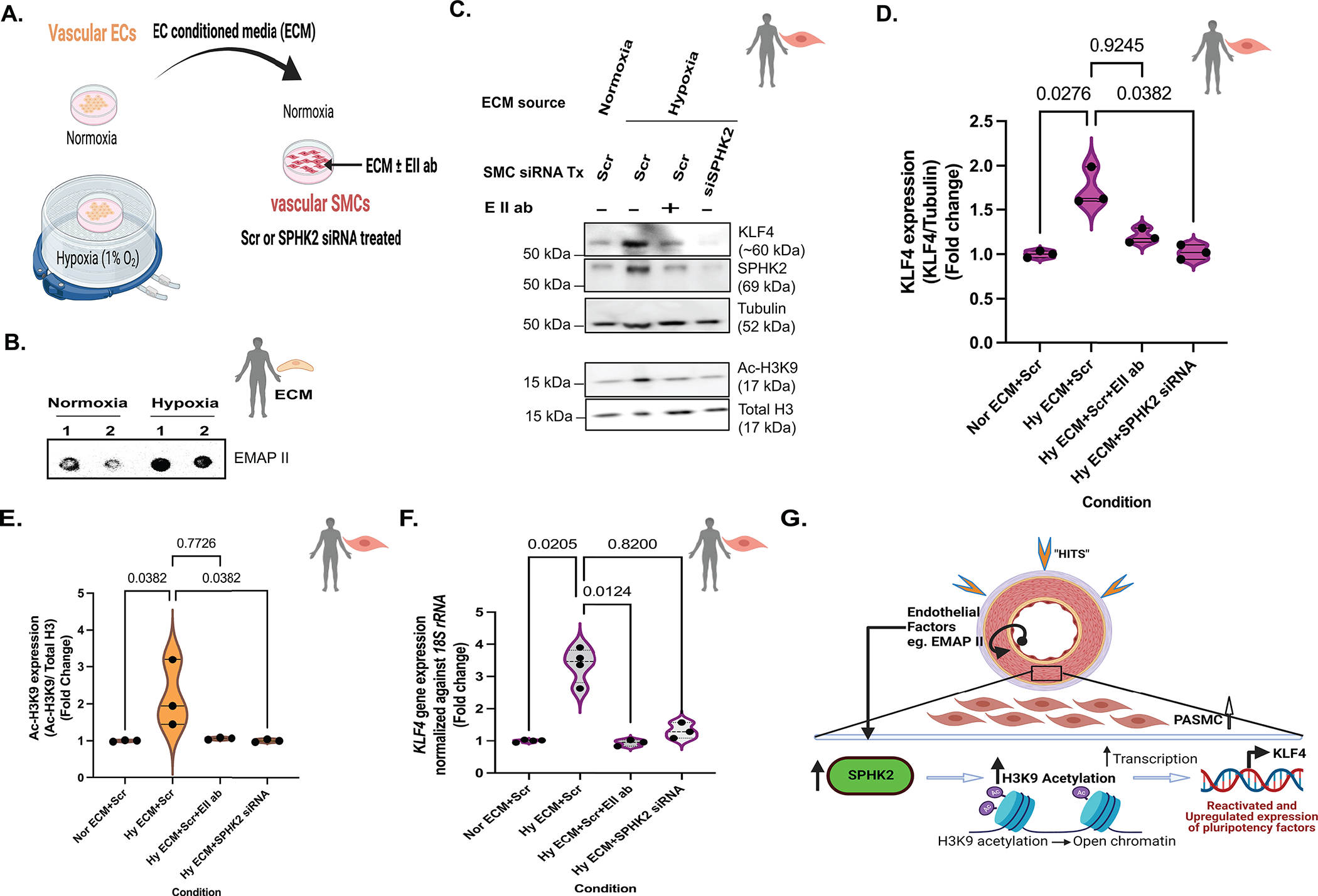
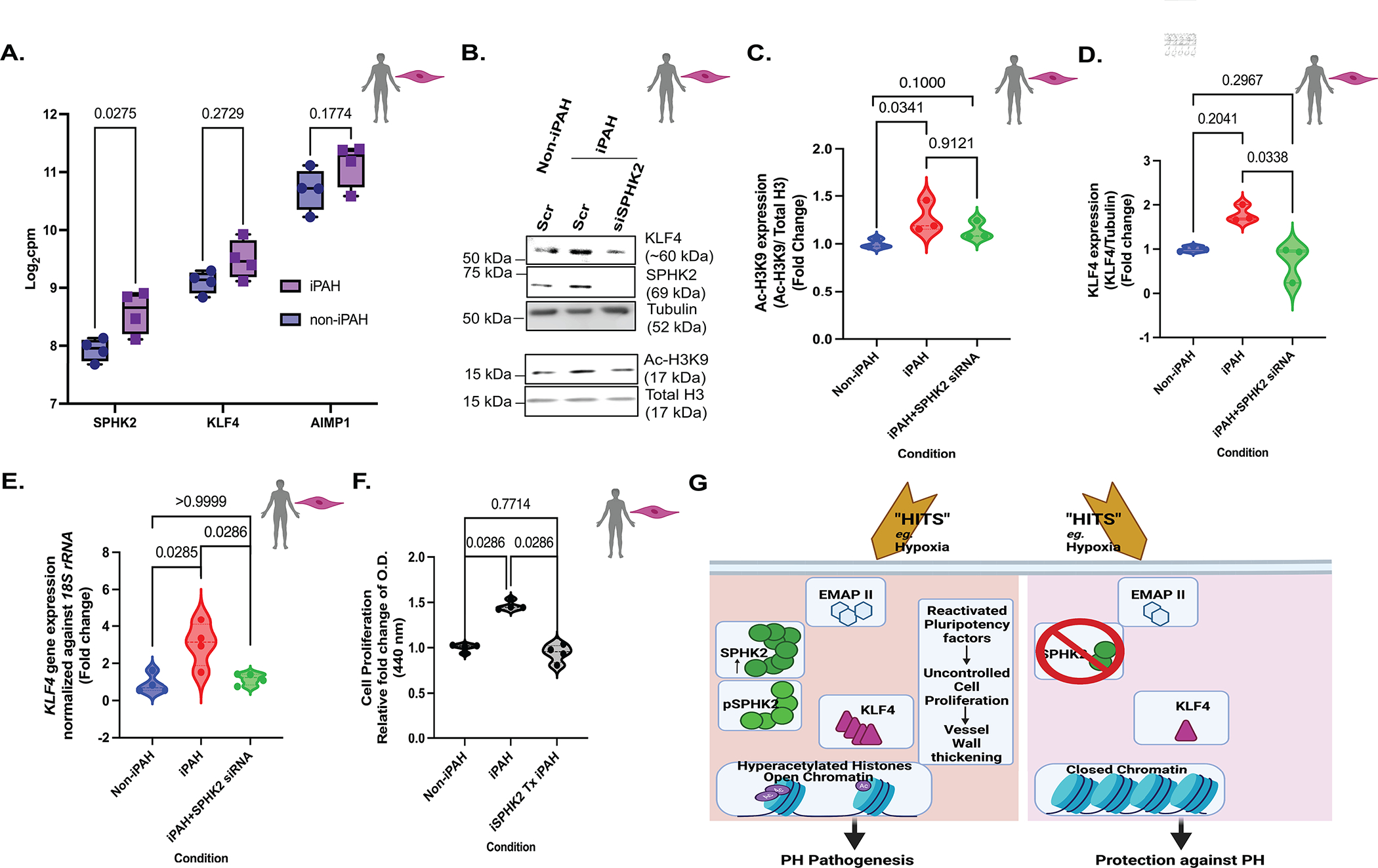
Results: We identified that Human PASMCs are vulnerable to the transcription-promoting epigenetic mediator histone acetylation resulting in alterations in transcription machinery and confirmed its pathological existence in PH:PASMC cells. We report that SPHK2 is elevated as much as 20-fold in iPAH lung tissue and is elevated in iPAH:PASMC cells. During PH pathogenesis, nuclear SPHK2 activates nuclear bioactive lipid S1P (sphingosine 1-phosphate) catalyzing enzyme and mediates transcription regulating histone H3K9 acetylation (acetyl histone H3 lysine 9 [Ac-H3K9]) through EMAP (endothelial monocyte activating polypeptide) II. In iPAH lungs, we identified a 4-fold elevation of the reversible epigenetic transcription modulator Ac-H3K9:H3 ratio. Loss of SPHK2 inhibited hypoxic-induced PH and Ac-H3K9 in mice. We discovered that pulmonary vascular endothelial cells are a priming factor of the EMAP II/SPHK2/S1P axis that alters the acetylome with a specificity for PASMC, through hyperacetylation of histone H3K9. Using cleavage under targets and release using nuclease, we further show that EMAP II-mediated SPHK2 has the potential to modify the local transcription machinery of pluripotency factor KLF4 (Krüppel-like factor 4) by hyperacetylating KLF4 Cis-regulatory elements while deletion and targeted inhibition of SPHK2 rescues transcription altering Ac-H3K9.

Conclusions: SPHK2 expression and its activation of the reversible histone H3K9 acetylation in human pulmonary artery smooth muscle cell represent new therapeutic targets that could mitigate PH vascular remodeling.

Keywords: chromatin; endothelial cells; epigenomics; hypertension, pulmonary.


Conflict of interest statement

Disclosures The authors declare that they have no known competing financial interests or personal relationships or conflicts of interests that could have appeared to influence the work reported in this article.

Figures

Figure 1.
Figure 1.. H3K9 acetylation and SPHK2 expression show a potential correlation in PAH patients’ lungs.
(A) Representative immunoblot probed for Ac-H3K9, total H3, tubulin and Ponceau S staining in protein lysates of human idiopathic pulmonary arterial hypertension (iPAH: type of Group 1 PH) lung or failed donor lung (FDL) tissue specimens and (B) quantitation of Ac-H3K9/Total H3, n=19–20 (C) quantitation of Ac-H3K9/Tubulin in protein lysates of human iPAH (n=11) or FDL (n=9). (D) SPHK2 expression levels normalized against 18S rRNA in iPAH lung and FDL tissues. n=20 (E) Representative immunoblot probed for SPHK2 and Tubulin in protein lysates of human iPAH (type of Group 1 PH) lung or FDL tissue specimens and (F) quantitation of SPHK2/Tubulin in protein lysates of human iPAH lung or FDL, n=20. P values are calculated using unpaired t-test and results are shown as means ± SEM.
Figure 2.
Figure 2.. SPHK2 ablation confers protection against experimental PH and hyperacetylation of H3K9 in hypoxia-induced experimental PH mouse model.
SPHK2 KO or wild type (WT) control mice (C57BL/6NJ) were subjected to 3 wks of hypoxia (10% O2) or normoxia (room air). (A) Pulmonary vascular resistance (the maximum velocity of tricuspid regurgitation/the velocity time integral of the right ventricular outflow tract, TRmax velocity/VTIRVOT) n=8–10/group. (B) Pulmonary acceleration time (PAT), n=8–10/group. (C) RV hypertrophy/Fulton Index (the weight ratio of the right ventricle divided by the sum of left ventricle and septum, RV/(LV + S)) n=8–10/group. (D) Representative images of elastin-stained distal pulmonary vessels and, wall thickness of distal pulmonary vessels in elastin-stained lung tissue sections (wall thickness (%) = (2 × medial wall thickness / external diameter) × 100) n= 3/group randomly selected mice per each group and n=15 images of distal pulmonary vessels, scale bar is 10 μm, n=5–6/group. (E) Representative immunoblot probed for Ac-H3K9, total H3, Tubulin and ponceau S staining in whole tissue lysates from WT or SPHK2 KO in normoxia or hypoxia on same blot n=5–7/group and, quantitation of Ac-H3K9/Total H3. P values are calculated using one-way ANOVA following Tukey’s multiple comparisons test, and results are shown as means ± SEM.
Figure 3.
Figure 3.. EMAP II has the potential to be a key pathogenic mediator in PH through modulating epigenetic equilibrium via histone H3K9 acetylation uniquely in vascular SMCs.
(A) Representative immunoblot probed for AIMP1 (precursor form of EMAP II) and Tubulin in protein lysates of human iPAH or FDL, n=19–20/group and, (B) quantitation of AIMP1 (AIMP1/Tubulin) in protein lysates of human iPAH or FDL, n=19–20/group. (C) Representative immunoblot probed for Ac-H3K9, total H3 or tubulin in hPASMCs following EMAP II treatment for 0, 1, 2, 4 and 6 hours and (D) quantitation of Ac-H3K9 expression levels normalized against total H3 in hPASMCs, n=4. (E) Representative immunoblot probed for Ac-H3K9, total H3 or tubulin in hPMVECs following EMAP II treatment for 0, 1, 2, 4 and 6 hours and (F) quantitation of Ac-H3K9 expression levels normalized against total H3 in hPMVECs, n=3. P values are calculated using unpaired t-test or Kolmogorov-Smirnov non-parametric testing and results are shown as means ± SEM or median and inter-quartile range.
Figure 4.
Figure 4.. EMAP II promotes nuclear activation of SPHK2 that in turn generates nuclear lipid, S1P in vascular SMCs.
(A) Representative immunocytochemistry images of pSPHK2 (pink), actin (green, cytoplasmic marker) and DAPI (blue, nuclear) coimmunostaining in EMAP II treated (2 hr) or vehicle treated fixed hPASMCs, scale bar is 20 μm, n=3. (B) Representative immunoblot probed for pSPHK2, tubulin and lamin B in cytoplasmic and nuclear fractions of hPASMCs following EMAP II treatment for 0, 2 and 4 hours, n=3. (C) Representative immunoblot probed for pSPHK2 and lamin B in nuclear fractions of hPASMCs following EMAP II treatment (150 minutes) with or without SPHK2 inhibitor (D) quantification of nuclear pSPHK2/lamin B, n=3. (E) ELISA-nuclear C18-S1P levels normalized against 1 μg of nuclear proteins in the nuclear fractions of hPASMCs following EMAP II for 15 or 150 minutes with or without SPHK2 inhibitor, n=3 or 4/group. P values are calculated using Kruskal-Wallis against control or Kolmogorov-Smirnov non-parametric test and results are shown as median and inter-quartile range.
Figure 5.
Figure 5.. EMAP II mediated SPHK2 signaling promotes global hyperacetylation of histone H3K9 in vascular SMCs.
(A) Representative immunoblot probed for Ac-H3K9, total H3, SPHK2 and tubulin in whole cell lysates of hPASMCs following siRNA mediated SPHK2 silencing and post-transfection EMAP II treatment for 4 hours and (B) quantitation of Ac-H3K9/total H3 and (C) quantification of SPHK2/tubulin, n=4. (D) Volcano plot showed the log2-fold changes and statistical significance of hyperacetylated H3K9 regions calculated after differential binding analysis of EMAP II treated vs control hPASMCs. Pink points indicate significantly hyperacetylated H3K9 regions in EMAP II (right to 0) or in control (left to 0). FDR=0.05, n=2 (E) Genome wide distribution of differentially enriched hyperacetylated H3K9 peaks (log2-fold change > 1, p value < 0.05) n=2. (F) Number of peaks of Ac-H3K9 normalized to IgG in with or without SPHK2 inhibitor and EMAP II treated (2–3 hours) hPASMCs, n=2. (G) Gene Ontology results using differentially enriched Ac-H3K9 peaks in EMAP II treated hPASMCs, n=2. (H) Cell proliferation rate in hPASMCs treated with vehicle or EMAP II following SPHK2 inhibitor treatment for 24 hours, n=3. P values are calculated using Kruskal-Wallis against control or Kolmogorov-Smirnov non-parametric test and results are shown as means ± SEM or median and inter-quartile range.
Figure 6.
Figure 6.. EMAP II mediated SPHK2 signaling promotes local hyperacetylation of histone H3K9 of KLF4 enhancers and alters the local transcription machinery of KLF4 in vascular SMCs.
(A) The Venn’s diagram of differential acetylated sites in control vs EMAP II (total) (purple), EMAP II vs iSPHK2+EMAP II (yellow) and control vs EMAPII only in 5’UTR and upstream with fold enrichment greater than 2 (green). The red circle indicates the potential gene set with potential upstream candidate regulatory elements that would be differentially acetylated by EMAP II through SPHK2 in hPASMCs. Venn diagram is created using Venny 2.1 (an online interactive tool), n=2/group (B) Snapshot of IGV view of KLF4 gene in Ac-H3K9 CUT&RUN data of with or without SPHK2 inhibitor and EMAP II treated (2–3 hours) hPASMCs. (cCRE= candidate Cis-Regulatory Elements) n=2/group (C) Representative immunoblot probed for KLF4, SPHK2 and tubulin in whole cell lysates of hPASMCs following siRNA mediated SPHK2 silencing and post-transfection EMAP II treatment for 6–8 hours, and (D) quantitation of KLF4/tubulin and (E) KLF4 expression levels normalized against 18S rRNA in hPASMC cells following siRNA mediated SPHK2 silencing and EMAP II treatment for 6 hours, n=4. P values are calculated using Kolmogorov-Smirnov non-parametric testing and results are shown as median and inter-quartile range.
Figure 7.
Figure 7.. EMAP II/SPHK2/Ac-H3K9 mediated KLF4 signaling is a novel pathway that exists in PH disease pathogenesis.
(A) Schematic diagram representing the collection of vascular endothelial cells (ECs) conditioned media (ECM) from ECs grown in 1%O2 or room air to treat vascular smooth muscle cells (SMCs) and, (B) representative dot blot probed for secreted EMAP II expression in ECM. (C) Representative immunoblot probed for KLF4, SPHK2, tubulin, Ac-H3K9 and total histone H3 in whole cell lysates of normoxia or hypoxia ECM with or without EMAP II neutralizing antibody treated hPASMCs pre-transfected with siRNA mediated SPHK2 or scramble silencing and, (D) quantification of KLF4/Tubulin, n=3 and (E) quantification of Ac-H3K9/total histone H3, n=3. (F) KLF4 expression levels normalized against 18S rRNA in normoxia or hypoxia ECM with or without EMAP II neutralizing antibody treated hPASMCs pre-transfected with siRNA mediated SPHK2 or scramble silencing, n=3–4. (G) EMAP II secreted by vascular ECs promote SPHK2/Ac-H3K9/KLF4 signaling in vascular SMCs that may promote PASMCs proliferation. P values are calculated using Kruskal-Wallis against Hy ECM+Scr or Kolmogorov-Smirnov non-parametric test if not mentioned otherwise, and results are shown as median and inter-quartile range.
Figure 8.
Figure 8.. EMAP II/SPHK2/Ac-H3K9 mediated KLF4 signaling is a novel pathway that exists in PH disease pathogenesis.
(A) RNA-seq data of SPHK2, KLF4 and AIMP1 in iPAH: PASMCs and non-iPAH:PASMCs in log2-fold of count per million (cpm). Following two-way ANOVA, Sidak’s multiple comparisons test for logarithmic values, n=4. (B) Representative immunoblot probed for KLF4, SPHK2, tubulin, Ac-H3K9 and total histone H3 in whole cell lysates of non: iPAH or iPAH PASMCs with scramble or SPHK2 siRNA transfection and, quantification of (C) Ac-H3K9/total histone H3 and, (D) KLF4/Tubulin, n=3 (E) KLF4 expression levels normalized against 18S rRNA in non: iPAH or iPAH PASMCs with scramble or SPHK2 siRNA transfection, n=4. (F) Cell proliferation rate of non: iPAH or iPAH PASMC with or without iSPHK2 pretreatment for 24 hours, n=4. (G) The proposed model: Endothelial monocyte activating polypeptide II (EMAP II) plays a key role in reawakening pluripotency factor, KLF4 in human pulmonary artery smooth muscle cells (PASMCs) through stimulation of the nuclear SPHK2/S1P epigenetic modulating axis, suggesting that cooperation between SPHK2 and EMAP II could be a major driving force for epigenetic-mediated vascular PASMCs reprogramming and remodeling in PH. Ablation of SPHK2 expression confers protection against PH by rescuing the global and local transcription machinery from histone acetylation and activation of the pluripotency factor, KLF4. P values are calculated using Kruskal-Wallis against iPAH or Kolmogorov-Smirnov non-parametric test if not mentioned otherwise, and results are shown as median and inter-quartile range.

References

    1. Rabinovitch M Molecular pathogenesis of pulmonary arterial hypertension. J Clin Invest 2012;122:4306–4313. doi:10.1172/JCI60658. - DOI - PMC - PubMed
    1. Wang L, Moonen JR, Cao A, Isobe S, Li CG, Tojais NF, Taylor S, Marciano DP, Chen PI, Gu M, Li D, Harper RL, El-Bizri N, Kim YM, Stankunas K, Rabinovitch M. Dysregulated Smooth Muscle Cell BMPR2–ARRB2 Axis Causes Pulmonary Hypertension. Circ Res 2023;132:545–564. doi:10.1161/CIRCRESAHA.121.320541. - DOI - PMC - PubMed
    1. Bisserier M, Mathiyalagan P, Zhang S, Elmastour F, Dorfmüller P, Humbert M, David G, Tarzami S, Weber T, Perros F, Sassi Y, Sahoo S, Hadri L. Regulation of the Methylation and Expression Levels of the BMPR2 Gene by SIN3a as a Novel Therapeutic Mechanism in Pulmonary Arterial Hypertension. Circulation 2021;144:52–73. doi:10.1161/CIRCULATIONAHA.120.047978. - DOI - PMC - PubMed
    1. Benincasa G, Maron BA, Affinito O, D’Alto M, Franzese M, Argiento P, Schiano C, Romeo E, Bontempo P, Golino P, Berrino L, Loscalzo J, Napoli C. Association Between Circulating CD4+ T Cell Methylation Signatures of Network-Oriented SOCS3 Gene and Hemodynamics in Patients Suffering Pulmonary Arterial Hypertension. J Cardiovasc Transl Res 2023;16:17–30. doi:10.1007/S12265-022-10294-1/FIGURES/6. - DOI - PMC - PubMed
    1. Benincasa G, Napoli C, Loscalzo J, Maron BA. Pursuing functional biomarkers in complex disease: Focus on pulmonary arterial hypertension. Am Heart J 2023;258:96–113. doi:10.1016/J.AHJ.2022.12.009. - DOI - PubMed

Publication types