. 2025 Mar;15(2):554-563.
doi: 10.1177/21925682231200783. Epub 2023 Sep 12.

An Automatized Deep Segmentation and Classification Model for Lumbar Disk Degeneration and Clarification of Its Impact on Clinical Decisions

Affiliations

An Automatized Deep Segmentation and Classification Model for Lumbar Disk Degeneration and Clarification of Its Impact on Clinical Decisions

Zafer Soydan et al. Global Spine J. 2025 Mar.

Abstract

Study design: Cross-sectional database study.

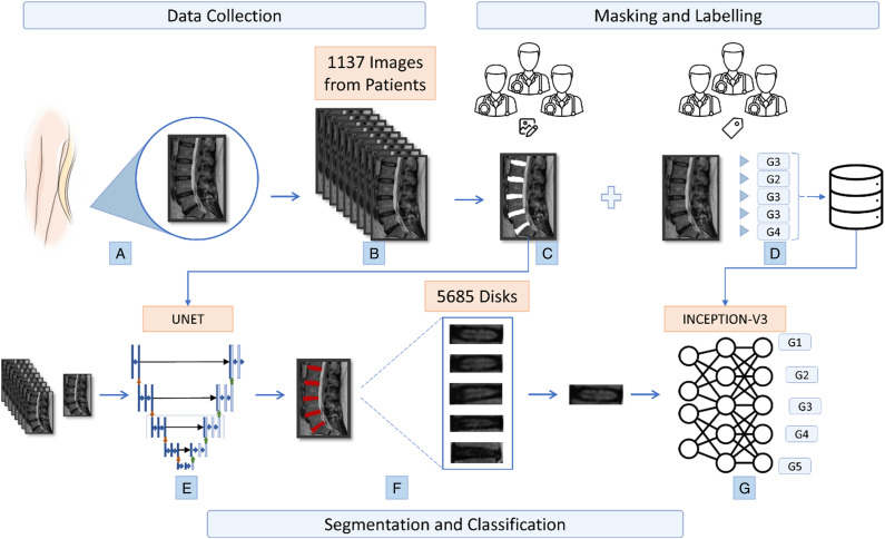
Objective: The purpose of this study was to develop a successful, reproducible, and reliable convolutional neural network (CNN) model capable of segmentation and classification for grading intervertebral disc degeneration (IVDD), as well as quantify the network's impact on doctors' clinical decision-making.

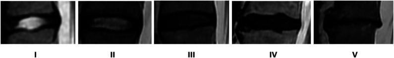
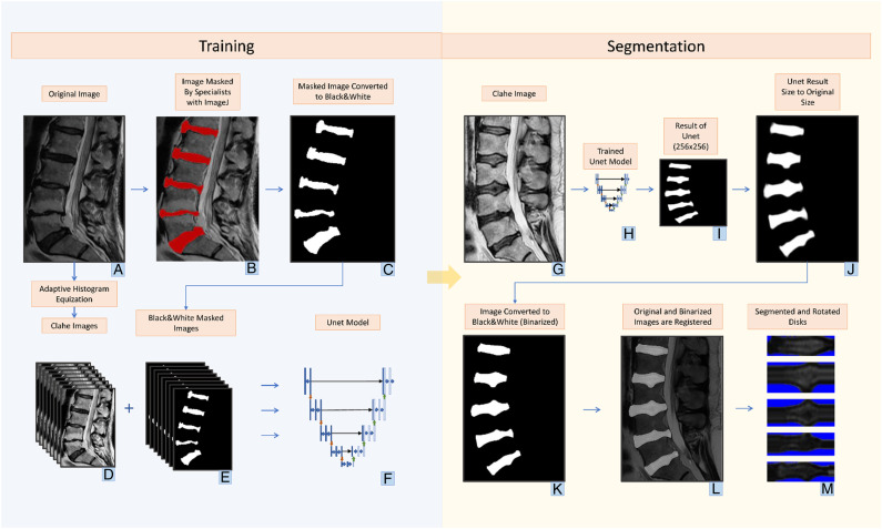
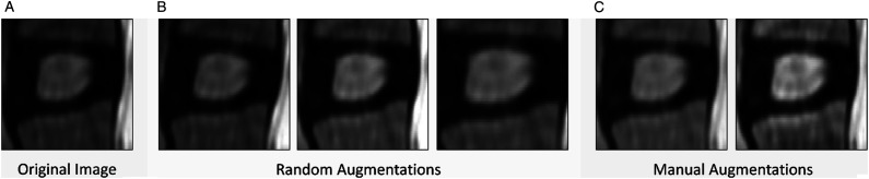
Methods: 5685 discs from 1137 patients were graded separately by four experienced doctors according to the Pfirrmann classification. A ground truth (GT) was established for each disc in accordance with the decision of the majority of doctors. The U-net model is used for segmentation. 1815 discs from 363 patients were used to train and test the U-net. The Inception V3 model is employed for classification. All discs were separated into two distinct sets: 90% in a training set and 10% in a test set. The performance metrics of these models were measured. Reliability tests were performed. The impact of CNN assistance on doctors was assessed.

Results: Segmentation accuracy was .9597 with a .8717 Jaccard Index and a .9314 Sorensen Dice coefficient. Classification accuracy is .9346, and the F1 score is .9355. The intraclass correlation coefficient (ICC) and kappa values between CNN and GT were .95-.97. With CNN's assistance, the success rates of doctors increased by 7.9% to 22%.

Conclusions: The fully automated network outperformed doctors markedly in terms of accuracy and reliability. The results of CNN were comparable to those of other recent studies in the literature. It was determined that CNN's assistance had a substantial positive effect on the doctor's decision.

Keywords: convolutional neural network; degenerative disc disease; pfirrmann classification; segmentation.


Figures

Figure 1.
Figure 1.
Pfirrmann classification(6) from grade 1 to 5.
Figure 2.
Figure 2.
Flowchart of study (a) obtaining lumbar discs (b) collection of disc images from 1137 patients (c) masking of images by specialists with imageJ program for segmentation (d) labeling of 5 discs in each image for classification (e) creation of the U-net model (f) obtaining discs from images according to the U-net model (g) classification of segmented discs according to the generated CNN inception-v3 model.
Figure 3.
Figure 3.
Training phase (a) original Images (b) masked images for training phase with imageJ program by doctors (c) image converted to black and white to focus only on disc areas (d) images converted to contrast limited adaptive histogram equalization (CLAHE) by adaptive histogram equalization method (e) masked versions of CLAHE images (f) generating the U-net model from CLAHE and masked images. segmentation phase CLAHE image for segmentation (h) trained U-net model (i) image created with the Unet model in 256 × 256 dimensions (j) converting the image from U-net to its original size (k) changing the image to black and white with thresholding (l) determining the location of the discs in the white areas by overlaying the original image and the image converted to black and white as a result of U-net (m) saving the images taken as a result of cropping and rotating horizontally the region of interests. Discs are ready for classification.
Figure 4.
Figure 4.
Utilized augmentation methods (a) original image (b) random augmentations: brightness, rotation, zoom variations (c) CLAHE and gamma augmentations.

Similar articles

Cited by

References

    1. Hoy DG, Smith E, Cross M, et al. Reflecting on the global burden of musculoskeletal conditions: lessons learnt from the global burden of disease 2010 study and the next steps forward. Ann Rheum Dis. 2015;74(1):4-7. doi:10.1136/annrheumdis-2014-205393 - DOI - PubMed
    1. Matsuyama Y, Chiba K, Iwata H, Seo T, Toyama Y. A multicenter, randomized, double-blind, dose-finding study of condoliase in patients with lumbar disc herniation. J Neurosurg Spine. 2018;28(5):499-511. doi:10.3171/2017.7.SPINE161327 - DOI - PubMed
    1. Le Maitre CL, Hoyland JA, Freemont AJ. Catabolic cytokine expression in degenerate and herniated human intervertebral discs: IL-1beta and TNFalpha expression profile. Arthritis Res Ther. 2007;9(4):R77. doi:10.1186/ar2275 - DOI - PMC - PubMed
    1. Thorpe AA, Binch AL, Creemers LB, Sammon C, Le Maitre CL. Nucleus pulposus phenotypic markers to determine stem cell differentiation: fact or fiction? Oncotarget. 2016;7(3):2189-2200. doi:10.18632/oncotarget.6782. - DOI - PMC - PubMed
    1. Binch ALA, Fitzgerald JC, Growney EA, Barry F. Cell-based strategies for IVD repair: clinical progress and translational obstacles. Nat Rev Rheumatol. 2021;17(3):158-175. doi:10.1038/s41584-020-00568-w - DOI - PubMed

LinkOut - more resources