. 2023 Nov 1;325(5):L580-L593.
doi: 10.1152/ajplung.00135.2023. Epub 2023 Sep 12.

Electronic cigarette exposure disrupts airway epithelial barrier function and exacerbates viral infection

Affiliations

Electronic cigarette exposure disrupts airway epithelial barrier function and exacerbates viral infection

Andjela Raduka et al. Am J Physiol Lung Cell Mol Physiol. .

Abstract

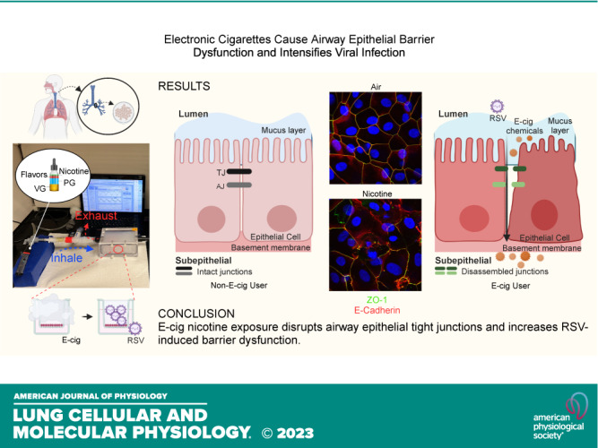
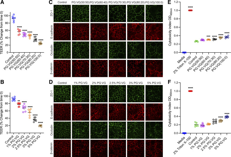
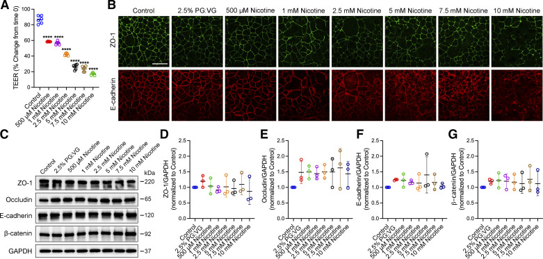
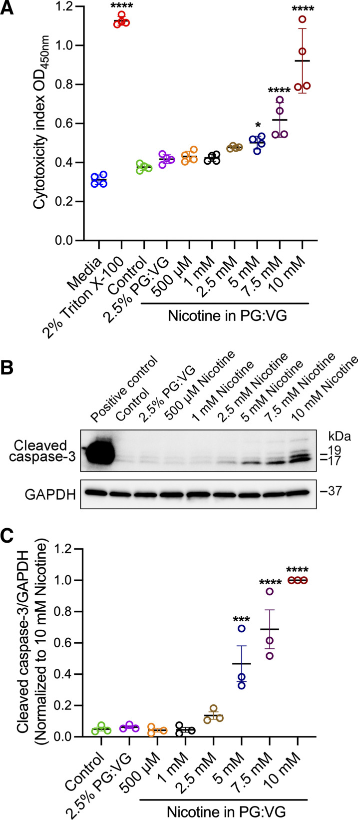
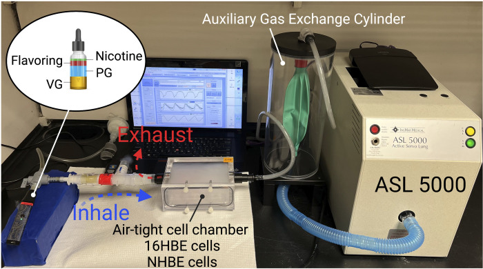
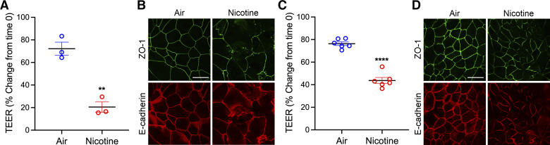
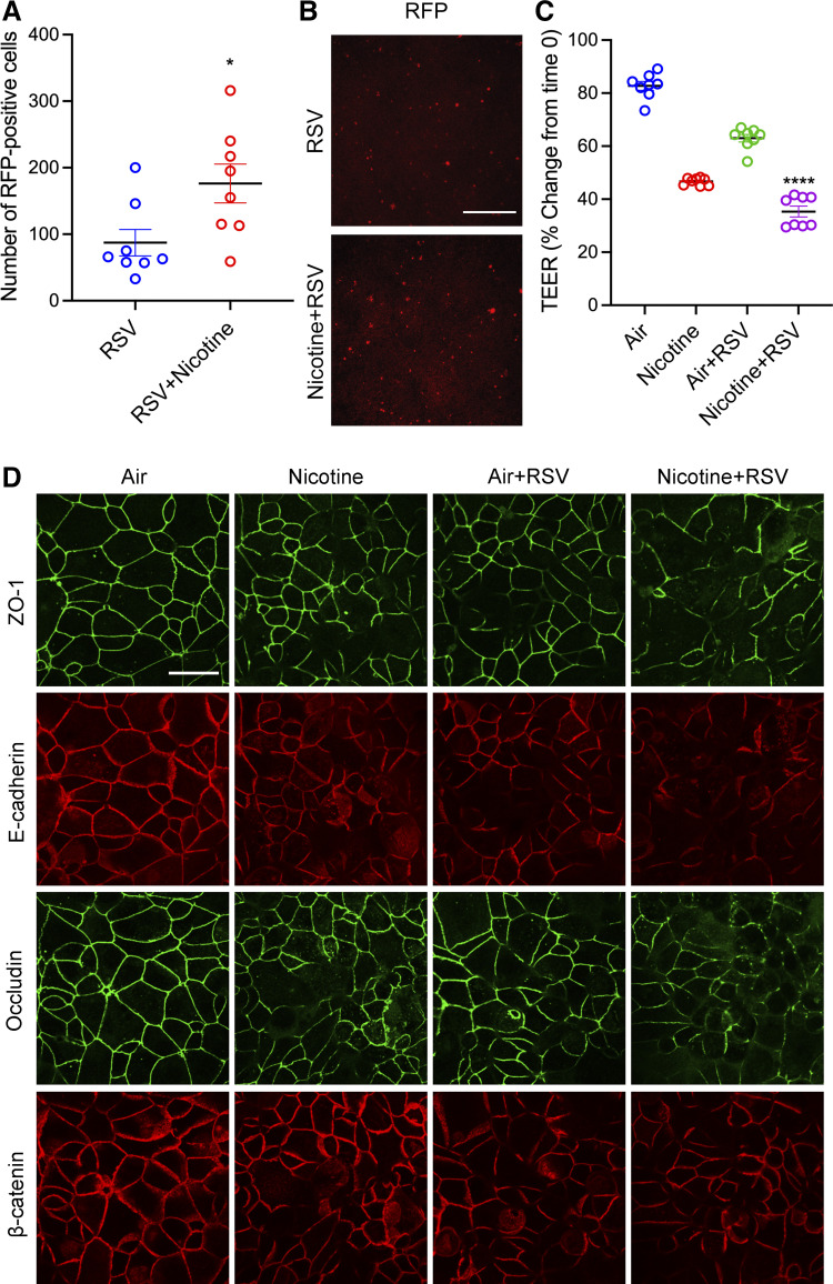
The use of electronic cigarettes (e-cigs), especially among teenagers, has reached alarming and epidemic levels, posing a significant threat to public health. However, the short- and long-term effects of vaping on the airway epithelial barrier are unclear. Airway epithelial cells are the forefront protectors from viruses and pathogens. They contain apical junctional complexes (AJCs), which include tight junctions (TJs) and adherens junctions (AJs) formed between adjacent cells. Previously, we reported respiratory syncytial virus (RSV) infection, the leading cause of acute lower respiratory infection-related hospitalization in children and high-risk adults, induces a "leaky airway" by disrupting the epithelial AJC structure and function. We hypothesized chemical components of e-cigs disrupt airway epithelial barrier and exacerbate RSV-induced airway barrier dysfunction. Using confluent human bronchial epithelial (16HBE) cells and well-differentiated normal human bronchial epithelial (NHBE) cells, we found that exposure to extract and aerosol e-cig nicotine caused a significant decrease in transepithelial electrical resistance (TEER) and the structure of the AJC even at noncytotoxic concentrations. Western blot analysis of 16HBE cells exposed to e-cig nicotine extract did not reveal significant changes in AJC proteins. Exposure to aerosolized e-cig cinnamon or menthol flavors also induced barrier disruption and aggravated nicotine-induced airway barrier dysfunction. Moreover, preexposure to nicotine aerosol increased RSV infection and the severity of RSV-induced airway barrier disruption. Our findings demonstrate that e-cig exposure disrupts the airway epithelial barrier and exacerbates RSV-induced damage. Knowledge gained from this study will provide awareness of adverse e-cig respiratory effects and positively impact the mitigation of e-cig epidemic.NEW & NOTEWORTHY Electronic cigarette (e-cig) use, especially in teens, is alarming and at epidemic proportions, threatening public health. Our study shows that e-cig nicotine exposure disrupts airway epithelial tight junctions and increases RSV-induced barrier dysfunction. Furthermore, exposure to aerosolized flavors exaggerates e-cig nicotine-induced airway barrier dysfunction. Our study confirms that individual and combined components of e-cigs deleteriously impact the airway barrier and that e-cig exposure increases susceptibility to viral infection.

Keywords: airway epithelial cells; electronic cigarette; epithelial barrier; respiratory syncytial virus (RSV); vaping.


Conflict of interest statement

No conflicts of interest, financial or otherwise, are declared by the authors.

Figures

None
Graphical abstract
Figure 1.
Figure 1.
Exposure to PG:VG induced barrier dysfunction and AJC disassembly. Confluent 16HBE cells were exposed to control medium or various PG:VG ratios (2.5% vol/vol at 50:50, 60:40, 70:30, 80:20, and 100:0) and concentrations (1%, 2%, 2.5%, 3%, and 5% vol/vol at 50:50 ratio) for 24 h. A: at 24 h postexposure to various PG:VG ratios, TEER (Ω × cm2) was measured by the volt-ohm meter and plotted as percentage versus time 0 for each group. B: at 24 h postexposure to various PG:VG (50:50) concentrations, TEER was measured and plotted as a percentage versus time 0 for each group. C: at 24 h postexposure, cells were fixed with methanol and immunolabeled for tight junction proteins (ZO-1 and occludin) and adherens junction proteins (E-cadherin and β-catenin) and imaged by confocal microscopy. D: at 24 h postexposure, cells were fixed with methanol and immunolabeled for tight junction proteins (ZO-1 and occludin) and adherens junction proteins (E-cadherin and β-catenin) and imaged by confocal microscopy. The cytotoxicity index of cells exposed to different PG:VG ratios (E) and concentrations (F) for 24 h. The culture medium served as the negative control, and the medium from cells treated with 2% Triton X-100 served as the positive control. The cytotoxicity index was calculated and compared with untreated cells (control). Data are shown as means ± SE, n = 5-6 independent experiments; one-way ANOVA followed by Dunnett’s multiple comparisons test. **P < 0.01, ***P < 0.001, ****P < 0.0001. The images are representative of at least three independent experiments. Scale bar, 30 μm. AJC, apical junctional complex; PG, propylene glycol; TEER, transepithelial electrical resistance; VG, vegetable glycerin.
Figure 2.
Figure 2.
Exposure to e-cig nicotine extract disrupted the airway epithelial barrier in a dose-dependent manner. Confluent 16HBE cells were exposed to a control medium or multiple concentrations of nicotine extract (0.5–10 mM) prepared in 2.5% PG:VG (50:50). A: at 24 h postexposure, TEER (Ω × cm2) was measured by the volt-ohm meter and plotted as a percentage change from time 0. B: cells were fixed with methanol and immunolabeled for tight junction protein (ZO-1) and adherens junction protein (E-cadherin) and imaged by confocal microscopy. C: representative Western blot of whole cell lysates of three independent experiments from 16HBE cells exposed to control medium, 2.5% PG:VG (50:50), and various nicotine extract concentrations for 24 h. GAPDH served as the control for protein loading. Densitometric qualification of three independent experiments for AJC components ZO-1 (D), occludin (E), E-cadherin (F), and β-catenin (G) Western blotting. Data are shown as means ± SE, n = 3–6, one-way ANOVA followed by Dunnett’s multiple comparisons test. ****P < 0.0001. Images are representative of three independent experiments. Scale bar, 30 μm. AJC, apical junctional complex; PG, propylene glycol; TEER, transepithelial electrical resistance; VG, vegetable glycerin; 16HBE, 16HBE14o- human bronchial epithelial.
Figure 3.
Figure 3.
Exposure to lower concentrations of e-cig extract did not cause cell cytotoxicity or apoptosis. Confluent 16HBE cells exposed to control medium, 2.5% PG:VG (50:50), or various concentrations of nicotine (0.5–10 mM) prepared in 2.5% PG:VG (50:50) for 24 h. A: results show the cytotoxicity index of cells exposed to various nicotine extract concentrations for 24 h compared with untreated cells. The culture medium served as a negative control, and cells lysed with 2% Triton X-100 served as a positive control. B: representative Western blot of whole cell lysates from 16HBE cells of three independent experiments probed with antibodies against cleaved caspase-3. Cell lysate from cytochrome c-treated Jurkat cells served as the positive control. GAPDH served as the control for protein loading. Total protein levels of cleaved caspase-3 (C) were analyzed by densitometry and plotted as normalized as indicated. Data are presented as means ± SE, n = 3 or 4, one-way ANOVA followed by Dunnett’s multiple comparisons test. *P < 0.05, ***P < 0.001, ****P < 0.0001. Images are representative of three independent experiments. 16HBE, 16HBE14o- human bronchial epithelial.
Figure 4.
Figure 4.
Schematic diagram of experiment model for e-cig aerosol exposure. IngMar ASL 5000 breathing simulator that generates realistic patient breathing patterns is connected to a laptop with specialized software that uses preprogrammed lung models with various tidal volumes and breath rates. A custom-built air-tight cell chamber is connected to the ASL 5000 to represent the upper airways. An anesthesia bag in a rigid cylinder (IngMar Auxiliary Gas Exchange Cylinder) connects the cell chamber and the ASL 5000 to keep vaping aerosol from contaminating the breathing simulator. A three-way valve connects the e-cig (Vaporesso XROS vaping pen) and HEPA filter to the air-tight cell chamber. The annotation of the photograph was created with a licensed version of BioRender.com.
Figure 5.
Figure 5.
Exposure of differentiated NHBE and polarized 16HBE cells to e-cig nicotine aerosol disrupted the airway barrier. A: NHBE cells were grown on permeable membranes, differentiated under an air-liquid interface, and exposed to 36 mg/mL aerosolized e-cig nicotine or HEPA-filtered air at 0, 8, 24, 32, and 48 h. TEER (Ω × cm2) was measured by the volt-ohm meter at 48 h postexposure and plotted as a percentage change from time 0. B: after 48 h of aerosolized e-cig nicotine exposure, NHBE cells were fixed with methanol and immunolabeled for tight junction protein (ZO-1) and adherens junction protein (E-cadherin) and imaged by confocal microscopy. C: 16HBE cells were grown to confluent and exposed to 36 mg/mL aerosolized e-cig nicotine or HEPA-filtered air at 0, 8, and 24 h. TEER was measured at 24 h postexposure and plotted as a percentage change from time 0. D: after 24 h postexposure of 16HBE cells to aerosolized e-cig nicotine, cells were fixed with methanol and immunolabeled for tight junction protein (ZO-1) and adherens junction protein (E-cadherin) and imaged by confocal microscopy. Data are shown as means ± SE, n = 3 or 6, unpaired t test. **P < 0.01, ****P < 0.0001. The images are representative of at least three independent experiments. Scale bar, 30 μm. NHBE, normal human bronchial epithelial; TEER, transepithelial electrical resistance; 16HBE, 16HBE14o- human bronchial epithelial.
Figure 6.
Figure 6.
Exposure to flavor in e-cig liquid induced barrier dysfunction and exaggerated e-cig nicotine-induced airway barrier dysfunction. A: confluent 16HBE cells were exposed to aerosolized e-cig nicotine, cinnamon, or e-cig nicotine mixed with cinnamon or HEPA-filtered air three times for 24 h. TEER (Ω × cm2) was measured 24 h postexposure and plotted as a percentage change from time 0. B: at 24 h postexposure, 16HBE cells were fixed with methanol and immunolabeled for tight junction proteins (ZO-1 and occludin) and adherens junction proteins (E-cadherin and β-catenin) and imaged by confocal microscopy. C: confluent 16HBE cells were exposed to aerosolized e-cig nicotine, menthol, or e-cig nicotine mixed with menthol or HEPA-filtered air three times for 24 h. TEER was measured at 24 h postexposure and plotted as a percentage change from time 0. D: at 24 h postexposure, 16HBE cells were fixed with methanol and immunolabeled for tight junction proteins (ZO-1 and occludin) and adherens junction proteins (E-cadherin and β-catenin) and imaged by confocal microscopy. Data are presented as means ± SE, n = 6–10, one-way ANOVA followed by Dunnett’s multiple comparisons test. ****P < 0.0001. The images are representative of at least three independent experiments. Scale bar, 30 μm. TEER, transepithelial electrical resistance; 16HBE, 16HBE14o- human bronchial epithelial.
Figure 7.
Figure 7.
Pre-exposure to aerosolized e-cig nicotine increased RSV-induced airway epithelial barrier disruption. Confluent 16HBE cells were exposed to aerosolized e-cig nicotine and HEPA-filtered air three times for 24 h, followed by RSV infection (rrRSV derived from the RSV A2 strain) at a multiplicity of infection (MOI) of 0.25 for 24 h. A and B: at 24 h post-RSV infection, the expression of RFP (red fluorescent protein, a representative of viral replication) in infected 16HBE cells was visualized by an inverted fluorescence microscope and quantified by Image J. C: TEER (Ω × cm2) was measured by the volt-ohm meter and plotted as a percentage change from time 0. D: 16HBE cells were fixed with methanol and immunolabeled for tight junction proteins (ZO-1 and occludin) and adherens junction proteins (E-cadherin and β-catenin) and imaged by confocal microscopy. Data are presented as means ± SE, n = 8, one-way ANOVA followed by Dunnett’s multiple comparisons test. *P < 0.05, ****P < 0.0001. The images are representative of at least three independent experiments. Scale bar, in B = 100 μm and D = 30 μm. RSV, respiratory syncytial virus; TEER, transepithelial electrical resistance.

References

    1. Zhu S-H, Sun JY, Bonnevie E, Cummins SE, Gamst A, Yin L, Lee M. Four hundred and sixty brands of e-cigarettes and counting: implications for product regulation. Tob Control 23, Suppl 3: iii3–iii9, 2014. doi: 10.1136/tobaccocontrol-2014-051670. - DOI - PMC - PubMed
    1. Ma S, Qiu Z, Yang Q, Bridges JFP, Chen J, Shang C. Expanding the e-liquid flavor wheel: classification of emerging e-liquid flavors in online vape shops. Int J Environ Res Public Health 19, 13953, 2022. doi: 10.3390/ijerph192113953. - DOI - PMC - PubMed
    1. CDC. Outbreak of Lung Injury Associated with the Use of E-Cigarette, or Vaping, Products (Online). https://www.cdc.gov/tobacco/basic_information/e-cigarettes/severe-lung-d... [2023 Apr 25].
    1. Corcoran A, Carl JC, Rezaee F. The importance of anti-vaping vigilance-EVALI in seven adolescent pediatric patients in Northeast Ohio. Pediatr Pulmonol 55: 1719–1724, 2020. doi: 10.1002/ppul.24872. - DOI - PMC - PubMed
    1. CDC. Burden of Cigarette Use in the U.S. (Online). https://www.cdc.gov/tobacco/campaign/tips/resources/data/cigarette-smoki... [2023 Sep 26].

Publication types