. 2023 Jan-Dec:37:3946320231193832.
doi: 10.1177/03946320231193832.

Activation of GPR81 aggravated intestinal ischemia/reperfusion injury-induced acute lung injury via HMGB1-mediated neutrophil extracellular traps formation

Affiliations

Activation of GPR81 aggravated intestinal ischemia/reperfusion injury-induced acute lung injury via HMGB1-mediated neutrophil extracellular traps formation

Sun Yili et al. Int J Immunopathol Pharmacol. 2023 Jan-Dec.

Abstract

Introduction: Intestinal ischemia/reperfusion (II/R) injury is a life-threatening situation accompanied by severe organ injury, especially acute lung injury (ALI). A great body of evidence indicates that II/R injury is usually associated with hyperlactatemia. G-protein-coupled receptor 81 (GPR81), a receptor of lactate, has been recognized as a regulatory factor in inflammation, but whether it was involved in II/R injury-induced ALI is still unknown.

Methods: To establish the II/R injury model, the superior mesenteric artery of the mice was occluded gently by a microvascular clamp for 45 min to elicit intestinal ischemia and then a 90-min reperfusion was performed. Broncho-alveolar lavage fluid (BALF) and lung tissues were obtained to evaluate the lung injury after II/R. The pulmonary histopathological alteration was evaluated by H&E staining. The concentration of proteins, the number of infiltrated cells, and the level of IL-6 were measured in BALF. The formation of neutrophil extracellular traps (NETs) was evaluated by the level of double-stranded DNA (dsDNA) and myeloperoxidase- double-stranded DNA (MPO-dsDNA) complex in BALF, and the content of citrullinated histone H3 (Cit-H3) in lung tissue. The level of HMGB1 in the BALF and plasma was measured by enzyme linked immunosorbent assay (ELISA).

Results: Administration of the GPR81 agonist 3,5-dihydroxybenzoic acid (DHBA) aggravated II/R injury-induced lung histological abnormalities, upregulated the concentration of proteins, the number of infiltrated cells, and the level of IL-6 in BALF. In addition, DHBA treatment increased the level of dsDNA and MPO-dsDNA complex in BALF, and promoted the elevation of Cit-H3 in lung tissue and the release of HMGB1 in BALF and plasma.

Conclusion: After induction of ALI by II/R, the administration of DHBA aggravated ALI through NETs formation in the lung.

Keywords: GPR81; acute lung injury; inflammation; intestinal ischemia/reperfusion; neutrophil extracellular traps.


Conflict of interest statement

The author(s) declared no potential conflicts of interest with respect to the research, authorship and/or publication of this article.

Figures

Figure 1.
Figure 1.
DHBA exacerbated II/R-induced lung histologic abnormalities. Mice were treated with DHBA (60 mg/kg) intraperitoneally 10 min before removing the clamp and subjected to intestinal ischemia for 45 min and reperfusion for 90 min. The histological analysis of the lung was performed with (a) H&E staining. The morphological abnormalities of the lung were observed under microscope (original magnification ×200). (b) Histological injury scores of the lungs were determined as described in the methods. The data were expressed as median and IQR *p < .05, **p < .01, ns: no significant difference.
Figure 2.
Figure 2.
DHBA elevated the level of total protein concentration in the BALF. Mice were treated with DHBA (60 mg/kg) intraperitoneally 10 min before removing the clamp and subjected to intestinal ischemia for 45 min and reperfusion for 90 min. The samples of alveolar lavage fluid were collected. BALF protein concentration was measured by BCA. All data are expressed as mean ± SD (n = 8). **p < .01, ns: no significant difference.
Figure 3.
Figure 3.
DHBA elevated the level of total protein concentration in the BALF. Mice were treated with DHBA (60 mg/kg) intraperitoneally 10 min before removing the clamp and subjected to intestinal ischemia for 45 min and reperfusion for 90 min. The samples of alveolar lavage fluid were collected. BALF total cells were counted a cell counting chamber. All data are expressed as mean ± SD (n = 8). **p < .01, ns: no significant.
Figure 4.
Figure 4.
DHBA elevated the level of total protein concentration in the BALF. Mice were treated with DHBA (60 mg/kg) intraperitoneally 10 min before removing the clamp and subjected to intestinal ischemia for 45 min and reperfusion for 90 min. The samples of alveolar lavage fluid were collected. The level of cytokine IL-6 was detected by ELISA. All data are expressed as mean ± SD (n = 8). **p < .01, ns: no significant difference.
Figure 5.
Figure 5.
DHBA elevated the level of total protein concentration in the BALF. Mice were treated with DHBA (60 mg/kg) intraperitoneally 10 min before removing the clamp and subjected to intestinal ischemia for 45 min and reperfusion for 90 min. The samples of alveolar lavage fluid and plasma were collected. (a) dsDNA levels in the (b) BALF and plasma were tested, n = 8. All the data expressed as mean ± SD (n = 8). **p < .01, ns: no significant difference.
Figure 6.
Figure 6.
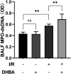
DHBA elevated the level of total protein concentration in the BALF. Mice were treated with DHBA (60 mg/kg) intraperitoneally 10 min before removing the clamp and subjected to intestinal ischemia for 45 min and reperfusion for 90 min. The samples of alveolar lavage fluid were collected. The level of BALF MPO-dsDNA complex was tested. All the data expressed as mean ± SD (n = 4), **p < .01, ns: no significant difference.
Figure 7.
Figure 7.
DHBA elevated the level of total protein concentration in the BALF. Mice were treated with DHBA (60 mg/kg) intraperitoneally 10 min before removing the clamp and subjected to intestinal ischemia for 45 min and reperfusion for 90 min. The samples of the lung tissues were collected. The expression of (a, b) Cit-H3 in the lung tissues were determined by immunoblotting. All the data expressed as mean ± SD (n = 4), **p < .01, ns: no significant difference.
Figure 8.
Figure 8.
DHBA elevated the level of total protein concentration in the BALF. Mice were treated with DHBA (60 mg/kg) intraperitoneally 10 min before removing the clamp and subjected to intestinal ischemia for 45 min and reperfusion for 90 min. The samples of alveolar lavage fluid and plasma were collected. The levels of (a, b) HMGB-1 in the BALF and plasma were tested. The data were expressed as means ± SD (n = 8). **p < .01, ns: no significant difference.

References

    1. Jia Y, Cui R, Wang C, et al. (2020) Metformin protects against intestinal ischemia-reperfusion injury and cell pyroptosis via TXNIP-NLRP3-GSDMD pathway. Redox Biology 32: 101534. - PMC - PubMed
    1. Cen C, McGinn J, Aziz M, et al. (2017) Deficiency in cold-inducible RNA-binding protein attenuates acute respiratory distress syndrome induced by intestinal ischemia-reperfusion. Surgery 162(4): 917–927. - PubMed
    1. Ito K, Ozasa H, Horikawa S. (2005) Edaravone protects against lung injury induced by intestinal ischemia/reperfusion in rat. Free Radical Biology and Medicine 38(3): 369–374. - PubMed
    1. Jin B, Li G, Zhou L, et al. (2022) Mechanism involved in acute liver injury induced by intestinal ischemia-reperfusion. Frontiers in Pharmacology 13: 924695. - PMC - PubMed
    1. Wang J, Qiao L, Li S, et al. (2013) Protective effect of ginsenoside Rb1 against lung injury induced by intestinal ischemia-reperfusion in rats. Molecules 18(1): 1214–1226. - PMC - PubMed