. 2023 Oct;43(11):1450-1458.
doi: 10.1002/pd.6436. Epub 2023 Sep 12.

Biometric magnetic resonance imaging analysis of fetal brain development in Down syndrome

Affiliations

Biometric magnetic resonance imaging analysis of fetal brain development in Down syndrome

Rie Kitano et al. Prenat Diagn. 2023 Oct.

Abstract

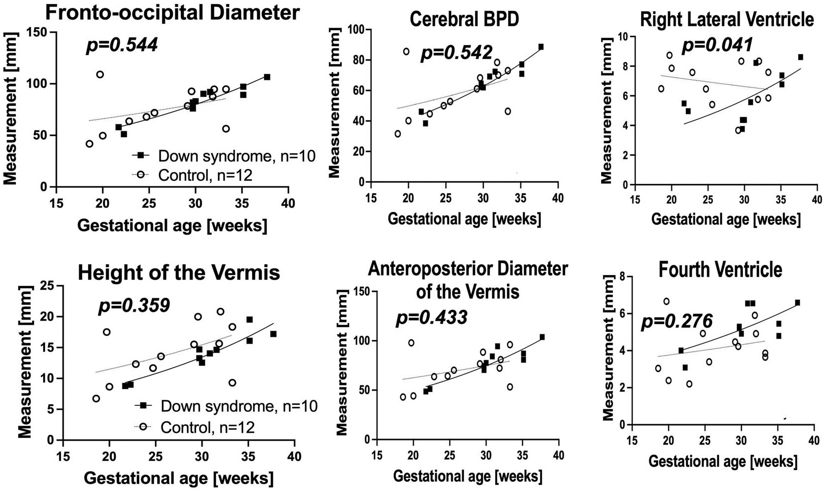
Objectives: To assess brain development in living fetuses with Down syndrome (DS) by biometric measurements on fetal brain magnetic resonance images (MRI).

Methods: We scanned 10 MRIs of fetuses with confirmed trisomy 21 at birth and 12 control fetal MRIs without any detected anomalies. Fetal brain MRIs were analyzed using 14 fetal brain and skull biometric parameters. We compared measures between DS and controls in both raw MRIs and motion-corrected and anterior-posterior commissure-aligned images.

Results: In the reconstructed images, the measured values of the height of the cerebellar vermis (HV) and anteroposterior diameter of the cerebellar vermis (APDV) were significantly smaller, and the anteroposterior diameter of the fourth ventricle (APDF) was significantly larger in fetuses with DS than controls. In the raw MRIs, the measured values of the right lateral ventricle were significantly larger in fetuses with DS than in controls. Logistic regression analyses revealed that a new parameter, the cerebellar-to-fourth-ventricle ratio (i.e., (APDV * Height of the vermis)/APDF), was significantly smaller in fetuses with DS than controls and was the most predictive to distinguish between fetuses with DS and controls.

Conclusions: The study revealed that fetuses with DS have smaller cerebellums and larger fourth ventricles compared to the controls.


Conflict of interest statement

CONFLICT OF INTEREST STATEMENT

The authors declare that they have no conflicts of interest.

Figures

FIGURE 1
FIGURE 1
Biometric measurements of fetal brain. Illustration of each biometric measurement as indicated by a solid line and a dotted line. APD, anteroposterior distance; APDF, Anteroposterior diameter of the fourth ventricles; APDM, Anteroposterior diameter of the medulla; APDP, Anteroposterior diameter of the pons; APDV, Anteroposterior diameter of the vermis; BPD, Biparietal diameter; FOD, Fronto-occipital diameter; HV, Height of the vermis; LCC, Length of the corpus callosum; LDT, Lateral diameter of the third ventricle.
FIGURE 2
FIGURE 2
Biometric measurements from the ACPC images. Non-linear regression model of biometric measurements in ACPC images. Height of the vermis (HV) and APDV were significantly smaller and APDF was significantly larger in fetuses with Down syndrome (DS) (n = 10) than in controls (n = 12).
FIGURE 3
FIGURE 3
Biometric measurements from the RAW images. Non-linear regression model of biometric measurements in Raw MRI. The right lateral ventricle was significantly larger in fetuses with Down syndrome (DS).
FIGURE 4
FIGURE 4
(APDV * Height of the vermis (HV))/APDF index. As shown in the figure, the distribution of measures from the fetuses with Down syndrome (DS) was different from that from the control fetuses. (APDV * HV)/APDF indices were significantly smaller in fetuses with DS (p < 0.05, area under the curve (AUC) = 1).

References

    1. DeGraaf G, Buckley F, Skotko BG. Estimates of the live births, natural losses, and elective terminations with Down syndrome in the United States. Am J Med Genet. 2015;167A(4):756–767. 10.1002/ajmg.a.37001 - DOI - PubMed
    1. DeGraaf G, Buckley F, Dever J, Skotko BG. Estimation of live birth and population prevalence of Down syndrome in nine U.S. states. Am J Med Genet. 2017;173(10):2710–2719. 10.1002/ajmg.a.38402 - DOI - PubMed
    1. Guihard-Costa A-M, Khung S, Delbecque K, Ménez F, Delezoide A-L Biometry of face and brain in fetuses with trisomy 21. Pediatr Res. 2006;59(1):33–38. 10.1203/01.pdr.0000190580.88391.9a - DOI - PubMed
    1. Guidi S, Bonasoni P, Ceccarelli C, et al. Neurogenesis impairment and increased cell death reduce total neuron number in the hippocampal region of fetuses with Down syndrome. Brain Pathol. 2008;18(2):180–197. 10.1111/j.1750-3639.2007.00113.x - DOI - PMC - PubMed
    1. Winter TC, Reichman JA, Luna JA, et al. Frontal lobe shortening in second-trimester fetuses with trisomy 21: usefulness as a US marker. Radiology. 1998;207(1):215–222. 10.1148/radiology.207.1.9530318 - DOI - PubMed