

SARS-CoV-2 spread to endocrine organs is associated with obesity: an autopsy study of COVID-19 cases
- PMID: 37698811
- PMCID: PMC10806201
- DOI: 10.1007/s12020-023-03518-0
SARS-CoV-2 spread to endocrine organs is associated with obesity: an autopsy study of COVID-19 cases
Abstract
Purpose: SARS-CoV-2 infection may be limited to the respiratory tract or may spread to multiple organs. Besides disease severity, factors associated with virus spread within the host are elusive. Here, we tried to identify features associated with SARS-CoV-2 spread to endocrine organs.
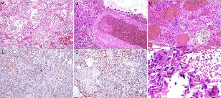
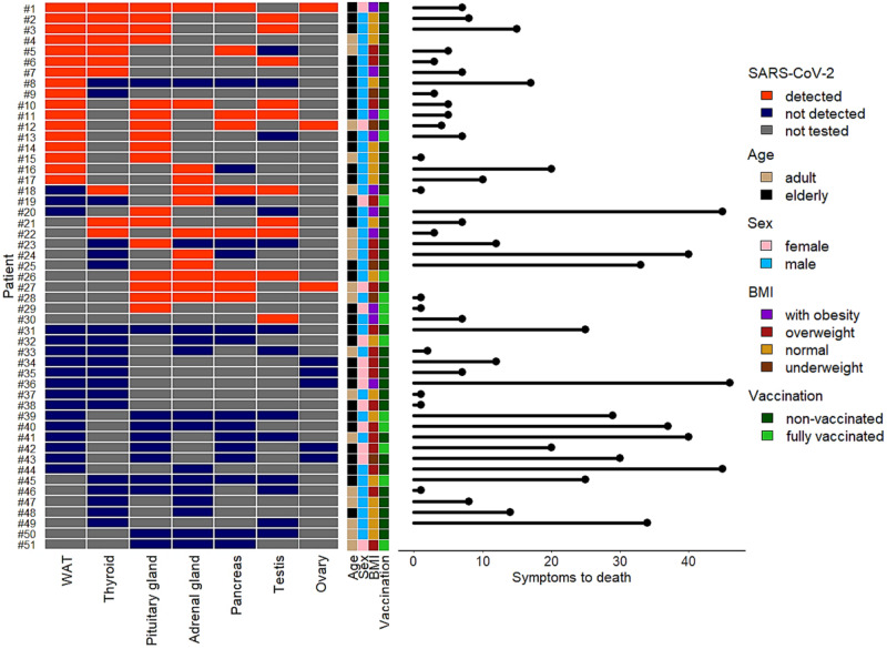
Methods: In a retrospective autoptic cohort of 51 subjects who died because of COVID-19, we analyzed the severity and type of lung pathology, patients' features and the detection of virus in thyroid, testis, adrenal gland, pancreas, anterior pituitary, and the white adipose tissue (WAT).
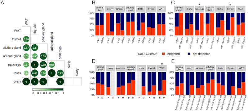
Results: The SARS-CoV-2 genome was detected in endocrine organs of 30/51 cases. The anterior pituitary and WAT were most frequently positive for virus. While pathological features of lung were not associated with the presence of virus in endocrine organs, obesity (BMI > 30) was significantly associated to virus detection in pancreas (p = 0.01) and thyroid (p = 0.04). WAT infection was detected more frequently in males (p = 0.03).
Conclusion: In subject with obesity dying of COVID-19, the virus frequently spreads to endocrine organs. The findings emphasize the need for optimal treatment of patients with obesity at the very onset of COVID-19. Since post-COVID conditions remain a major issue worldwide, a rigorous follow-up of endocrine function-especially of thyroid and pancreas-is advocated in subjects with obesity.
Keywords: COVID-19; Endocrine; Obesity; SARS-CoV-2.
© 2023. The Author(s).
Conflict of interest statement
The authors declare no competing interests.
Figures
Similar articles
-
COVID-19 autopsy cases: detection of virus in endocrine tissues.J Endocrinol Invest. 2022 Jan;45(1):209-214. doi: 10.1007/s40618-021-01628-y. Epub 2021 Jun 30. J Endocrinol Invest. 2022. PMID: 34191258 Free PMC article.
-
Transcriptional changes in multiple endocrine organs from lethal cases of COVID-19.J Mol Med (Berl). 2023 Aug;101(8):973-986. doi: 10.1007/s00109-023-02334-3. Epub 2023 May 29. J Mol Med (Berl). 2023. PMID: 37246981 Free PMC article.
-
SARS-CoV-2 infection impairs the insulin/IGF signaling pathway in the lung, liver, adipose tissue, and pancreatic cells via IRF1.Metabolism. 2022 Aug;133:155236. doi: 10.1016/j.metabol.2022.155236. Epub 2022 Jun 8. Metabolism. 2022. PMID: 35688210 Free PMC article.
-
Insights into the possible impact of COVID-19 on the endocrine system.Arch Physiol Biochem. 2023 Dec;129(4):998-1006. doi: 10.1080/13813455.2021.1890131. Epub 2021 Mar 3. Arch Physiol Biochem. 2023. PMID: 33653188 Review.
-
Coronaviruses and Endocrine System: A Systematic Review on Evidence and Shadows.Endocr Metab Immune Disord Drug Targets. 2021;21(7):1242-1251. doi: 10.2174/1871530320666200905123332. Endocr Metab Immune Disord Drug Targets. 2021. PMID: 32888287
Cited by
-
SARS-CoV-2 Infection Alters the Phenotype and Gene Expression of Adipocytes.Int J Mol Sci. 2024 Feb 8;25(4):2086. doi: 10.3390/ijms25042086. Int J Mol Sci. 2024. PMID: 38396763 Free PMC article.
-
Changes in nonfunctional adrenal incidentaloma after COVID-19 infection and a model for predicting benign and malignant adrenal incidentaloma.Front Endocrinol (Lausanne). 2024 Sep 2;15:1374282. doi: 10.3389/fendo.2024.1374282. eCollection 2024. Front Endocrinol (Lausanne). 2024. PMID: 39286271 Free PMC article.
-
The interplay of aging, adipose tissue, and COVID-19: a potent alliance with implications for health.Geroscience. 2024 Jun;46(3):2915-2932. doi: 10.1007/s11357-023-01058-z. Epub 2024 Jan 8. Geroscience. 2024. PMID: 38191833 Free PMC article. Review.
-
COVID19 infection and vaccination and the risk of pituitary apoplexy: an entangled yarn.Endocrine. 2025 Feb;87(2):459-467. doi: 10.1007/s12020-024-04078-7. Epub 2024 Oct 21. Endocrine. 2025. PMID: 39433700
References
-
- E.G. Bentley, A. Kirby, P. Sharma et al. SARS-CoV-2 Omicron-B.1.1.529 variant leads to less severe disease than Pango B and Delta variants strains in a mouse model of severe COVID-19. Microbiology (2021). 10.1101/2021.12.26.474085
MeSH terms
Grants and funding
LinkOut - more resources
Full Text Sources
Medical
Miscellaneous
