. 2023 Oct 23;8(20):e162100.
doi: 10.1172/jci.insight.162100.

The human channel gating-modifying A749G CACNA1D (Cav1.3) variant induces a neurodevelopmental syndrome-like phenotype in mice

Affiliations

The human channel gating-modifying A749G CACNA1D (Cav1.3) variant induces a neurodevelopmental syndrome-like phenotype in mice

Nadine J Ortner et al. JCI Insight. .

Abstract

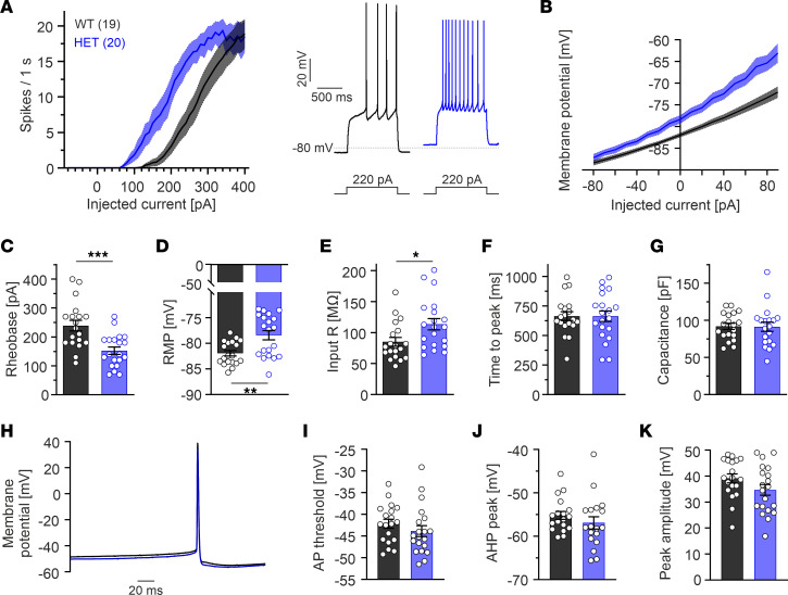
Germline de novo missense variants of the CACNA1D gene, encoding the pore-forming α1 subunit of Cav1.3 L-type Ca2+ channels (LTCCs), have been found in patients with neurodevelopmental and endocrine dysfunction, but their disease-causing potential is unproven. These variants alter channel gating, enabling enhanced Cav1.3 activity, suggesting Cav1.3 inhibition as a potential therapeutic option. Here we provide proof of the disease-causing nature of such gating-modifying CACNA1D variants using mice (Cav1.3AG) containing the A749G variant reported de novo in a patient with autism spectrum disorder (ASD) and intellectual impairment. In heterozygous mutants, native LTCC currents in adrenal chromaffin cells exhibited gating changes as predicted from heterologous expression. The A749G mutation induced aberrant excitability of dorsomedial striatum-projecting substantia nigra dopamine neurons and medium spiny neurons in the dorsal striatum. The phenotype observed in heterozygous mutants reproduced many of the abnormalities described within the human disease spectrum, including developmental delay, social deficit, and pronounced hyperactivity without major changes in gross neuroanatomy. Despite an approximately 7-fold higher sensitivity of A749G-containing channels to the LTCC inhibitor isradipine, oral pretreatment over 2 days did not rescue the hyperlocomotion. Cav1.3AG mice confirm the pathogenicity of the A749G variant and point toward a pathogenetic role of altered signaling in the dopamine midbrain system.

Keywords: Calcium channels; Calcium signaling; Mouse models; Neuroscience.


Figures

Figure 1
Figure 1. Delayed gain of body weight and no major endocrine dysfunctions in HET Cav1.3AG mice.
(A and B) Curves represent mean ± SEM (7-day bins) body weights of male (A) or female (B) WT, HET and HOM mice. For statistics at different ages, see Supplemental Figure 2, A and B. (C) Plasma aldosterone levels were similar in male WT and mutants (~15 wk) but significantly increased in female HETs compared with WT (~13 wk; 2-way ANOVA; genotype F2,48 = 5.06, P = 0.0101) with Šídák’s multiple-comparison test). (D and E) Blood glucose values of male (D; ~6 months) and female (E; ~13 wk) mice during an i.p. glucose test (1 mg/kg glucose i.p. injection after 6-hour fasting). Data are shown as mean ± SEM. Time point 0 represent the fasting basal blood glucose level before glucose injection. Mixed-effects model (D; males, time: F3.8,193.2 = 71.7, P < 0.001; genotype: F2,52 = 15.7, P < 0.001; interaction: F10,252 = 2.8; P = 0.0027) with Dunnett’s multiple-comparison post hoc test or 2-way ANOVA (E; females, time: F5,95 = 25.8, P < 0.001; no differences among genotypes). (F) Fasting basal blood glucose levels were significantly reduced in male HOMs only (2-way ANOVA; interaction F2,68 = 5.08, P = 0.0088) with Dunnett’s multiple-comparison test. ***P < 0.001, **P < 0.01.
Figure 2
Figure 2. Cav1.3AG mutant mice display increased locomotor activity and a social deficit.
Data are shown as mean ± SEM. One-way ANOVA (A and E) or Kruskal-Wallis (BD) with Dunnett’s or Dunn’s multiple-comparison post hoc test, respectively, were used, as was a paired Student’s t test (F). (A) In the open field (150 lux), compared with WT the distance traveled (left) and average speed (middle) was significantly increased in mutants, associated with less time spent immobile (right). Anxiety-related parameters (center/periphery time) were unchanged (Supplemental Figure 3E). (B) In the light-dark box (400 lux), mutant mice spent significantly less time in the light compartment. (C) HOM mutants did not bury any marble (out of 20) within a 30-minute time period. (D and E) Mice showed a significantly reduced ratio of rearing (D) or grooming (E) time over frequency in a novel environment in HET (rearing) and HOM (rearing and grooming) mice. A similar significant reduction for HOMs was observed in a familiar environment (data not shown). (F) Three-chamber social test. Quantification of the time spent in a chamber containing a grid with a unfamiliar mouse (“social”) or an empty grid (“nonsocial”) as well as direct nose-to-grid interaction time revealed a social deficit in HOM mutants. ***P < 0.001, **P < 0.01, *P < 0.05.
Figure 3
Figure 3. Similar brain morphology in Cav1.3AG mutant mice.
Data are shown as mean ± SEM. (A) Representative pictures of Nissl-stained brain sections from adult (13–15 wk) male mice of the cerebellum (n = 3–7/genotype), hippocampus (n = 5–10/genotype), and olfactory bulb (n = 5–7/genotype). Scale bars: 1 mm (cerebellum; top), 500 μm (hippocampus, middle; olfactory bulb, bottom). (B) Brain weight and respective body weight of male and female animals for the indicated age and number of animals. KO, Cav1.3-KO animals. Statistical analyses were performed with 1-way ANOVA with Dunnett’s multiple-comparison post hoc test. Due to the limited availability of homozygous mutants and respective experiments, we have excluded homozygous animals of the 5 wk cohort from statistical analysis due to the low n. (C) Left: Representative pictures of Nissl-stained sagittal brain sections from male WT and HET mice (olfactory bulb was not captures at the same level). Right: Comparable volumes of individual brain regions (Cavalieri principle; unpaired Student’s t test). Scale bar: 2 mm. (DG) No statistically significant differences (1-way ANOVA) of the striatal volume (D, dorsal: caudate putamen [CPu]; E, ventral: nucleus accumbens [NAc]) or TH+ neuron number within the SN (F) and VTA (G) between adult male WT and mutant mice determined in serial TH-stained brain sections (Supplemental Figure 4, A and B). ***P < 0.001, **P < 0.01, *P < 0.05.
Figure 4
Figure 4. Gating changes and altered firing in acutely isolated MCCs from adult male HET Cav1.3AG mice.
(A) Voltage dependence of steady-state activation (normalized conductance-voltage curves; circles) and inactivation (squares) of WT (n = 7) and HET (n = 7) LTCC currents. Bottom, window Ca2+ current (Iw) was increased for HET MCCs between −50 and −30 mV (shaded area). (B) Representative sets of pharmacologically isolated LTCC Ca2+ currents from a WT and HET MCC. (C) Similar current densities among genotypes (inward current at +10mV normalized to the cell size; WT, n = 30; HET, n = 34). (D) Averaged normalized LTCC currents during 600 ms depolarizations to +10 mV. Inactivation kinetics followed a double exponential time course: WT Aslow 38%, τslow 390 ms, Afast 19%, τfast 18 ms, plateau (C) = 44%; HET Aslow 65%, τslow 904 ms, Afast 31%, τfast 36 ms, C = 4%. (E) Compared with WT (n = 14), the resting membrane potential of HET MCCs (n = 12) was significantly decreased (P < 0.01, unpaired Student’s t test). (F) Percentage of spontaneously firing versus silent cells over a total of n = 19 WT or HET MCCs. Significance testing on categorical data was performed by RxC contingency tables and a χ2 test (P < 0.01). (G) Mean frequency of spontaneously firing HET MCCs (n = 11) was significantly decreased compared with WT (n = 10; P < 0.01, unpaired Student’s t test). (H) Current-clamp traces of representative WT and HET MCCs without current injection. Dashed lines indicate baseline; dotted line indicates 0 mV. Blue arrows indicate spontaneous subthreshold oscillations often observed in HET MCCs. (I) Representative AP traces at increasing current injections (4, 10, 16 pA) from a HP of −70 mV. (J and K) Plot of fo (first interspike interval frequency) and fss (last interspike interval frequency) against the injected current. fo is unaltered (J), while fss (K) exhibits a marked decrease with current injections above 10 pA in HET MCCs. Paired Student’s t test was used. **P < 0.01, *P < 0.05.
Figure 5
Figure 5. Projection-specific alterations of SN DA neuron firing in brain slices from HET Cav1.3AG mice.
Whole-cell patch-clamp recordings from DLS-projecting lSN (“lateral SN”) or DMS-projecting mSN DA neurons (“medial SN”) were performed in brain slices from adult male WT and HET mice. Data are shown as mean ± SEM. (A) Projection specificity was achieved by infusion of red beads into the DLS or DMS retrogradely labeling DLS- or DMS-projecting SN DA neurons (41). Scale bar: 1,000 μm. (B) Only DA cells (TH+; blue) that contained red beads (RB; red) and were filled with neurobiotin (NB; green) during the recording were included into the analysis. Scale bar: 100 μM, 50 μm (zoom). (C) Representative traces of autonomous pacemaking of both investigated SN DA neuron populations. DMS-projecting mSN DA neurons from HETs had a significantly higher firing frequency in the on-cell (D, P = 0.0069) and whole-cell configuration (E; P < 0.001) compared with WT (Mann-Whitney U test; Supplemental Table 2). (F) Upon hyperpolarization to approximately –80 mV (by 2-second current injection, gray rectangle), HET DMS-projecting mSN DA neurons showed a significantly increased sag component (G, P = 0.0395, unpaired Student’s t test) and faster rebound spiking (H; P = 0.0027, Mann-Whitney U test) (Supplemental Table 2). ***P < 0.001, **P < 0.01, *P < 0.05.
Figure 6
Figure 6. Striatal medium spiny neurons (MSNs) from HET Cav1.3AG mice are hyperexcitable.
Whole-cell patch-clamp recordings in acute brain slices from adult male and female WT and HET mice. Parameters of male and female mice did not differ significantly; therefore, data were pooled. Data are shown as mean ± SEM for the indicated number of cells. (A) Current-response curves (left, injected current versus number of elicited AP spikes) derived from 1-second current injections from –80 to 400 pA show that HET MSNs require less current stimulation to fire APs. Right, representative traces at 220 pA. (B) In MSNs from HETs, current injections elicited a stronger depolarization of the membrane potential compared with WT. (CG) HET MSNs had a decreased rheobase current (current at which the first sweep with APs occurred, C; P < 0.001), more depolarized resting membrane potential (RMP, D; P = 0.0020), and increased input resistance in HETs (E; P = 0.0193) without changes of the time to peak measured at the rheobase (F) and cell size estimated by the capacitance (G). (H) Mean of the first AP at the rheobase sweep of WT (n = 19) and HET (n = 20) MSNs. (IK) Manual analysis revealed no statistically significant differences for the AP threshold (I), afterhyperpolarization (AHP) peak (J), or AP peak amplitude (K). Statistical analyses were performed with unpaired Student’s t test. ***P < 0.001, *P < 0.05.
Figure 7
Figure 7. Variant-specific isradipine sensitivity in vitro and effects on locomotion by oral in vivo isradipine administration.
(A and B) Human Cav1.3 α1-subunits were coexpressed with β3 and α2δ-1 in tsA-201 cells (HEK293 cells that stably express a SV40 temperature-sensitive T antigen) (15 mM Ca2+). Isradipine sensitivity was determined during 100 ms square pulses from a HP of –89.3 mV to Vmax (0.1 Hz). Data are shown as mean ± SEM. (A) Concentration-response curves for the human C-terminally full-length Cav1.3 channel (hCav1.3L) WT, A749G, and G407R steady-state Ca2+ current (ICa) inhibition by isradipine. IC50 values (means ± 95% CI) were obtained by fitting the curves using a Hill slope = 1 and top-bottom fixed (bottom = 0; top = 100). Statistical analyses were performed with extra sum-of-squares F test (P < 0.001). (B) Representative current traces for inhibition by 30 nM or 3 μM isradipine (full block). (C and F) Experimental design for the pharmacological rescue experiment in adult males (C) or females (F; 2 independent cohorts each). (D and E) Male mice received twice daily 0.5–1 mg isradipine (ISR) mixed into yogurt (1–2 mg/day; WT n = 10; HET n = 13). Three WT and 6 HET mice received yogurt only (vehicle group). On day 3, drug effects were tested in the EPM ~4–5 hours after the morning dose, and immediately afterward, plasma was isolated. Distance traveled (D) and immobility time (E) of vehicle- or isradipine-treated WT and HET animals. Statistical analyses were performed with unpaired Student’s t test (P = 0.041, D; P = 0.046, E). (G and H) Female mice received yogurt (vehicle group, WT n = 13; HET n = 7) or 0.1 mg ISR mixed into yogurt (0.3 mg/day; WT n = 8; HET n = 6) 3 times daily. On day 3, mice were tested in the EPM ~2 hours after the morning dose, and immediately afterward, plasma was taken. Distance traveled (G) and immobility time (H) of vehicle- or isradipine-treated WTs and HETs. Statistical analyses were performed with unpaired Student’s t test (P = 0.0447, G; P = 0.0389, H). Statistical analyses were performed with 2-way ANOVA; a significant comparison was only observed for the genotype (D, P = 0.0173; E, P = 0.0128; G, P = 0.0216; H, 0.0224). *P < 0.05.

Similar articles

Cited by

References

    1. Catterall WA. Voltage-gated calcium channels. Cold Spring Harb Perspect Biol. 2011;3(8):a003947. doi: 10.1101/cshperspect.a003947. - DOI - PMC - PubMed
    1. Baig SM, et al. Loss of Ca(v)1.3 (CACNA1D) function in a human channelopathy with bradycardia and congenital deafness. Nat Neurosci. 2011;14(1):77–84. doi: 10.1038/nn.2694. - DOI - PubMed
    1. Platzer J, et al. Congenital deafness and sinoatrial node dysfunction in mice lacking class D L-type Ca2+ channels. Cell. 2000;102(1):89–97. doi: 10.1016/S0092-8674(00)00013-1. - DOI - PubMed
    1. Ortner NJ, et al. Lower affinity of isradipine for L-type Ca2+ channels during substantia nigra dopamine neuron-like activity: implications for neuroprotection in Parkinson’s disease. J Neurosci. 2017;37(28):6761–6777. doi: 10.1523/JNEUROSCI.2946-16.2017. - DOI - PMC - PubMed
    1. Marschallinger J, et al. The L-type calcium channel Cav1.3 is required for proper hippocampal neurogenesis and cognitive functions. Cell Calcium. 2015;58(6):606–616. doi: 10.1016/j.ceca.2015.09.007. - DOI - PubMed

Publication types