. 2023 Sep 12;9(1):94.
doi: 10.1038/s41421-023-00588-2.

CCL2-mediated inflammatory pathogenesis underlies high myopia-related anxiety

Affiliations

CCL2-mediated inflammatory pathogenesis underlies high myopia-related anxiety

Xiangjia Zhu et al. Cell Discov. .

Abstract

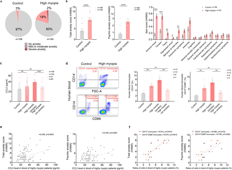
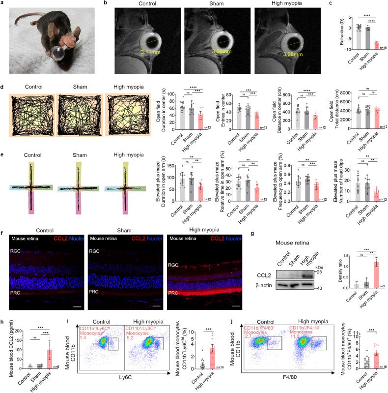
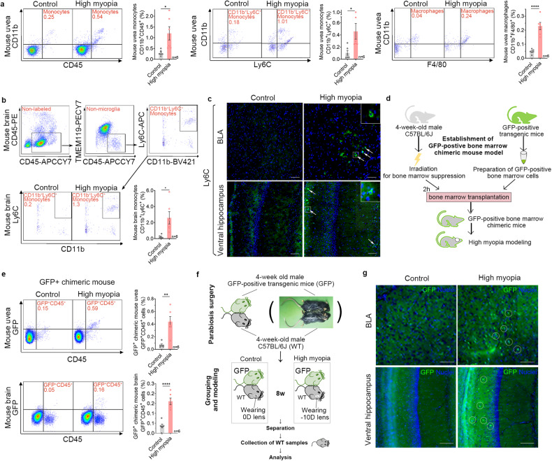
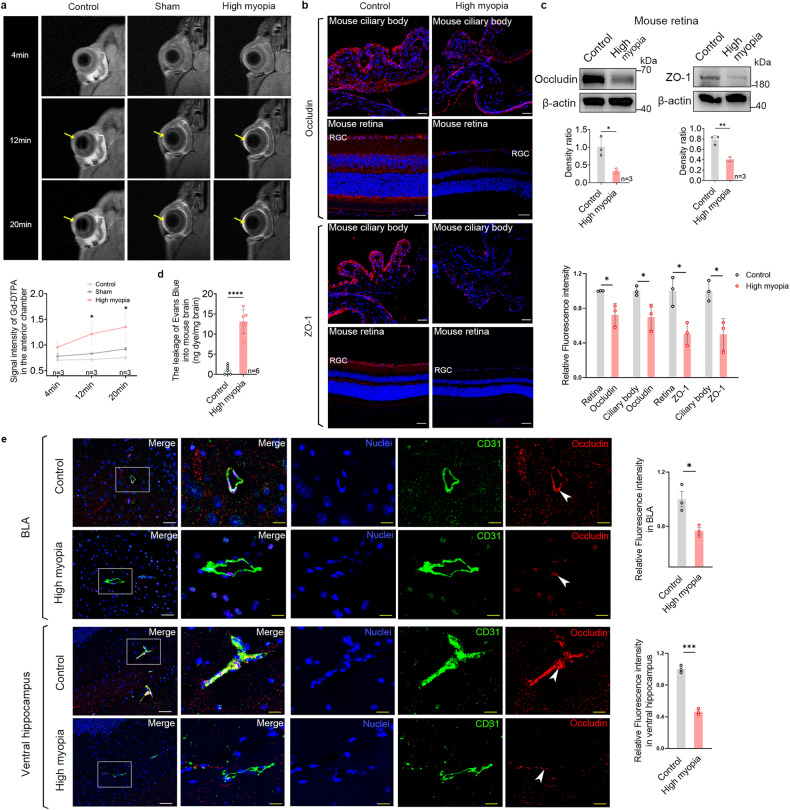
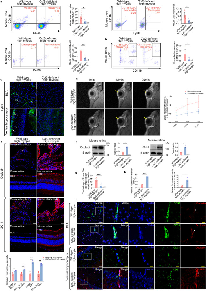
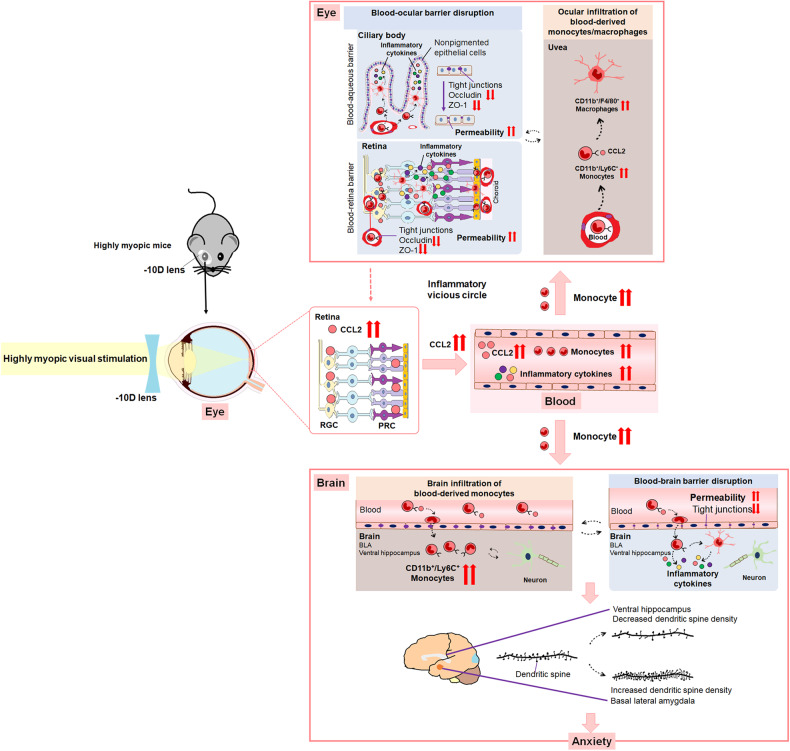
High myopia is a leading cause of blindness worldwide. It may lead to emotional defects that rely closely on the link between visual sensation and the central nervous system. However, the extent of the defects and its underlying mechanism remain unknown. Here, we report that highly myopic patients exhibit greater anxiety, accompanied by higher CC chemokine ligand 2 (CCL2) and monocyte levels in the blood. Similar findings are found in the mouse model of high myopia. Mechanistic evaluations using GFP-positive bone marrow chimeric mice, parabiotic mouse model, enhanced magnetic resonance imaging, etc., show that highly myopic visual stimulation increases CCL2 expression in eyes, aggravates monocyte/macrophage infiltration into eyes and brains, and disrupts blood-ocular barrier and blood-brain barrier of mice. Conversely, Ccl2-deficient highly myopic mice exhibit attenuated ocular and brain infiltration of monocytes/macrophages, reduced disruption of the blood-ocular barrier and blood-brain barrier, and less anxiety. Substantial alleviation of high myopia-related anxiety can also be achieved with the administration of CCL2-neutralizing antibodies. Our results establish the association between high myopia and anxiety, and implicate the CCL2-mediated inflammatory pathogenesis as an underlying mechanism.


Conflict of interest statement

The authors declare no competing interests.

Figures

Fig. 1
Fig. 1. Highly myopic patients exhibit higher anxiety scores and elevated CCL2 and monocyte levels in the blood.
a Proportions of no anxiety, mild to moderate anxiety, and severe anxiety in the control and highly myopic groups. b Total anxiety scores, psychic anxiety scores, and item scores on the HAMA. c Measurement of CCL2 concentration in human blood with a cytokine array. n = 106 in the control group, n = 104 in the highly myopic group, n = 21 in the highly myopic group with anxiety (total anxiety score ≥ 7), and n = 83 in the highly myopic group with no anxiety (ac). d Flow-cytometric quantification of monocytes (CD14+) and CD14+/CD68+ monocytes in human blood. n = 12 in the control and highly myopic groups, n = 5 in the highly myopic group with anxiety, and n = 7 in the highly myopic group with no anxiety. e Correlations between total and psychic anxiety scores and CCL2 levels in the blood of highly myopic patients (n = 104). f Correlations between total and psychic anxiety scores and ratios of monocytes (CD14+) and CD14+/CD68+ monocytes in the blood of highly myopic patients (n = 12). Data are mean ± SD. Level of significance was detected with χ2 test (a), Mann–Whitney U test (b), Student’s t-test (c, d), and Pearson’s analysis (e, f). ****P < 0.0001, ***P < 0.001, **P < 0.01, ns P > 0.05.
Fig. 2
Fig. 2. Highly myopic mouse model exhibits increased anxiety-like behaviors, upregulated CCL2 expression in the eye, and higher CCL2 and monocyte levels in the blood.
a Representative photograph of a mouse wearing a −10 D lens to induce high myopia in the right eye. b Representative images of ocular MRI (sagittal) showing an obviously longer AL in the highly myopic eye than in the control and sham eyes. Yellow lines indicate the measurement of AL. c The refraction of the control, sham, and highly myopic eyes (n = 15 in each group). d Representative maps of animal tracking (black lines) in the dark showing increased anxiety-like behaviors in the mouse model of high myopia, manifested as shorter duration, fewer entries, and shorter distance traveled in the center (yellow square) of the open field (n = 12 in each group). e Representative maps of animal tracking (black lines) in the dark showing increased anxiety-like behaviors in the mouse model of high myopia, manifested as shorter duration, fewer entries, and fewer head dips in the open arms (pink and yellow area) of the elevated plus maze (n = 12 in each group). The blue and green areas refer to closed arms and the orange area refers to the center of the maze. f Immunofluorescent images of CCL2 staining in the mouse retina showing upregulated CCL2 expression in the RGC layer and PRC layer of highly myopic mice (n = 3 in each group). Scale bars: 25 μm. g Examination of CCL2 expression in mouse retina with western blotting. The band density of CCL2 was normalized to the loading control for statistical analysis (n = 3 in each group). h Measurement of CCL2 concentration in mouse blood with a proinflammatory biomarker array (n = 6 in each group). i, j Flow-cytometric quantification showed higher ratios of CD11b+/Ly6Chi monocytes and CD11b+/F4/80+ monocytes in the blood of highly myopic mice than in the controls (n = 16 in each group). Data are mean ± SD. Levels of significance were detected with one-way ANOVA (ce, g, h) and Student’s t-test (i, j). ****P < 0.0001, ***P < 0.001, **P < 0.01, *P < 0.05, ns P > 0.05.
Fig. 3
Fig. 3. Elevated CCL2 expression increases infiltration of blood-derived monocytes/macrophages into the eyes and brains of highly myopic mice.
a Flow-cytometric quantification showed higher ratios of monocytes (CD11b+/CD45+), CD11b+/Ly6C+ monocytes, and macrophages (CD11b+/F4/80+) in the uvea of highly myopic mice than in the controls. b Flow-cytometric gating strategy showed exclusion of CD45-PE labeled cells and microglia (TMEM119+) before quantification of infiltrated monocytes. A significantly higher ratio of CD11b+/Ly6C+ monocytes was identified in the brain of highly myopic mice than in the controls. c Immunofluorescent images showed increased Ly6C+ monocyte infiltration (arrows) into the BLA and ventral hippocampus of highly myopic mice. Enlarged images in white squares are shown on the top right. Scale bars: 100 μm. d Schematic representation of the establishment of a model of GFP-positive bone marrow chimeric mice. e Flow-cytometric quantification showed higher ratios of GFP+CD45+ cells in both the uvea and brain of GFP-positive bone marrow chimeric mice with high myopia than in the chimeric mice without high myopia. f Schematic representation of the establishment of the parabiotic model. g Immunofluorescent images showed increased GFP+ cell infiltration (circles) into the BLA and ventral hippocampus of parabiotic wild-type mice with high myopia than in the controls. Scale bars: 100 μm. Data are mean ± SD. n = 6 in each group (a, b, e); n = 3 in each group (c, g). The level of significance was detected using Student’s t-test. ****P < 0.0001, **P < 0.01 and *P < 0.05.
Fig. 4
Fig. 4. Increased monocyte/macrophage infiltration further disrupts the BOB/BBB in highly myopic mice.
a Representative enhanced MRI images (coronal) showed faster leakage of contrast agents into the anterior chamber (yellow arrows) of highly myopic eyes than those into eyes of the control or sham eyes at both 12 min and 20 min after Gd-DTPA injection. b Immunofluorescent images of tight junction proteins occludin and ZO-1 in the ciliary body and retina of mice. Scale bars: 25 μm. Fluorescence intensity normalized to the averaged value of the control for quantification analyses. c Examination of occludin and ZO-1 expression in mouse retina with western blotting. Band density normalized to that of the loading control for statistical analysis. d Leakage of EB into the mouse brain (ng dye/mg brain tissue). e Immunofluorescent images of tight junction protein occludin (red) and vessel marker CD31 (green) in the BLA and ventral hippocampus of mice. Enlarged images of CD31-labeled vessels (square) showing downregulated occludin (arrowheads) in highly myopic mice. Scale bars: 40 μm in white and 20 μm in yellow. Fluorescence intensity normalized to the averaged value of the control for quantification analyses. Data are mean ± SD. n = 3 in each group (ac, e); n = 6 in each group (d). Level of significance was detected with one-way repeated measures ANOVA (a) or Student’s t test (be). ****P < 0.0001, ***P < 0.001, **P < 0.01, *P < 0.05.
Fig. 5
Fig. 5. Ccl2 deficiency substantially attenuates anxiety and reverses anxiety-related neuronal changes in highly myopic mice.
a Representative maps of animal tracking (black lines) in the dark showed significantly reduced anxiety levels in Ccl2-deficient highly myopic mice than in wild-type highly myopic mice in OFT. b Representative maps of animal tracking (black lines) in the dark showed significantly reduced anxiety levels in Ccl2-deficient highly myopic mice than in wild-type highly myopic mice in the EPM test. c Representative Golgi images of the dendritic spines in BLA and ventral hippocampus of mice. Scale bars: 5 μm. d High myopia modeling increased the spine density of BLA pyramidal neurons and decreased the spine density of ventral hippocampus neurons, which were partially reversed by Ccl2 deficiency. Data are mean ± SD. n = 12 in each group (a, b); n = 4 in each group (c, d) . The level of significance was detected with Student’s t-test (a, b) or one-way ANOVA (d). ****P < 0.0001, ***P < 0.001, **P < 0.01, *P < 0.05, ns P > 0.05.
Fig. 6
Fig. 6. Ccl2 deficiency reduces ocular and brain infiltration of monocytes/macrophages and BOB/BBB disruption in highly myopic mice.
a Flow-cytometric quantification showed decreased ratios of monocytes (CD11b+/CD45+), CD11b+/Ly6C+ monocytes, and macrophages (CD11b+/F4/80+) in the uvea of Ccl2-deficient highly myopic mice than in those of wild-type highly myopic mice. b Flow-cytometric quantification showed decreased ratios of CD11b+/Ly6C+ monocytes in the brain of Ccl2-deficient highly myopic mice than in the controls. c Immunofluorescent images showed decreased infiltration of Ly6C+ monocytes (arrows) in the BLA and ventral hippocampus of Ccl2-deficient highly myopic mice. Enlarged images in white squares are shown on the top right. Scale bars: 100 μm. d Representative images of enhanced MRI (coronal) showed slower leakage of contrast agent into the anterior chamber (yellow arrows) of Ccl2-deficient highly myopic mice than into those of wild-type highly myopic mice at both 12 min and 20 min after Gd-DTPA injection. e Immunofluorescent images of tight junction proteins occludin and ZO-1 in the ciliary body and retina of mice. Fluorescence intensity normalized to the averaged value of the control for quantification analyses. Scale bars: 25 μm. f Examination of occludin and ZO-1 expression in mouse retina with western blotting. The band density was normalized to that of the loading control for statistical analysis. The lane of ZO-1 for the wild-type highly myopic group was the same as that for the highly myopic group. g Leakage of EB into the mouse brain. h Relative fluorescence intensity of occludin in the BLA and ventral hippocampus area. Fluorescence intensity was normalized to the averaged value of the wild-type for quantification analyses. i Immunofluorescent images of occludin and CD31 in the BLA and ventral hippocampus of mice. Enlarged images of CD31-labeled vessels (square) showed upregulated occludin (arrowheads) in the Ccl2-deficient highly myopic mice. Scale bars: 40 μm in white and 20 μm in yellow. Data are mean ± SD. n = 6 in each group (a, b, g); n = 3 in each group (cf, h). The level of significance was detected with one-way repeated measures ANOVA (d) or Student’s t-test (a, b, eh). ****P < 0.0001, ***P < 0.001, **P < 0.01, *P < 0.05.
Fig. 7
Fig. 7. Schematic illustration of CCL2-mediated inflammatory pathogenesis that underlies high myopia-related anxiety.
Highly myopic visual stimulation upregulates ocular CCL2 expression and then increases blood CCL2 and monocyte levels, which increases the ocular and brain infiltration of monocytes/macrophages. Ocular infiltration further exacerbates BOB disruption and CCL2 release into the circulation, aggravating an inflammatory vicious circle. Brain infiltration further disrupts BBB, exacerbates local inflammation and alters dendritic spines, eventually leading to anxiety.

References

    1. Ruiz-Medrano J, et al. Myopic maculopathy: current status and proposal for a new classification and grading system (ATN) Prog. Retin. Eye Res. 2019;69:80–115. - PubMed
    1. Hayashi K, et al. Long-term pattern of progression of myopic maculopathy: a natural history study. Ophthalmology. 2010;117:1595–1611. - PubMed
    1. Ikuno Y. Overview of the complications of high myopia. Retina. 2017;37:2347–2351. - PubMed
    1. Zhu X, et al. Aberrant TGF-β1 signaling activation by MAF underlies pathological lens growth in high myopia. Nat. Commun. 2021;12:2102. - PMC - PubMed
    1. Yokoi T, et al. Predictive factors for comorbid psychiatric disorders and their impact on vision-related quality of life in patients with high myopia. Int. Ophthalmol. 2014;34:171–183. - PubMed