Review
. 2023 Sep 13;8(1):345.
doi: 10.1038/s41392-023-01569-3.

Amino acid metabolism in health and disease

Affiliations
Review

Amino acid metabolism in health and disease

Zhe-Nan Ling et al. Signal Transduct Target Ther. .

Abstract

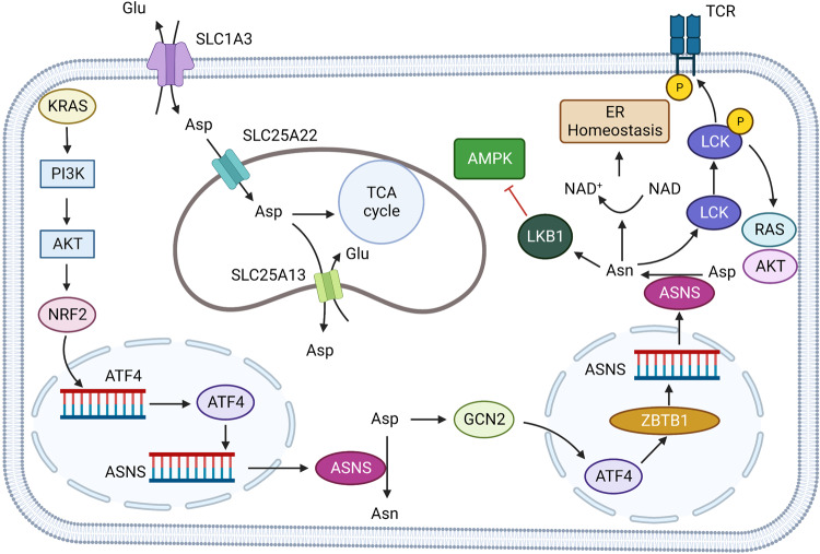
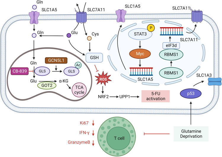
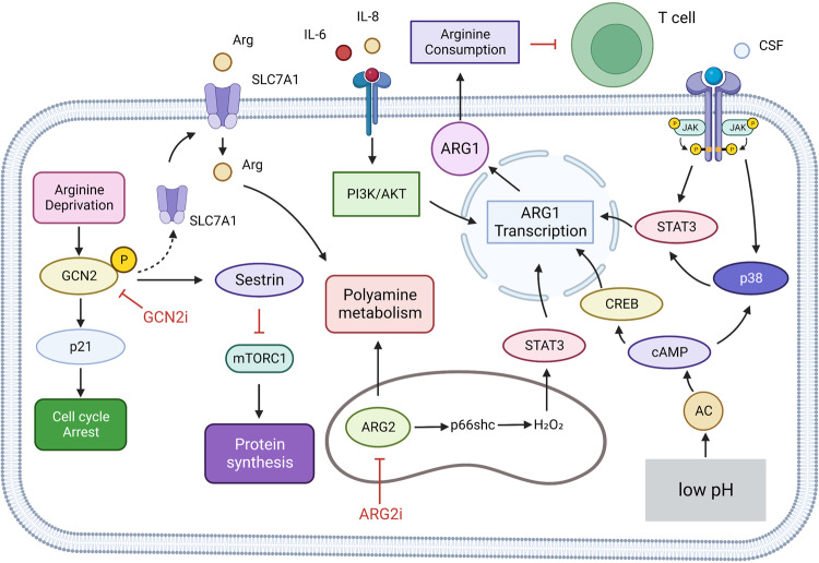
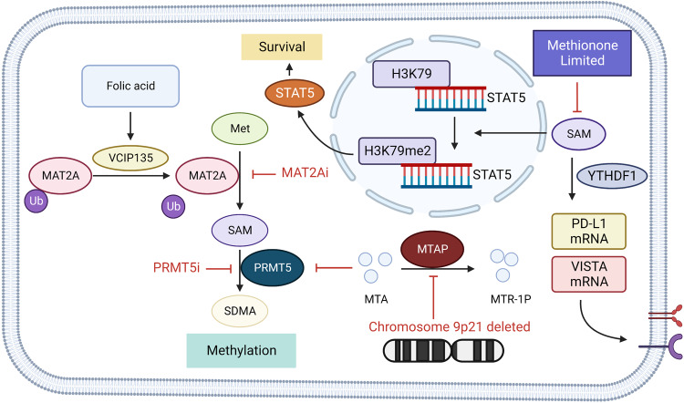
Amino acids are the building blocks of protein synthesis. They are structural elements and energy sources of cells necessary for normal cell growth, differentiation and function. Amino acid metabolism disorders have been linked with a number of pathological conditions, including metabolic diseases, cardiovascular diseases, immune diseases, and cancer. In the case of tumors, alterations in amino acid metabolism can be used not only as clinical indicators of cancer progression but also as therapeutic strategies. Since the growth and development of tumors depend on the intake of foreign amino acids, more and more studies have targeted the metabolism of tumor-related amino acids to selectively kill tumor cells. Furthermore, immune-related studies have confirmed that amino acid metabolism regulates the function of effector T cells and regulatory T cells, affecting the function of immune cells. Therefore, studying amino acid metabolism associated with disease and identifying targets in amino acid metabolic pathways may be helpful for disease treatment. This article mainly focuses on the research of amino acid metabolism in tumor-oriented diseases, and reviews the research and clinical research progress of metabolic diseases, cardiovascular diseases and immune-related diseases related to amino acid metabolism, in order to provide theoretical basis for targeted therapy of amino acid metabolism.


Conflict of interest statement

The authors declare no competing interests.

Figures

Fig. 1
Fig. 1
Overview of amino acid metabolism. The human body can obtain amino acids through food digestion and absorption, tissue decomposition, internal synthesis of three ways. Amino acids in amino acid metabolism pool can be deacidified to produce amino and carbon dioxide. Or participate in the synthesis of purine, pyrimidine and other nitrogenous compounds in the transformation of metabolites; Or deamination produces α-ketoacid and NH3. According to different enzymes and pathways, α-ketoacid can produce keto bodies, or participate in oxidative energy supply or sugar and lipid synthesis; NH3 enters the urea cycle. Created with BioRender.com
Fig. 2
Fig. 2
Glutamine and BCAA metabolism. BCAAs can be absorbed by the cell through L-type amino acid transporter (LATs), and L-type amino acid transporter 1(LAT1) can also exchange intracellular glutamine with extracellular leucine. In cells, BCAAs are catalyzed to formα-ketoisocaproate (KIC), α-ketoisovalerate (KIV), and α-keto-β-methylvalerate (KMV). The three substances are collectively known as branched alpha-ketoacids (BCKAs). Further, BCKAs produce acetyl-CoA through an irreversible rate-limiting reaction catalyzed by branched alpha-ketoate dehydrogenase (BCKDH) and subsequent reactions. Acetyl-CoA may be involved in the TCA cycle or other amino acid synthesis. Glutamine can be transported by SLC1A5 (ASCT2), LAT1 (L-type amino acid transporter), and xCT (SLC7A11). Glutamine is involved in glutathione (GSH) synthesis and cell REDOX homeostasis regulation in cytoplasm. In the mitochondria, glutamine produces Glutamate through a reaction catalyzed by glutaminase (GLS), which participates in the TCA cycle by producing α-KG by aminotransferase (ATs) and Glutamate dehydrogenase (GLUD). Created with BioRender.com. (The red blunt line represents inhibition)
Fig. 3
Fig. 3
BCAAs metabolism in Cancer. In pancreatic ductal adenocarcinoma (PDAC), KRAS can inhibit the ubiquitination of BCAT2 by spleen tyrosine kinase (SYK) and E3 ubiquitination ligase TRIM21, thereby stabilizing the expression level of BCAT2 in PDAC cells and promoting the proliferation of tumor cells. BCAAs promote Ubiquitin Specific Peptidase 1 (USP 1) through the GCN2-eIF2a pathway and inhibit the degradation of BCAT2 by deubiquitination of the K299 site of BCAT2. This process is inhibited during the BCAAs deprivation. cAMP-responsive Elin-Binding (CREB)-binding protein (CBP) and SIRT4 compete to bind the K44 site of BCAT2, regulating the acetylation level of this site and the degradation of BCAT2. In triple-negative breast cancer, tumor cells can activate MAPK and PI3K/AKT signaling through IGF-1 and insulin signaling, and PI3K/AKT signaling can go on to activate Foxo3a, mTOR signaling, BCAT in the cytoplasm of tumor cells can also promote mitochondrial genesis and mitochondrial function by activating Foxo3a, AKT, mTOR, and Nrf2 to gain survival advantages. Created with BioRender.com. (The red blunt line represents inhibition; The dotted line indicates that the middle step is omitted)
Fig. 4
Fig. 4
BCAAs metabolism in tumor microenvironment. In triple-negative breast cancer, tumor cells can activate MAPK and PI3K/AKT signaling through IGF-1 and insulin signaling, and PI3K/AKT signaling can go on to activate Foxo3a, mTOR signaling, BCAT in the cytoplasm of tumor cells can also promote mitochondrial genesis and mitochondrial function by activating Foxo3a, AKT, mTOR, and Nrf2 to gain survival advantages. In Leukemia, the RNA-binding protein Musashi 2 (MSI2) binds to BCAT1 mRNA to promote the translation of BCAT1. BCAT1 containing CXXC motif has strong reductive and antioxidant properties, and in wild-type BCAT1 leukemia cells with CXXC motif, The number of cell surface markers CD11b, CD14, CD68, and CD36 decreased. BCKAs excretion in glioblastoma is heavily mediated by monocarboxylate transporter 1 (MCT 1), and the excreted BCKAs are phagocytic and resynthesized into BCAAs by tumor-related macrophages (TAM), but phagocytic activity of macrophages exposed to BCKAs is significantly reduced. BCAT 1 is selectively upregulated in isocitrate dehydrogenase (IDH) wild-type (WT) GBM, alpha-ketoglutaric acid (α-KG) mediates cell death in BCAT 1-deprived IDH WT GBM, and the combination of BCAT 1 inhibitor Gabapentin and α-KG induces tumor cell death.In the tumor microenvironment, CAFs upregulate the transcription of BCAT1 through SMAD5 under the influence of transforming growth factor β (TGF-β) signal, significantly increase the catabolism of BCAAs and secrete BCKAs. PDAC uses BCKAs secreted by CAFs as substrates for BCAAs synthesis or in a BCKDH-dependent mode to promote the increase of BCKA oxidative metabolic flux. Created with BioRender.com. (The red blunt line represents inhibition)
Fig. 5
Fig. 5
Aspartate, Arginine and Methionine metabolism. Aspartate aminotransferase (ASAT) catalyzes the transfer of amino groups from glutamine to oxaloacetic acid to produce aspartate and α-ketoglutaric acid. Aspartic acid is catalyzed by aspartic synthase (ASNS), and the amino group is provided by glutamine to form asparagine. Aspartic acid can participate in NAD biosynthesis by aspartic oxidase (AO). Aspartate is also involved in the synthesis of Tyrosine and Phenylalanine through its conversion to Arogenate. Aspartic acid can be transformed into Aspartate semialdehyde through aspartic kinase (AK), which further catalyzes o-phospho-l-homoserine (OPLH) to participate in Lysine, Methionine, Threonine, Synthesis of Isoleucine. Arginine in cells is catalyzed by Arginase to produce Ornithine and enter the ornithine cycle. Ornithine transcarbamylase (OTC) catalyzes the production of citrulline in mitochondria. In the cytoplasm, arginine produces citrulline and nitric oxide by nitric oxide synthase (NOS), the first step in the urea cycle. Citrulline is produced by Argininosuccinate synthase (ASS) to arginine, which is catalyzed by Argininosuccinate Lythase (ASL) to produce arginine, and the resultant fumaric acid enters the TCA cycle. In addition, ornithine in mitochondria can be converted from glutamic acid and proline. Methionine can be catalyzed by methionine adenosine transferase (MAT) to produce S-adenosine methionine (SAM). As a methyl donor, SAM participates in the methylation of histones, nucleic acids and proteins under the catalysis of methyltransferase, and produces S-homocysteine (SAH). SAH is catalyzed to produce HOMOcysteine by Adenosylhomocysteinase (AHCY), which may participate in glutathione synthesis (GSH) or in folate recycling and resynthesis of methionine via methionine synthase (MS). In the methionine remedial synthesis pathway, SAM participates in polyamine metabolism via Adenosylmethionine decarboxylase 1 (AMD1), 5,-methylthioadenosine (MTA) is produced and then phosphorylase is re-synthesized through 5-methylthioadenosine (MTAP) and the subsequent reaction. Created with BioRender.com. (The dotted line represents the intermediate process omission)
Fig. 6
Fig. 6
Aspartate metabolism in solid tumor. The high expression of SLC1A3 in tumor cells promoted the absorption of aspartate, supplemented the low aspartate state caused by ASNase, and produced resistance to ASNase therapy. SLC25A22 expressed on mitochondria can increase the intake of mitochondrial aspartate, promote mitochondrial function and reduce oxidative stress. KRAS activates NRF2-ATF4 axis through PI3K/AKT signaling pathway, promotes ASNS transcription and increases intracellular asparagine concentration. Asparagine (Asn) can bind to SRC family tyrosine kinase LCK to assist in phosphorylation of LCK at Tyr394. Enhance LCK activity and T cell receptor signaling, and promote AKT, RAS activation. Asparagine can inhibit AMPK signaling pathway activity by binding to LKB1. In T lymphocytic leukemia cells, ATF4 binds to ASNS gene promoter through ZBTB1 (Zinc Finger and BTB domain-containing protein 1), promotes ASNS transcription, increases intracellular Asparagine concentration. Created with BioRender.com. (The red blunt line represents inhibition)
Fig. 7
Fig. 7
Glutamine metabolism in tumor. GCN5L1 (general control of amino acid synthesis 5 like 1) in mitochondria can promote the acetylation and inactivation of GLS, thus inhibiting the activation of mTORC1 and cell proliferation. GOT2 catalyzes the production of α-KG (α-ketoglutaric acid) from glutamate. When the expression level of GOT2 is decreased, the participation of Glu in the synthesis of GSH increases and Glu is sensitive to the glutaminase inhibitor CB-839. Treatment with CB-839 increased ROS (reactive oxygen species) levels and promoted the activation of 5-FU through the NRF2 (Nuclear factor erythroid 2-related factor 2)-UPP1 (Uridine phosphorylase 1) axis. SASP(Sulfasalazine) reduces intracellular glutamate and extracellular cystine exchange by inhibiting SLC7A11. Glutamine deprivation increases the expression of SLC1A3 on the surface of colon cancer cells by stimulating p53. In glutamine depletion environment, T cells secreted less Granzyme B and IFN-γ, and their function was inhibited. Acute myeloid leukemia (AML) cells promote SLC1A5 transcription via the STAT3-MYC axis. RNA-binding protein RBMS1 in lung cancer promotes SLC7A11 translation by binding to eIF3d. Created with BioRender.com. (The red blunt line represents inhibition)
Fig. 8
Fig. 8
Arginine metabolism in tumor cells. Arginine depletion can increase the phosphorylation level of GCN2 in hepatocellular cancer cells, activate GCN, increase the expression level of SLC7A11 and increase the uptake of arginine. Activated GCN2 can also be mediated by p21 cell cycle arrest; GCN2 also increases protein synthesis by activating mTORC1 via sestrin. ARG2 in the mitochondria of melanoma cells increases transfer-promoting gene transcription via the p66SC-H2O2-Stat3 axis. Myeloid cells can promote intracellular p38 and ARG1 transcription by receiving tumor cell-derived CSF and activation of STAT3. In addition, low pH of tumor microenvironment also promoted ARG1 transcription through H+ activation of intracellular cAMP-CREB axis. IL-6 and IL-8 promote ARG1 transcription by activating the PI3K/AKT pathway. The arginine metabolism of myeloid cells with high expression of ARG1 was enhanced, and the arginine metabolism of T cells was inhibited, and the tumor immunity was inhibited. Created with BioRender.com. (The red blunt line represents inhibition)
Fig. 9
Fig. 9
Methionine metabolism in tumor cells. In hepatocellular carcinoma, valosin-containing protein P97/P47 complex-interacting protein 1 (VCIP135) responded to folic acid signals to bind and stabilize MAT2A. In MTAP deficient cells, the MTAP substrate, MTA, accumulates and inhibits PRMT5 activity. Tumor cells can increase methionine intake through high expression of SLC43A2, competitive consumption of methionine in the environment, resulting in methionine deficiency in T cells. T cell methionine restriction can inhibit the normal methylation in cells, resulting in the transcription of STAT5 gene obstruction, affecting T cell survival and function. On the other hand, methionine metabolism inhibited PD-L1 and V-domain Ig suppressor of T cell activation (VISTA) immune checkpoint translation. Created with BioRender.com. (The red blunt line represents inhibition)

Similar articles

Cited by

References

    1. Horton, H. R. et al. (eds) Principles of Biochemistry (Pearson Press, 2006).
    1. Latham, M. C. (ed) Human Nutrition in the Developing World (Food & Agriculture Org. Press, 1997).
    1. Vettore L, Westbrook RL, Tennant DA. New aspects of amino acid metabolism in cancer. Br. J. Cancer. 2020;122:150–156. doi: 10.1038/s41416-019-0620-5. - DOI - PMC - PubMed
    1. Lieu EL, Nguyen T, Rhyne S, Kim J. Amino acids in cancer. Exp. Mol. Med. 2020;52:15–30. doi: 10.1038/s12276-020-0375-3. - DOI - PMC - PubMed
    1. Sun L, Sadighi Akha AA, Miller RA, Harper JM. Life-span extension in mice by preweaning food restriction and by methionine restriction in middle age. J. Gerontol. A Biol. Sci. Med. Sci. 2009;64:711–722. doi: 10.1093/gerona/glp051. - DOI - PMC - PubMed

Publication types