. 2023 Sep 12;14(1):5603.
doi: 10.1038/s41467-023-41376-6.

A Lassa virus mRNA vaccine confers protection but does not require neutralizing antibody in a guinea pig model of infection

Affiliations

A Lassa virus mRNA vaccine confers protection but does not require neutralizing antibody in a guinea pig model of infection

Adam J Ronk et al. Nat Commun. .

Abstract

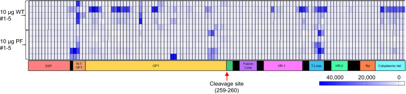
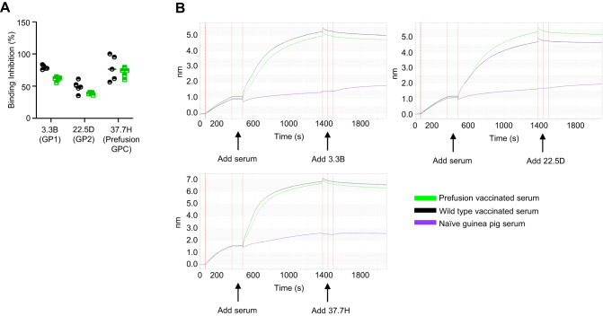
Lassa virus is a member of the Arenaviridae family, which causes human infections ranging from asymptomatic to severe hemorrhagic disease with a high case fatality rate. We have designed and generated lipid nanoparticle encapsulated, modified mRNA vaccines that encode for the wild-type Lassa virus strain Josiah glycoprotein complex or the prefusion stabilized conformation of the Lassa virus glycoprotein complex. Hartley guinea pigs were vaccinated with two 10 µg doses, 28 days apart, of either construct. Vaccination induced strong binding antibody responses, specific to the prefusion conformation of glycoprotein complex, which were significantly higher in the prefusion stabilized glycoprotein complex construct group and displayed strong Fc-mediated effects. However, Lassa virus-neutralizing antibody activity was detected in some but not all animals. Following the challenge with a lethal dose of the Lassa virus, all vaccinated animals were protected from death and severe disease. Although the definitive mechanism of protection is still unknown, and assessment of the cell-mediated immune response was not investigated in this study, these data demonstrate the promise of mRNA as a vaccine platform against the Lassa virus and that protection against Lassa virus can be achieved in the absence of virus-neutralizing antibodies.


Conflict of interest statement

S.H. and A.C. are employees of Moderna. The remaining authors declare no competing interests.

Figures

Fig. 1
Fig. 1. Generation of mRNA-based vaccine.
A The structure of LASV particle. B LASV L (green and yellow) and S (purple and multi-color for the GPC) segments. C SDS PAGE gel image of in vitro transcribed prefusion and wild-type LASC GPC vaccine constructs. This was run once. D Animal study outline: schedule for two doses of vaccine, challenge, and end of study. Each group included 5 animals, for a total of 25 animals in the study. The animals were 4–6 weeks old and had a starting weight of 350–400 g. The control group received two doses of sterile phosphate-buffered saline. Graphics were generated by N. Lloyd with Biorender.com and Microsoft PowerPoint.
Fig. 2
Fig. 2. Antibody response to vaccination with WT of prefusion-stabilized GPC mRNA.
A Quantification of IgG binding to postfusion LASV GPC determined by ELISA. Controls: HMAF from LASV-infected mice, 37.7H prefusion GPC-specific mAb, 22.5D GP2 specific mAb, and 3.3B a GP1 specific mAb. Pooled serum from 10 guinea pigs per vaccine. B Quantification of IgG binding to prefusion-stabilized LASV GPC determined by ELISA. Each symbol represents the average values of one group. N = 5. For (A) and (B), the error bars represent the standard deviation of the means of the 5 animals per group. C ELISA binding antibody titers, endpoint dilution, with prefusion GPC antigen from panel (B) (Two-way ANOVA), ns P-value = 0.998. Controls: HMAF and 37.7H. D Binding of IgG to prefusion stabilized Clade 2 (strain NIGA08-41) LASV GPC determined by ELISA. Ten animals shown individually. E Binding of IgG to prefusion stabilized Clade 5 (strain Soromba-R) LASV GPC determined by ELISA. Ten animals shown individually. For (D) and (E), the error bars represent the standard deviation between the duplicate values run. F Mean neutralization curves for both vaccine groups on day 54. N = 5. The error bars represent the standard deviation of the means of the 5 animals per group. G Individual neutralization curves for each animal on day 54—N = 5 per vaccine or control group. For all assays in this figure, samples were run in duplicates, and each assay was performed at least twice. Throughout, WT GPC vaccinated animals are represented by a filled-in black circle. Prefusion GPC vaccinated animals are represented by a filled-in green triangle. Antibody control, 37.7H is represented by a mustard-colored diamond. Serum control, HMAF, is represented by a red hexagon. Pre-immune serum control is represented by a filled-in orange square. Source data are provided as a Source Data file.
Fig. 3
Fig. 3. Peptide array mapping of linear epitopes associated with the vaccine-induced humoral response in vaccinated animals.
Array comprised 120 peptides spanning the length of the 491 aa LASV GPC. Each 15-mer peptide overlaps the next by 11 amino acids. Stable signal peptide (SSP) in peach, N-terminal domain (NTD) of GP1 in tangerine, GP1 in yellow, fusion loop in violet, HR-1 in pink, T-loop in light blue, HR-2 in mint green, TM in orange, cytoplasmic tail in cyan. Each sample was run in triplicate. Source data are provided as a Source Data file.
Fig. 4
Fig. 4. Mapping of the antibody response by biolayer interferometry competition binding assay with mAbs of known epitope specificity.
A Inhibition of binding of the immune sera by the indicated mAbs. The antigen (GPmperP) is the non-prefusion stabilized, uncleaved GPC that contains a linker between GP1 and GP2. This protein adopts both the pre-fusion and the post-fusion forms. Values normalized to 100%. B Representative data curves for mAbs 37.7H, 3.3B, and 22.5D with vaccinated and naïve guinea pig sera. Samples were run in duplicate, and the assay was performed twice. N = 5 animals per study group. Source data are provided as a Source Data file.
Fig. 5
Fig. 5. Fc-mediated effector functions of the immune sera after vaccination with wild type or prefusion-stabilized mRNA constructs.
A, B Antibody-dependent neutrophil phagocytosis (ADNP). C, D Antibody-dependent cellular phagocytosis (ADCP). E, F Percentages of NK cells positive for CD107a. G, H Percentages of NK cells positive for MIP-1β. I, J Antibody-dependent complement deposition (ADCD). Antibody response to wild-type LASV GPC vaccine (A, C, E, G, I) and prefusion stabilized LASV GPC vaccine (B, D, F, H, J). Ns, not significant; * P < 0.05; ** P < 0.01; *** P < 0.001. One-way ANOVA with Tukey’s post-hoc test. A ns, P = 0.1145. B ns, P = 0.3029. C Between WT and PreF ns, P = 0.8455, between WT and control ns, P = 0.0955. D ns, P = 0.966. E Between WT and PreF ns, P = 0.9949. Between WT and control ns, P = 0.1120. Between PreF and control ns, P = 0.1326. F Between WT and PreF ns, P = 0.8805. Between WT and control ns, P = 0.9325. Between PreF and control ns, P = 0.9881. G ns, P = 0.6163. H Between WT and PreF ns, P = 0.7593. Between WT and control ns, P = 0.7045. Between PreF and control ns, P = 0.2990. I ns, P = 0.6545. J ns, P = 0.3827. For (A, B, E, F, G, and H), assays were run with PBMCs isolated from two separate donors. On all charts, the mean of the 5 animals per group and the standard deviation of the 5 animals per group is represented with error bars. For all figures, WT GPC vaccinated animals represented by a filled-in black circle. Prefusion GPC vaccinated animals represented by a filled-in green triangle. All non-vaccinated control animals represented but a filled-in purple diamond. For all figures, N = 5 animals per group. Source data are provided as a Source Data file.
Fig. 6
Fig. 6. Protective efficacy in vaccinated guinea pigs.
A Disease score. B Weight change. Dashed line represents 20% weight loss (animal at 80% of the original weight) which was the weight cutoff for euthanasia. C Body temperature, D Viremia. Lower limit of detection is 50 PFU/mL. Only the control group is included, as no viremia was detected in either vaccinated groups. For (BD), error bars represent the standard deviation between the averages of the 5 animals per group. E Percent survival. Mean values based on 5 animals per group ± SD. P = 0.0014 for both WT GPC vaccine versus control and prefusion GPC vaccine versus control. Log-rank (Mantel-Cox) test. For all figures, WT GPC vaccinated animals represented by a filled-in black circle. Prefusion GPC vaccinated animals represented by a filled-in green triangle. All non-vaccinated control animals represented but a filled-in purple diamond. For all figures, each group is an average of the N = 5 animals per group. Source data are provided as a Source Data file.
Fig. 7
Fig. 7. Histopathology and LASV immunohistochemistry in organs of vaccinated and control animals.
Images of H&E stained (AI) and LASV nucleoprotein immunostained (JR) tissues of guinea pig lung (AC and JL), spleen (D, E and MO) and liver (GI and PR). Scale bars in bottom right corners of H&E images = 103 μM. IHC images magnified at 20x, scale bars added in ImageJ in bottom right corners = 72 μM. C The box indicates a representative area of viral interstitial pneumonia. D, E The arrows indicate germinal center activation, which is notably more robust in vaccinated animals compared to the unvaccinated control. I The arrow shows an area demonstrating steatosis in the liver of the control animal, which is not present in the vaccinated animals. Staining was performed once on duplicate slices of tissue.

References

    1. Hallam HJ, et al. Baseline mapping of Lassa fever virology, epidemiology and vaccine research and development. NPJ Vaccines. 2018;3:11. doi: 10.1038/s41541-018-0049-5. - DOI - PMC - PubMed
    1. Troup JM, White HA, Fom ALMD, Carey DE. An outbreak of Lassa fever on the Jos Plateau, Nigeria, in January–February 1970: a preliminary report. Am. J. Trop. Med. Hyg. 1970;19:695–696. doi: 10.4269/ajtmh.1970.19.695. - DOI - PubMed
    1. Robinson JE, et al. Most neutralizing human monoclonal antibodies target novel epitopes requiring both Lassa virus glycoprotein subunits. Nat. Commun. 2016;7:11544. doi: 10.1038/ncomms11544. - DOI - PMC - PubMed
    1. Hastie KM, et al. Structural basis for antibody-mediated neutralization of Lassa virus. Science. 2017;356:923–928. doi: 10.1126/science.aam7260. - DOI - PMC - PubMed
    1. Cross RW, et al. Treatment of Lassa virus infection in outbred guinea pigs with first-in-class human monoclonal antibodies. Antivir. Res. 2016;133:218–222. doi: 10.1016/j.antiviral.2016.08.012. - DOI - PMC - PubMed

Publication types