. 2023 Aug;59(7):486-504.
doi: 10.1007/s11626-023-00785-z. Epub 2023 Sep 12.

Effect of Wnt10a/β-catenin signaling pathway on promoting the repair of different types of dentin-pulp injury

Affiliations

Effect of Wnt10a/β-catenin signaling pathway on promoting the repair of different types of dentin-pulp injury

Yue Li et al. In Vitro Cell Dev Biol Anim. 2023 Aug.

Abstract

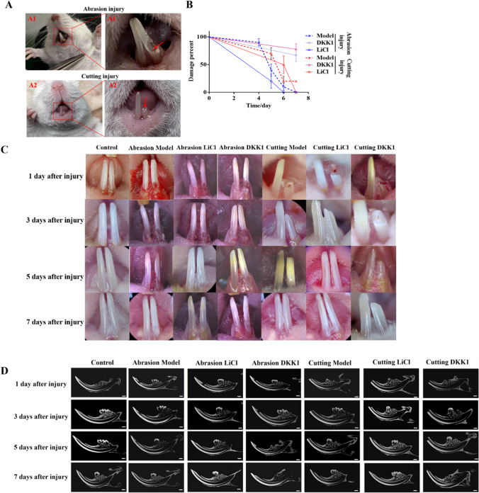
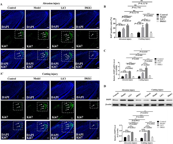
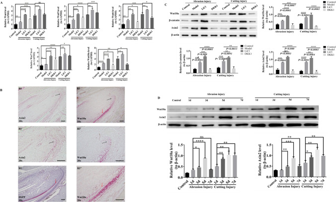
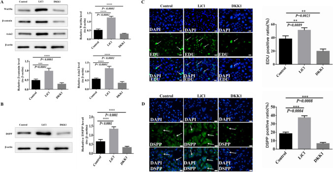
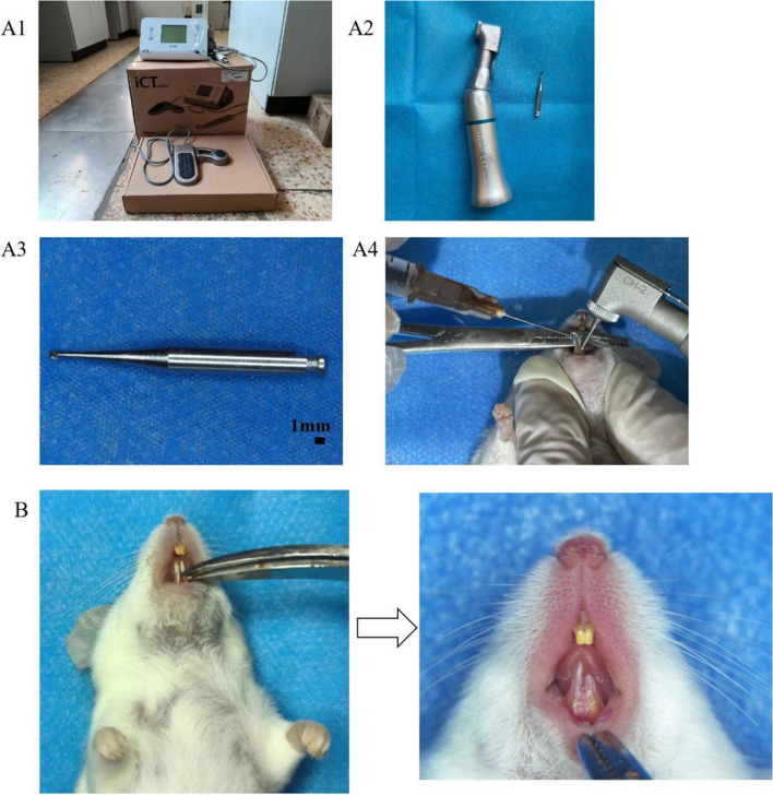
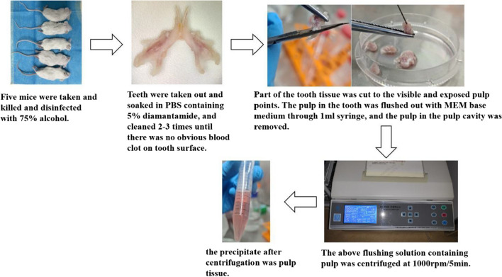
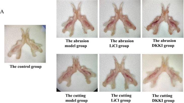
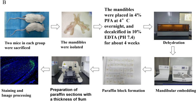
How to repair dentin-pulp injury effectively has always been a clinical problem, and the comparative study of repair process between different injuries is unknown. Dental pulp stem cells (DPSCs) often are selected as seed cells for the study of dentin-pulp injury repair due to excellent advantages in odontogenesis and pulp differentiation. Although many previous researches have indicated that the Wnt protein and Wnt/β-catenin signaling pathway were crucial for dental growth, development, and injury repair, the specific mechanism remained unknown. In this study, different dentine-pulp injury models of adult mice were established successfully by abrasion and cutting methods. The gross morphology and micro-CT were used to observe the repair of injured mice incisor in different groups. We found that the repair time of each group was different. The repair time of the cutting group was longer than the abrasion group and the qRT-PCR detection showed that the expression of DSPP in the cutting group was higher than that in the abrasion group, but there was no significant difference in proliferation among the groups. In vivo and cell experiments showed that activation of Wnt/β-catenin signaling pathway can promote the proliferation and odontoblast differentiation of DPSCs. In addition, by using RNAscope staining, we observed that Wnt10a was mainly expressed in the proliferative region and partially expressed in the odontoblast region. The Western blotting results showed that in the early stage of repair, the expression of Wnt10a increased with the extension of days after injury in both abrasion and cutting group and the increase of Wnt10a was tested obviously on the 5th day after injury. But on the 7th day after injury, the expression of Wnt10a was still obvious in the cutting group, while the expression of Wnt10a was significantly reduced in the abrasion group, which was close to the control group. It is suggested that Wnt10a acts as a repair-related protein and has an important role in tooth injury repair. Wnt10a was activated by R-spondin and LiCl, and Wnt10a-siRNA DPSCs were constructed to inhibit Wnt10a. The results showed that Wnt10a/β-catenin signaling pathway promoted the proliferation and odontoblast differentiation of DPSCs. It plays a crucial role in the repair process of different injuries. This study enriched the mechanisms of Wnt10a /β-catenin signaling pathways in different types of dentin-pulp injury repair, which could provide experimental evidences for the target gene screening and also give some new ideas for the subsequent research on the molecular mechanisms of tooth regeneration.

Keywords: DPSCs; Dentin-pulp complex injury; Wnt10a/β-catenin signaling pathway.


Conflict of interest statement

The authors declare no competing interests.

Figures

Figure. 1
Figure. 1
Observation and analysis of different injury models of mice incisor teeth. After successful establishment of the mice model and continuous injection of drugs for 7 d, observing the repair of dentin-pulp injury in the abrasion model, abrasion LiCl, abrasion DKK1, cutting model, cutting LiCl and cutting DKK1 groups at 1, 3, 5, and 7 d after injury. (A) The dentin-pulp injury model of abrasion and cutting methods was successfully established. (A1, A1') The dentin-pulp injury model was established in the abrasion method, and the red arrow indicated the red pulp point which was visible. (A2, A2') The dentin-pulp injury model was established in a cutting method, and the red pulp point which was indicated by the red arrow was visible. (B) The repair time and percentage of dentin-pulp injury in each group after the successful establishment of the mice model and continuous injection of drugs for 7 d. Percentage of injury = (number of mice with unrepaired incisor teeth in each group/10) × 100%. (C) Morphological observation. With the increase of time after injury, the defect area of the incisor tissue of mice in each group was gradually reduced and repaired to the same smoothness and length as the normal teeth on the opposite side. (D) Micro-CT observation. With the increase of time after injury, the defect area of the incisor tissue of mice in each group gradually decreased, and the defect at the dentin layer was gradually formed until it was completely repaired. The internal structure of repaired teeth were consistent with the control group. Scale bar, 1 mm.
Figure. 2
Figure. 2
The changes in proliferation and odontoblast differentiation of DPSCs were detected and analyzed in different injury models of mouse incisors. (A, A') The expression of Ki67 in incisor tissue was detected by immunofluorescence staining in the abrasion and cutting groups. The white arrows show Ki67 positive cells. The Ki67-positive cells were expressed in the cervical ring of the incisor teeth of mice in the control group. The expression of Ki67 in the model group was significantly higher than in the control group. And compared with the control group, Ki67 expression increased more significantly in the LiCl group and decreased prominently in the DKK1 group. Scale bars, 100 μm. (B) The ratio of Ki67 positive cells in the abrasion, cutting, control, model, LiCl, and DKK1 groups were analyzed respectively. (C) After 7 days of LiCl and DKK1 induction, the qRT-PCR analysis of DSPP mRNA (to GAPDH) in the abrasion model, abrasion LiCl, abrasion DKK1, cutting model, cutting LiCl, and cutting DKK1 groups. n = 70, *P < 0.05, **P < 0.01, ***P < 0.001. (D) Western blotting analysis of DSPP expression in the abrasion model, abrasion LiCl, abrasion DKK1, cutting model, cutting LiCl, and cutting DKK1 groups after 7 d of LiCl and DKK1 induction. n = 70, *P < 0.05, **P < 0.01, ***P < 0.001.
Figure. 3
Figure. 3
Detection of Wnt4, Wnt5, Wnt6, and Wnt7 and changes in the expression levels of Wnt10a and Wnt/β-catenin signaling pathways in mouse incisors of different injury models, as well as changes in Wnt10a and Wnt/β-catenin signaling pathways at different time points after injury. (A) The expressions of Wnt4, Wnt5, Wnt6, Wnt7, and Wnt10a in the incisor were detected by qRT-PCR. *P < 0.05, **P < 0.01, ***P < 0.001. Scale bars, 100 μm. (B) RNAscope in situ hybridization for the expression of Wnt10a and Axin2 in normal mouse incisors. The scale bars in the figure are all 100um. (B1) Under a 10 × microscope, Axin2 appeared high expression in the proliferative zone, and the black arrow and pink staining are positive for Axin2. (B1') was a magnification of (B1) at 20 × microscope in the black arrow region. (B2) DSPP appeared high expression in the odontoblast region, and the red arrow and pink staining are DSPP positive. (B3) Under a 10 × microscope, Wnt10a appeared high expression in the proliferative zone, with rarely expressed in the odontoblast zone. Black arrow, red arrow, and pink staining represent the presence of Wnt10a positivity in the proliferative and odontoblast regions, respectively. (B3') and (B3'') are (B3) at 20 × electron microscope with magnification of the black and red arrow areas, respectively. (C) Western blotting detected the expression of Wnt10a, β-catenin, and Axin2 in the control group, abrasion model group, abrasion LiCl group, abrasion DKK1 group, cutting model group, cutting LiCl group, and cutting DKK1 groups. (D) Western blotting detected changes in the expression of Wnt10a and Axin2 at 1, 3, 5, and 7 d after incisor abrasion and cutting injuries.
Figure. 4
Figure. 4
In the cell experiment, Western blotting detected the changes in the expression levels of Wnt10a, Wnt/β-catenin signaling pathway, and DSPP stimulated by LiCl and DKK1, and EDU and DSPP staining were used to analyze the proliferation and odontoblast differentiation of DPSCs and the differentiation of odontoblast cells. *P < 0.05, **P < 0.01, ***P < 0.001. Scale bars, 50um. (A) Western blotting analyzed expression changes of Wnt10a, β-catenin, and Axin2 proteins in the control, LiCl, and DKK1 groups under the stimulation of LiCl and DKK1. (B) Western blotting analyzed expression changes of DSPP protein in control, LiCl, and DKK1 groups under the stimulation of LiCl and DKK1. (C) EDU staining was used to observe the proliferative vitality of DPSCs in P3 generation mice stimulated by LiCl and DKK1. White arrows and green staining are positive cells. The proportion of positive cells in the control, LiCl, and DKK1 groups were analyzed. (D) DSPP immunofluorescence staining was used to identify mice DPSCs stimulated by LiCl and DKK1. White arrows and green staining are positive staining of DSPP. The positive proportion of DSPP in the control, LiCl, and DKK1 groups were analyzed.
Figure. 5
Figure. 5
Normal DPSCs and Wnt10a-siRNA DPSCs of mice were interfered by R-spondin and LiCl. Ki67 fluorescence staining, DSPP, and DMP-1 immunofluorescence staining were used to detect the changes of DPSC proliferation and odontogenesis differentiation of normal DPSCs and Wnt10A-SiRNA, and Western blotting was used to detect the changes of Wnt10a, Axin2, and β-catenin protein expression. *P < 0.05, **P < 0.01, ***P < 0.001. Scale bars, 50 µm. (A) Cell growth was observed in normal mice DPSCs and Wnt10a-siRNA DPSCs after R-spondin and LiCl intervention by microscope. (B) Ki67 fluorescence staining detected the proliferative vitality of DPSCs in control, R-spondin, LiCl, R-spondin + Wnt10a-siRNA, and LiCl + Wnt10a-siRNA groups. White arrows and green staining are positive cells, and analyzing the proportion of positive cells in each group. (C) Fluorescence staining detected DSPP in control, R-spondin, LiCl, R-spondin + Wnt10a-siRNA and LiCl + Wnt10a-siRNA groups. White arrows and green staining are DSPP positive cells, and analyzing the proportion of positive cells in each group. (D) Fluorescence staining detected DMP-1 in control, R-spondin, LiCl, R-spondin + Wnt10a-siRNA group, and LiCl + Wnt10a-siRNA group. White arrows and green staining are DMP-1 positive cells, and analyzing the proportion of positive cells in each group. (E) Western blotting analysis of Wnt10a, Axin2, and β-catenin protein expression in Control, R-spondin, LiCl, R-spondin + Wnt10a-siRNA, and LiCl + Wnt10a-siRNA group.
Figure. 6
Figure. 6
Model construction of mice incisor dentin-dental pulp damage. (A) The abrasion method: We used a dental slow-speed handpiece (EX-203C Set, NSK) connected to the planting machine power system (ICT motor Serial NO.B02DED1021, Dentium) and coupled with a 1-mm diameter tungsten steel slow bending ball drill (RA2, Dentsply Sirona) to abrade the root of one side incisor. During the abrasion process, the ball drill was stopped when the drill bit could fully enter the tooth tissue and the red pulp piercing point was visible to the naked eye. Cooling and washing with saline while grinding. The diameter of the defect was 1.0–1.5 mm and the depth was 1.0 mm. (A1) The planting machine power system (ICT motor Serial NO.B02DED1021, Dentium). (A2) Dental slow-speed handpiece (EX-203C Set, NSK). (A3) A 1-mm diameter tungsten steel slow bending ball drill (RA2, Dentsply Sirona). (A4) The process of abrasion. (B) The cutting method: One side incisor was cut directly with ophthalmic scissors until the broken end of the incisor was horizontal with the lingual gingiva.
Figure. 7
Figure. 7
Drug treatment in the abrasion groups and the cutting groups. For the periodontal ligament injection method, a needle was inserted into the gingival sulcus on the labial side of the injured incisor and normal incisors on one side of the control group, the inclined surface of the needle was close to the tooth surface, and the needle direction was at an angle of 45° to the long axis of the tooth, then slowly penetrated the periodontal membrane, and the drug was injected. The mice in the abrasion LiCl group and the cutting LiCI group were given LiCl solution (0.75 g/L) at a dose of 0.2 ml/kg and The mice in the abrasion DKK1 group and the cutting DKK1 group were injected with DKK1 protein (1ug/ml) at a dose of 0.2 μg/kg by local injection into the periodontal ligament, once every 2 d. (A) Drug treatment in the in the Abrasion LiCI group and the abrasion DKK1 group. (A1–A4) Process of drug treatment. (B) Drug treatment in the in the cutting LiCI group and the cutting DKK1 group. (B1–B4) Process of drug treatment.
Figure. 8
Figure. 8
Sampling method of mouse dental pulp. Five mice were sacrificed by excessive carbon dioxide and cervical dislocation. Then we put them in 75% alcohol to disinfect for 5 min. The mandibles were isolated and the soft tissues attached to the mandibles were removed and the mandibles were rinsed with PBS containing 5% diamantine for 2–3 times until there was no obvious blood clot on the tooth surface. Part of the tooth tissue of the tooth cusp and root tip was cut until the pulp point was visible and exposed. Then the pulp in the tooth was flushed out with MEM base medium through a 1-ml syringe and the pulp in the pulp cavity was removed, then rinsing again. The rinse solution was collected and centrifuged at 1000 rpm/5 min, and the precipitate after centrifugation was pulp tissue.
Figure. 9
Figure. 9
Preparation of paraffin sections. Two mice in each group were sacrificed, the mandibles were isolated and the soft tissues attached to the mandibles were removed and placed in 4% PFA overnight at 4°C and decalcified in 10% EDTA (pH 7.4) for about 4 weeks. The samples were dehydrated in ethanol at different concentrations of 50%, 70%, 80%, 90%, and 100%, each for 1 h. The samples were then mixed 1:1 with liquid paraffin, and the samples were placed in the mixture and placed in a 58°C incubator for 2 h. The samples were then placed in liquid paraffin and placed in a 58°C incubator for 2 h. The samples were then mixed 1:1 with liquid paraffin and placed in a 58°C incubator for 2 h. The samples were placed in a 58°C incubator for 2 h. Repeat 2 times. Paraffin embedding was performed and after the paraffin wax was completely hardened at room temperature, paraffin sections were prepared with a thickness of 9 μm and detailed labeling of section information. (A) The mandibles were isolated and the tissues are similar in size. (B) Paraffin slicing process.
Figure. 9
Figure. 9
Preparation of paraffin sections. Two mice in each group were sacrificed, the mandibles were isolated and the soft tissues attached to the mandibles were removed and placed in 4% PFA overnight at 4°C and decalcified in 10% EDTA (pH 7.4) for about 4 weeks. The samples were dehydrated in ethanol at different concentrations of 50%, 70%, 80%, 90%, and 100%, each for 1 h. The samples were then mixed 1:1 with liquid paraffin, and the samples were placed in the mixture and placed in a 58°C incubator for 2 h. The samples were then placed in liquid paraffin and placed in a 58°C incubator for 2 h. The samples were then mixed 1:1 with liquid paraffin and placed in a 58°C incubator for 2 h. The samples were placed in a 58°C incubator for 2 h. Repeat 2 times. Paraffin embedding was performed and after the paraffin wax was completely hardened at room temperature, paraffin sections were prepared with a thickness of 9 μm and detailed labeling of section information. (A) The mandibles were isolated and the tissues are similar in size. (B) Paraffin slicing process.
Figure. 10
Figure. 10
Cultivation and identification of mouse DPSCs. (A) The mesenchymal markers CD44, CD105, and CD146 and the hematopoietic markers CD45, CD34, and CD31 were detected by flow cytometry to identify the mouse DPSCs. (B) The cell was long spindle-shaped and grew in clusters, which were the typical morphology of mesenchymal stem cells in P3 generation of mouse DPSCs. (C) The adipogenic differentiation of P3 generation mouse DPSCs was obvious after 14 d of induction by oil Red O staining. (D) The osteogenic differentiation of P3 generation mouse DPSCs was significantly induced after 14 d induction by alizarin red staining. (E) The chondrogenic differentiation of P3 generation mouse DPSCs was significantly induced after 5 wk induction by Alsinland staining.

References

    1. About I. Dentin-pulp regeneration: the primordial role of the microenvironment and its modification by traumatic injuries and bioactive materials. Endo Top. 2013;28:61–89. doi: 10.1111/etp.12038. - DOI
    1. Almeida FM, Marques SA, Rodrigues RF, et al. Human dental pulp cells: a new source of cell therapy in a mouse model of compressive spinal cord injury. J Neurotrauma. 2011;28(9):1939–1949. doi: 10.1089/neu.2010.1317. - DOI - PubMed
    1. Anthony JS, Henry FD, Diogenes A, et al. Exploiting the bioactive properties of the dentin-pulp complex in regenerative endodontics. J Endod. 2016;42(1):47–56. doi: 10.1016/j.joen.2015.10.019. - DOI - PubMed
    1. Cao X, Wang X, Zhang W, et al. WNT10A induces apoptosis of senescent synovial resident stem cells through Wnt/calcium pathway-mediated HDAC5 phosphorylation in OA joints. Bone. 2021;150:116006. doi: 10.1016/j.bone.2021.116006. - DOI - PubMed
    1. Chai Y, Slavkin HC. Prospects for tooth regeneration in the 21st century: a perspective. Microsc Res Tech. 2003;60(5):469–479. doi: 10.1002/jemt.10287. - DOI - PubMed

LinkOut - more resources