. 2023 Sep 13;290(2006):20231158.
doi: 10.1098/rspb.2023.1158. Epub 2023 Sep 13.

Pre-mandibular pharyngeal pouches in early non-teleost fish embryos

Affiliations

Pre-mandibular pharyngeal pouches in early non-teleost fish embryos

Agata Horackova et al. Proc Biol Sci. .

Abstract

The vertebrate pharynx is a key embryonic structure with crucial importance for the metameric organization of the head and face. The pharynx is primarily built upon progressive formation of paired pharyngeal pouches that typically develop in post-oral (mandibular, hyoid and branchial) domains. However, in the early embryos of non-teleost fishes, we have previously identified pharyngeal pouch-like outpocketings also in the pre-oral domain of the cranial endoderm. This pre-oral gut (POG) forms by early pouching of the primitive gut cavity, followed by the sequential formation of typical (post-oral) pharyngeal pouches. Here, we tested the pharyngeal nature of the POG by analysing expression patterns of selected core pharyngeal regulatory network genes in bichir and sturgeon embryos. Our comparison revealed generally shared expression patterns, including Shh, Pax9, Tbx1, Eya1, Six1, Ripply3 or Fgf8, between early POG and post-oral pharyngeal pouches. POG thus shares pharyngeal pouch-like morphogenesis and a gene expression profile with pharyngeal pouches and can be regarded as a pre-mandibular pharyngeal pouch. We further suggest that pre-mandibular pharyngeal pouches represent a plesiomorphic vertebrate trait inherited from our ancestor's pharyngeal metameric organization, which is incorporated in the early formation of the pre-chordal plate of vertebrate embryos.

Keywords: evolution; mouth; pharyngeal pouch; pharynx; pre-oral gut; vertebrate head.


Conflict of interest statement

We declare we have no competing interests.

Figures
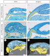
Figure 1.
Figure 1.
Early and late formation of the pre-oral gut (POG) and its connection with the pharynx. Bichir (a,c,e) and sturgeon (b,d,f) embryos, head to the left, showing the development of POG from late neurula stage (a,b), through pharyngula stage (b,c), until cement and hatching gland specification (e,f), endoderm yellow, POG marked by yellow arrowheads. Sagittal plastic sections (a–d) and whole-mount three-dimensional models based on micro-CT data from Minarik et al. [60]. ar, archenteron; ey, eye; fb, forebrain; yo, yolk; n, nose; nt, notochord; ot, otic; pp, pharyngeal pouches; white arrows mark mouth position.
Figure 2.
Figure 2.
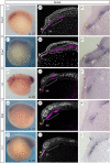
Bichir—gene expression patterns. Left column: whole-mount embryos, lateral views, head to the left. Mid column: sagittal sections showing POG (arrowheads) and pharynx (brackets), magenta shows ISH signal (see methods), white is DAPI. Right column: details of the original ISH signal in POG, dashed white line outlines the endodermal epithelium based on the segregation of nuclei visible with DAPI staining. fb, forebrain; som, somites; yo, yolk; asterisks point to primary gut cavity; arrowhead shows POG, arrows indicate placodal expression.
Figure 3.
Figure 3.
Sturgeon—early gene expression patterns. Left column: whole-mount embryos, dorsal views, head to the top. Mid column: medial sections, head to the left, magenta shows ISH signal (see methods), white is DAPI. Right column: details of the original ISH signal in POG, dashed white line outlines the endodermal epithelium based on the segregation of nuclei visible with DAPI staining. fb, forebrain; yo, yolk; asterisks point to primary gut cavity; arrowhead shows POG, arrows indicate placodal expression.
Figure 4.
Figure 4.
Sturgeon—late gene expression patterns. First column: whole-mount embryos with expression in POG, dorsal views, head to the top. Second column: medial sections, head to the left, magenta shows ISH signal (see methods), white is DAPI. Third column: details of the original ISH signal in POG, dashed white line outlines the endodermal epithelium based on the segregation of nuclei visible with DAPI staining. Fourth column: whole-mount embryos of slighter later stage showing expression in pharyngeal arches. fb, forebrain; pp, pharyngeal pouch; yo, yolk; asterisks point to primary gut cavity; arrowhead shows POG.
Figure 5.
Figure 5.
Serial homology of the pre-oral gut (POG) with pharyngeal pouches, and POG as an ancestral pharyngeal module in deuterostome embryos. (a) A cartoon of embryos with early primary gut cavity (endoderm, yellow) and later formation of pharyngeal pouches that share morphogenesis and gene expression profile in both the pre-oral and post-oral pharynx. (b) Late embryos with POG derivatives in clearly a pre-oral position, that disappear in later stages similar to the post-oral pharyngeal pouches. (c) POG-diverticula as stomochord (S) in hemichordates, as Hatschek's diverticula/club-shape gland (C) in amphioxus, with similar pre-oral pharyngeal domain supposedly also present early vertebrate yunnanozoan. M, mouth, G, gills.

Similar articles

References

    1. Hopwood N. 2015. Haeckel's embryos: images, evolution, and fraud. Chicago, IL: University of Chicago Press.
    1. Kuratani S, Nobusada Y, Horigome N, Shigetani Y. 2001. Embryology of the lamprey and evolution of the vertebrate jaw: insights from molecular and developmental perspectives. Phil. Trans. R. Soc. B 356, 1615-1632. (10.1098/rstb.2001.0976) - DOI - PMC - PubMed
    1. Irie N, Kuratani S. 2011. Comparative transcriptome analysis reveals vertebrate phylotypic period during organogenesis. Nat. Commun. 2, 248. (10.1038/ncomms1248) - DOI - PMC - PubMed
    1. Piotrowski T, Nüsslein-Volhard C. 2000. The endoderm plays an important role in patterning the segmented pharyngeal region in zebrafish (Danio rerio). Dev. Biol. 225, 339-356. (10.1006/dbio.2000.9842) - DOI - PubMed
    1. Couly G, Creuzet S, Bennaceur S, Vincent C, Le Douarin NM. 2002. Interactions between Hox-negative cephalic neural crest cells and the foregut endoderm in patterning the facial skeleton in the vertebrate head. Development 129, 1061-1073. (10.1242/dev.129.4.1061) - DOI - PubMed

Publication types

LinkOut - more resources