

Relationship between paramacular thinning, cerebral vasculopathy, and hematological risk factors in sickle cell disease
- PMID: 37700770
- PMCID: PMC10493280
- DOI: 10.3389/fmed.2023.1226210
Relationship between paramacular thinning, cerebral vasculopathy, and hematological risk factors in sickle cell disease
Abstract
Purpose: To identify risk factors for sickle cell maculopathy due to hematological parameters (especially anemia and hemolysis) or cerebral vasculopathy.
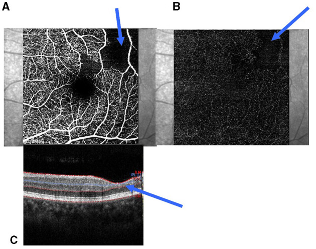
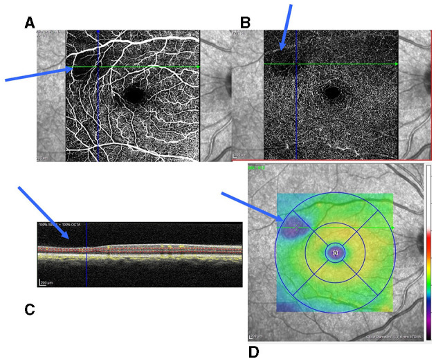
Methods: This retrospective study was conducted at a Referral Center. The follow-up included optical coherent tomography/optical coherent tomography angiography, neuro-radiological imaging, and a hematological assessment (hemoglobin, hemoglobin S level, reticulocytes, mean corpuscular volume, bilirubin, and lactate dehydrogenase).
Results: Hundred and thirty-two sickle cell patients were included. Maculopathy was observed in 127 eyes of SS patients and 10 eyes of SC patients (p < 0.001), unrelated to peripheral retinopathy. Cerebral vasculopathy was more frequent in SS patients (p < 0.001) and was also associated with the presence of maculopathy (p = 0.049), and it was related to peripheral retinopathy (p < 0.001). All biological parameters significantly differed according to the genotype (p < 0.001) but not according to the presence of cerebral vasculopathy or maculopathy. In the multivariate analysis, reticulocytes and bilirubin were associated with the presence of cerebral vasculopathy and maculopathy.
Conclusion: The data obtained were consistent with the role of anemia or hemolysis markers in cerebral vasculopathy and macular involvement. As a trend of hemolysis appears to be a risk factor for these complications, this validates the use of preventive plasmapheresis in these patients.
Keywords: OCT; cerebral vasculopathy; hemolysis (red blood cells); maculopathy; optical coherence tomography angiography (OCT-A); sickle cell disease.
Copyright © 2023 Orssaud, Flamarion, Michon, Ranque and Arlet.
Conflict of interest statement
The authors declare that the research was conducted in the absence of any commercial or financial relationships that could be construed as a potential conflict of interest.
Figures
Similar articles
-
Optical coherence tomography (OCT) and OCT angiography allow early identification of sickle cell maculopathy in children and correlate it with systemic risk factors.Graefes Arch Clin Exp Ophthalmol. 2020 Nov;258(11):2551-2561. doi: 10.1007/s00417-020-04764-y. Epub 2020 Jun 9. Graefes Arch Clin Exp Ophthalmol. 2020. PMID: 32518974
-
ATYPICAL FOVEAL AND PARAFOVEAL ABNORMALITIES IN SICKLE CELL DISEASE.Retina. 2024 Mar 1;44(3):506-514. doi: 10.1097/IAE.0000000000003987. Retina. 2024. PMID: 37948742
-
Sickle Cell Maculopathy: Microstructural Analysis Using OCTA and Identification of Genetic, Systemic, and Biological Risk Factors.Am J Ophthalmol. 2021 Apr;224:7-17. doi: 10.1016/j.ajo.2020.11.019. Epub 2021 Jan 4. Am J Ophthalmol. 2021. PMID: 33412123
-
Sickle cell retinopathy: What we now understand using optical coherence tomography angiography. A systematic review.Blood Rev. 2019 May;35:32-42. doi: 10.1016/j.blre.2019.03.001. Epub 2019 Mar 4. Blood Rev. 2019. PMID: 30852057
-
Sickle cell disease and the eye.Curr Opin Ophthalmol. 2017 Nov;28(6):623-628. doi: 10.1097/ICU.0000000000000423. Curr Opin Ophthalmol. 2017. PMID: 28984727 Review.
Cited by
-
Microvascular and Ultrastructural Changes of the Retina and Choroid in Patients with Sickle Cell Anemia.Turk J Ophthalmol. 2025 Apr 24;55(2):74-81. doi: 10.4274/tjo.galenos.2025.97792. Turk J Ophthalmol. 2025. PMID: 40272089 Free PMC article.
-
Correlations Between Visual Field Defects and Macular Thinning in Sickle Cell Disease.Invest Ophthalmol Vis Sci. 2025 Feb 3;66(2):67. doi: 10.1167/iovs.66.2.67. Invest Ophthalmol Vis Sci. 2025. PMID: 40014363 Free PMC article.
References
LinkOut - more resources
Full Text Sources
Research Materials