. 2023 Sep 6:16:5869-5885.
doi: 10.2147/IDR.S419892. eCollection 2023.

Clinical Efficiency of Metagenomic Next-Generation Sequencing in Sputum for Pathogen Detection of Patients with Pneumonia According to Disease Severity and Host Immune Status

Affiliations

Clinical Efficiency of Metagenomic Next-Generation Sequencing in Sputum for Pathogen Detection of Patients with Pneumonia According to Disease Severity and Host Immune Status

Can Chang et al. Infect Drug Resist. .

Abstract

Purpose: Severe pneumonia causes the highest mortality rate in immunocompromised patients. This study aimed to investigate the pathogen diagnostic efficacy of metagenomic next-generation sequencing (mNGS) using sputum sample in patients with pneumonia according to patients' disease severity and immune conditions.

Patients and methods: A total of 180 patients suffering from pneumonia were recruited, and sputum samples were collected in duplicate for pathogen detection by both conventional microbiological tests (CMT) and mNGS. Then, the performance of pathogen identification was examined between two methods, according to disease severity and patients' immune status.

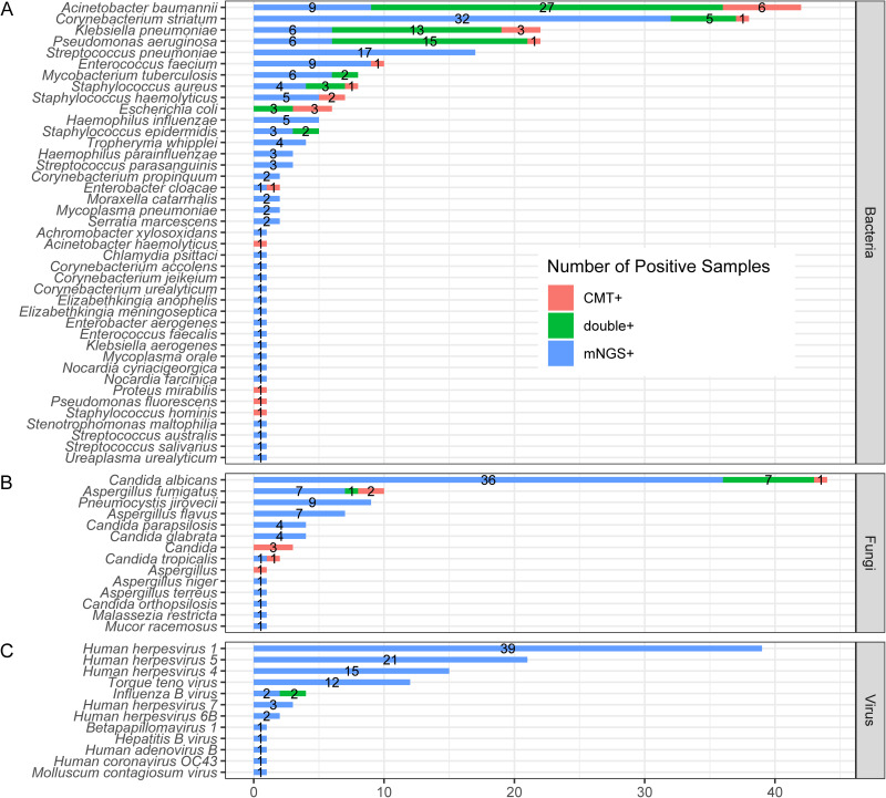
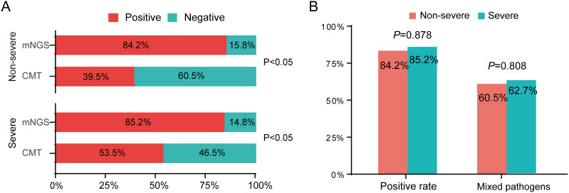
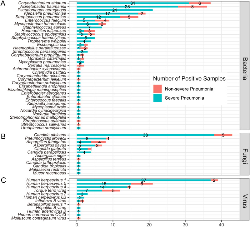
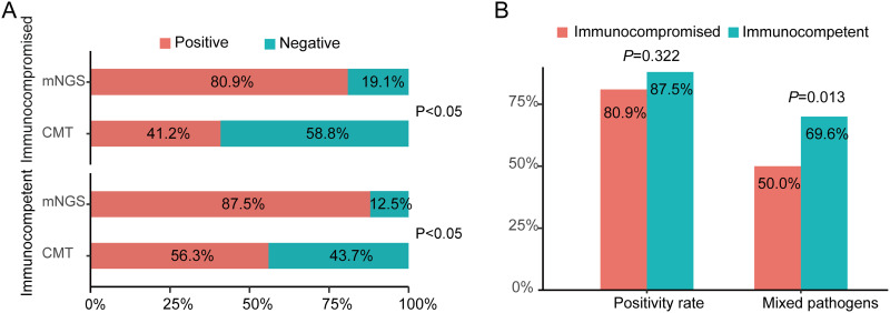
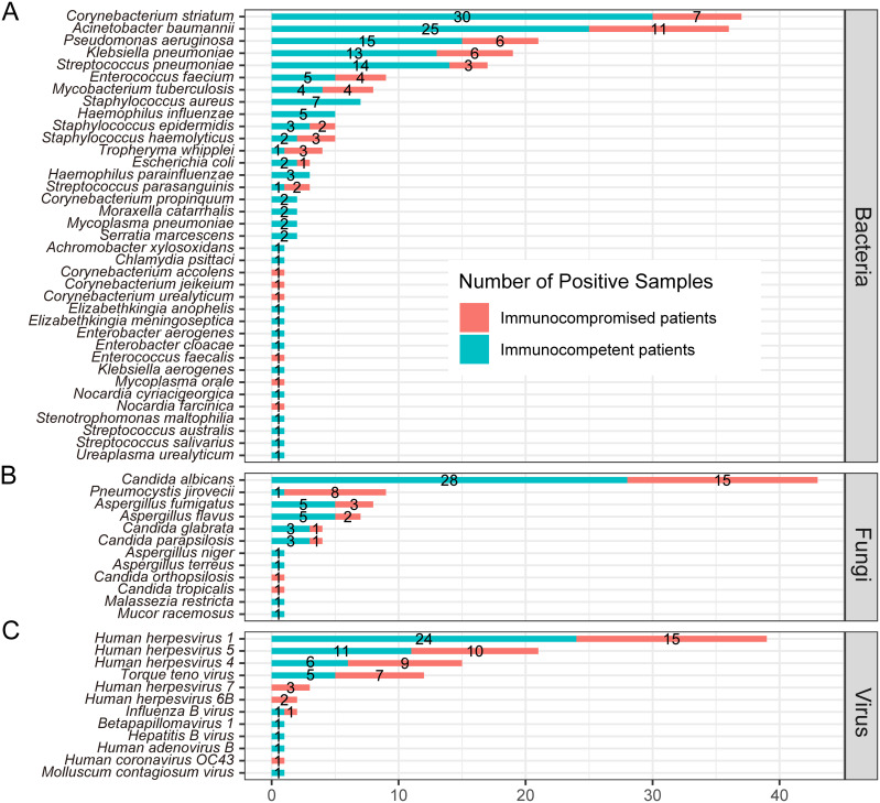
Results: In comparison to CMT, mNGS had higher positivity rates in all patients with pneumonia (85.0% vs 62.2%, P=9.445e-07). The most commonly detected microorganism in sputum of pneumonia patients was Acinetobacter baumannii (42/180, 23.3%) in bacterum level, Candida albicans in fungus level (44/180, 24.4%), and Human herpesvirus 1 (39/180, 27.5%) in virus level. However, for mNGS results, Candida albicans in 34.9% of positive patients, and Human herpesvirus 1 in 7.7% of positive cases were confirmed as pathogens causing pneumonia. Acinetobacter baumannii detected by mNGS in 75% of positive patients was diagnosed as pathogen of pneumonia. The microorganism profile of sputum mNGS differed according to disease severity and immune status of patients. Pneumocystis jirovecii was more likely to infect immunocompromised patients (P=0.002). Pseudomonas aeruginosa (14.8% vs 0.0%, P=0.008) and Human herpesvirus 1 (26.1% vs 5.3%, P=0.004) had higher infection rate in patients with severe pneumonia compared with non-severe cases. mNGS had overwhelming advantages over CMT in detecting a lot of microorganisms including Streptococcus pneumoniae, Enterococcus faecium, Pneumocystis jirovecii, and majority of viruses.

Conclusion: mNGS is a complementary tool of CMT for detecting suspected pathogens for patients with lower respiratory infections. The interpretation of opportunistic pathogens identified by mNGS is challenging, and needs comprehensive consideration of sequencing data and clinical factors.

Keywords: conventional microbiological test; immunocompromised patient; lower respiratory infections; metagenomic-next generation sequencing; severe pneumonia.


Conflict of interest statement

The authors report no conflicts of interest in this work.

Figures

Figure 1
Figure 1
The positive rate comparison of detected microorganisms by mNGS and CMT in patients with pneumonia. (A) Comparison of positive rate between pairwise mNGS and CMT in 180 patients with pneumonia, considering results of all sample types. (B) Comparison of positive rate between pairwise mNGS and CMT in 180 patients with pneumonia, only considering CMT results of sputum samples. (C) Pie chart demonstrates the positive and negative distribution of mNGS and CMT results. mNGS+: positive only by mNGS; CMT+: positive only by CMT; double+: both positive by CMT and mNGS; double-: both negative by mNGS and CMT. (D) For the double positive subgroup (grey in pattern c), 87 patients were divided into match (12/87), partial match (62/87) and mismatch (13/87). Match: both positive by CMT and mNGS and pathogens completely overlapped; mismatch: conflicts between mNGS and CMT; partial match: both positive by CMT and mNGS and partial overlapping of microorganisms.
Figure 2
Figure 2
The overlap and comparison of positive microorganisms between mNGS and CMT in 180 patients with pneumonia. (A) Bacteria levels; (B) fungi level; and (C) virus level.
Figure 3
Figure 3
The positive rate comparison of detected microorganisms by mNGS and CMT between non-severe and severe pneumonia patients. (A) Comparisons of positive rates for pairwise mNGS and CMT test in non-severe and severe pneumonia patients. (B) Comparison of positive rate and mixed infection (at least two) rate by mNGS between non-severe and severe patients.
Figure 4
Figure 4
Comparison of microorganisms detected by mNGS between non-severe and severe pneumonia patients in bacteria (A), fungi (B) and virus (C) levels.
Figure 5
Figure 5
The positive rate comparison of detected microorganisms by mNGS and CMT in immunocompetent or immunocompromised patients with pneumonia. (A) Comparisons of positive rates for pairwise mNGS and CMT test in immunocompetent and immunocompromised patients with pneumonia. (B) Comparison of positivity rate of mixed infection based on mNGS between immunocompetent and immunocompromised patients with pneumonia.
Figure 6
Figure 6
Comparison of microorganisms detected by mNGS between immunocompetent and immunocompromised patients with pneumonia in bacteria (A), fungi (B) and virus (C) levels.
Figure 7
Figure 7
The concordance between microorganisms detected by mNGS and the finally definitive pathogens confirmed by the clinicians. Dx-confirmed: the microorganisms detected by mNGS were finally confirmed as pathogens of pneumonia by clinicians; Dx-unsupported: the microorganisms detected by mNGS were not confirmed as pathogens of pneumonia by clinician. Dx-uncertain: the pathogen of pneumonia was unproven.

Similar articles

Cited by

References

    1. Feldman C, Shaddock E. Epidemiology of lower respiratory tract infections in adults. Expert Rev Respir Med. 2019;13(1):63–77. doi:10.1080/17476348.2019.1555040 - DOI - PubMed
    1. Bonell A, Azarrafiy R, Huong VTL, et al. A systematic review and meta-analysis of ventilator-associated pneumonia in adults in Asia: an analysis of national income level on incidence and etiology. Clin Infect Dis. 2019;68(3):511–518. doi:10.1093/cid/ciy543 - DOI - PMC - PubMed
    1. Collaborators GL. Estimates of the global, regional, and national morbidity, mortality, and aetiologies of lower respiratory tract infections in 195 countries: a systematic analysis for the Global Burden of Disease Study 2015. Lancet Infect Dis. 2017;17(11):1133–1161. doi:10.1016/S1473-3099(17)30396-1 - DOI - PMC - PubMed
    1. Collaborators GL. Age-sex differences in the global burden of lower respiratory infections and risk factors, 1990–2019: results from the Global Burden of Disease Study 2019. Lancet Infect Dis. 2022;22(11):1626–1647. doi:10.1016/S1473-3099(22)00510-2 - DOI - PMC - PubMed
    1. Azoulay E, Russell L, Van de Louw A, et al. Diagnosis of severe respiratory infections in immunocompromised patients. Intensive Care Med. 2020;46(2):298–314. doi:10.1007/s00134-019-05906-5 - DOI - PMC - PubMed