

Nail Whispers Revealing Dermatological and Systemic Secrets: An Analysis of Nail Disorders Associated With Diverse Dermatological and Systemic Conditions
- PMID: 37701161
- PMCID: PMC10494485
- DOI: 10.7759/cureus.45007
Nail Whispers Revealing Dermatological and Systemic Secrets: An Analysis of Nail Disorders Associated With Diverse Dermatological and Systemic Conditions
Abstract
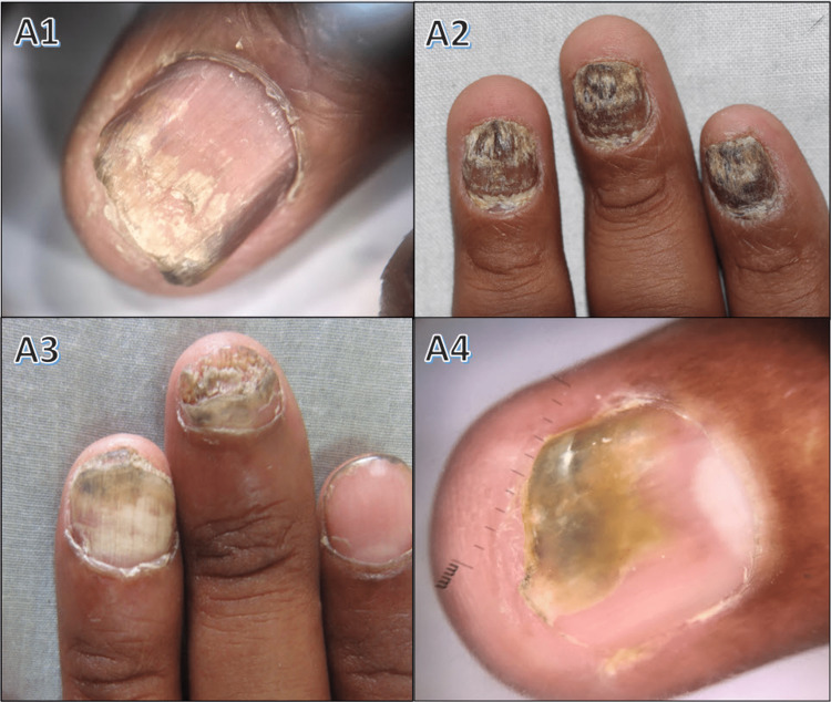
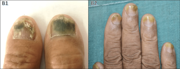
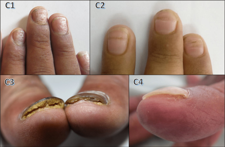
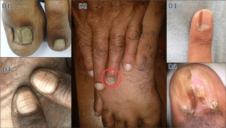
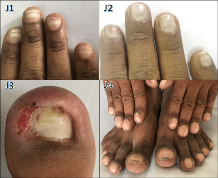
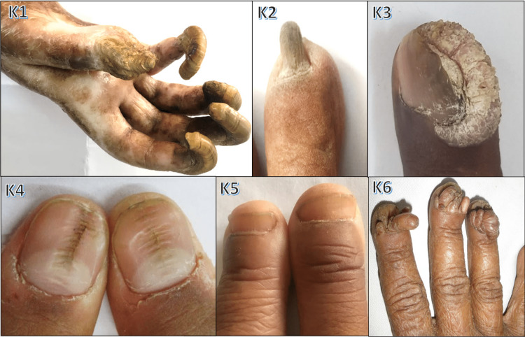
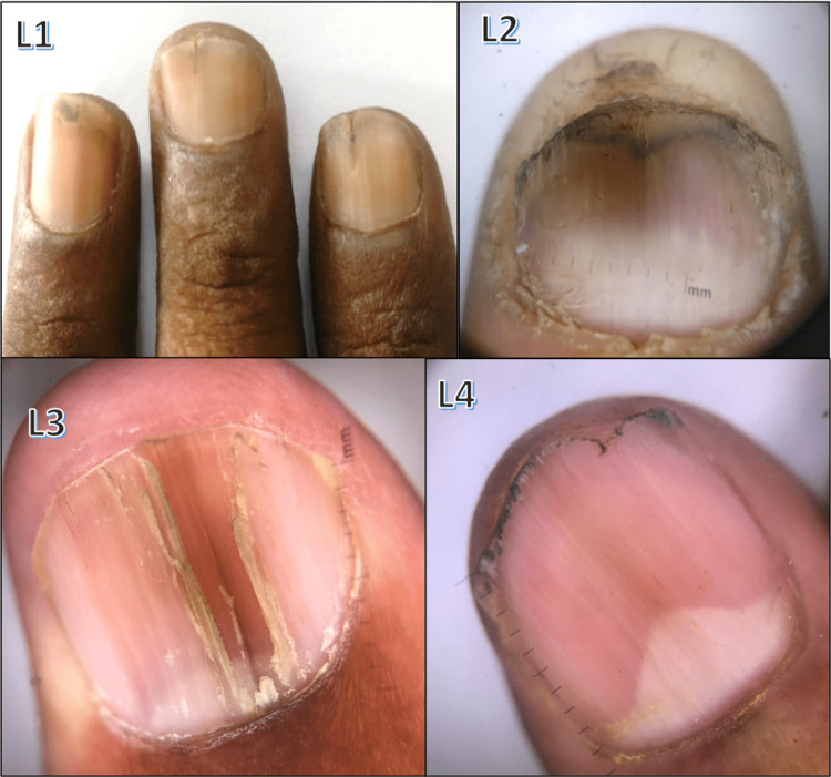
Background and objective Nail disorders encompass a wide spectrum of conditions, spanning congenital, developmental, infectious, neoplastic, degenerative, dermatological, and systemic diseases. A comprehensive exploration of their clinical manifestations, incidence, and associations is crucial for precise diagnosis and effective management. Methods This observational cross-sectional study conducted at B.J. Medical College and Civil Hospital, Ahmedabad involved 300 consecutive patients with nail changes from July 2017 to June 2019 reporting diverse dermatological and systemic conditions. The inclusion criteria involved patients of both genders and all age groups displaying nail changes associated with dermatological and systemic diseases. Data collection entailed a comprehensive clinical history, systemic and dermatological examinations, nail assessment using Dermoscope (DermLite 3, 10x), and supplementary tests. Analyses were performed on Microsoft Excel 2007 software. The study was approved by the Institute Ethics Committee. Results Among the 300 cases, females had a higher prevalence of nail involvement (57%), with a female-to-male ratio of 1.3:1. The most affected age group was 21-40 years, with 6-10 nails typically affected. Notably, housewives showed a higher prevalence. The most frequent nail condition was onychomycosis (24.33%) followed by psoriatic nail changes (20%). Less frequent nail changes involved eczema (5.7%), paronychia (5%), drug-induced (4.3%), lichen planus (3.7%), trauma-induced (3%), twenty nail dystrophy (2.33%), Darier's disease (2%), pemphigus vulgaris (2%), alopecia areata (1.67%), median Heller dystrophy (1.33%), atopic dermatitis (1%), epidermolysis bullosa (1%), racquet nail (1%), leprosy (1%), pityriasis rubra pilaris (0.67%), vitiligo (0.67%), secondary syphilis (0.67%), pachyonychia congenita (0.67%), as well as a case each of total leukonychia, subungual warts, Koenen tumor, and periungual fibroma(0.33%). Systemic autoimmune connective tissue disorders (CTD) accounted for 9%; the most common nail finding observed was nail fold erythema (48.1%) followed by nail fold telangiectasis (44.4%). In systemic sclerosis (SS), the most common finding was nail fold telangiectasia, and in systemic lupus erythematosus (SLE), the most common was nail fold erythema. Scleroderma capillary pattern on nail fold capillaroscopy was found in seven patients with SS, two patients with dermatomyositis, and only one patient with SLE. Nail changes observed in systemic diseases include onychomycosis in diabetes mellitus and chronic renal failure patients, splinter hemorrhages in ischemic heart disease and hypertension, longitudinal melanonychia in HIV, and koilonychia and platynychia in iron deficiency anemia. Other systemic diseases, such as Addison's disease and renal failure, also exhibited various nail changes. Conclusions Beyond their cosmetic importance, nails hold a vital pathologic role. Proficiency in nail terminology and classification is key for skillful evaluation. Understanding normal and abnormal nail variants, along with their disease associations, benefits diagnosis and tailored management. Nails, often overlooked but accessible, serve as a window into patients' general health and should be an integral part of thorough examinations. This study highlights an intricate clinical panorama of nail disorders, highlighting their significant role in both dermatological and systemic contexts.
Keywords: connective tissues disease; dermoscopy; epidemiology; nail diseases; nail psoriasis; onychomycosis; trachyonychia.
Copyright © 2023, Satasia et al.
Conflict of interest statement
The authors have declared that no competing interests exist.
Figures


Similar articles
-
Nail involvement in connective tissue diseases: an epidemiological, clinical, and dermoscopic study.Int J Dermatol. 2024 Jul;63(7):942-946. doi: 10.1111/ijd.17113. Epub 2024 Mar 1. Int J Dermatol. 2024. PMID: 38426318
-
Dermatologic diseases of the nail unit other than psoriasis and lichen planus.Dermatol Clin. 1985 Jul;3(3):401-7. Dermatol Clin. 1985. PMID: 3830503
-
Nail changes in connective tissue diseases: do nail changes provide clues for the diagnosis?J Eur Acad Dermatol Venereol. 2007 Apr;21(4):497-503. doi: 10.1111/j.1468-3083.2006.02012.x. J Eur Acad Dermatol Venereol. 2007. PMID: 17373977
-
Dermoscopy in General Dermatology: A Practical Overview.Dermatol Ther (Heidelb). 2016 Dec;6(4):471-507. doi: 10.1007/s13555-016-0141-6. Epub 2016 Sep 9. Dermatol Ther (Heidelb). 2016. PMID: 27613297 Free PMC article. Review.
-
Dermoscopic Evaluation of Inflammatory Nail Disorders and Their Mimics.Acta Derm Venereol. 2021 Sep 15;101(9):adv00548. doi: 10.2340/00015555-3917. Acta Derm Venereol. 2021. PMID: 34490472 Free PMC article. Review.
Cited by
-
[Nail changes in inflammatory dermatoses: recognition and treatment].Dermatologie (Heidelb). 2025 May;76(5):255-266. doi: 10.1007/s00105-025-05489-x. Epub 2025 Mar 11. Dermatologie (Heidelb). 2025. PMID: 40067500 Review. German.
References
-
- De Berker DAR, Baran R, Dawber RPR. Rooks Textbook of Dermatology, 7th Edition. Vol. 3. Hoboken, NJ: Blackwell; 2004. Disorders of nails; pp. 3137–3198.
-
- The rational clinical examination. Does this patient have clubbing? Myers KA, Farquhar DR. JAMA. 2001;286:341–347. - PubMed
-
- Guidelines of care for nail disorders. American Academy of Dermatology. https://experts.umn.edu/en/publications/guidelines-of-care-for-nail-diso.... J Am Acad Dermatol. 1996;34:529–533. - PubMed
-
- A clinico-epidemiological study of nail changes in various dermatoses. Nageswaramma S, Kumari DG, Vani T, Ragini P, Glory DG. http://www.iosrjournals.org/iosr-jdms/papers/Vol15-Issue%203/Version-5/A... IOSR J Dent Med Sci. 2016;15:1–6.
-
- Interdigital athlete's foot. The interaction of dermatophytes and resident bacteria. Leyden JJ, Kligman AM. https://jamanetwork.com/journals/jamadermatology/article-abstract/539336. Arch Dermatol. 1978;114:1466–1472. - PubMed
LinkOut - more resources
Full Text Sources