

Evaluation of imaging methods in cerebral toxoplasmosis
- PMID: 37701171
- PMCID: PMC10493861
- DOI: 10.5114/pjr.2023.130981
Evaluation of imaging methods in cerebral toxoplasmosis
Abstract
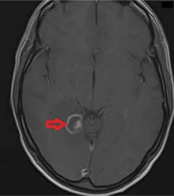
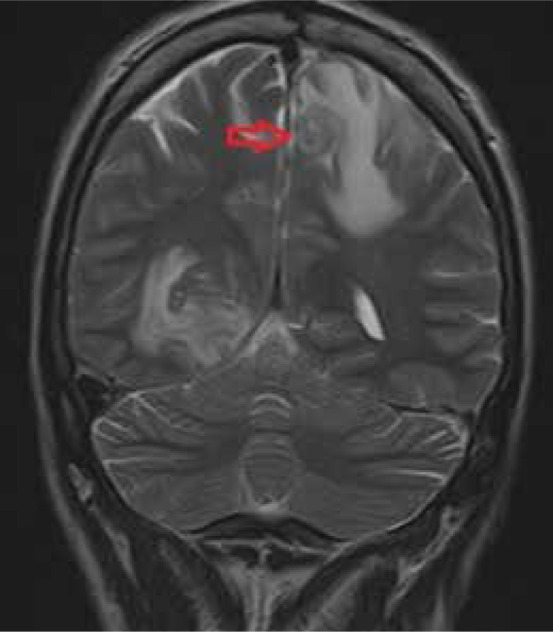
Cerebral toxoplasmosis is a parasitic disease resulting, in most cases, from a reactivation of a latent cyst with Toxoplasma gondii. The disease mainly affects immunosuppressed individuals, such as HIV (human immunodeficiency virus)-infected patients. Diagnosis is based on specialized antibody testing, clinical symptoms, neuroimaging methods, and histological examination. The gold standard for diagnosis is a brain biopsy, but more often the response to treatment seen in clinical symptoms and neuroimaging studies is sufficient. The imaging features support the diagnosis of cerebral toxoplasmosis and help assess the effectiveness of treatment.
Keywords: cerebral toxoplasmosis; computed tomography; encephalitis; magnetic resonance imaging; neuroimaging.
© Pol J Radiol 2023.
Conflict of interest statement
The authors report no conflict of interest.
Figures
Similar articles
-
IgG4 specific to Toxoplasma gondii excretory/secretory antigens in serum and/or cerebrospinal fluid support the cerebral toxoplasmosis diagnosis in HIV-infected patients.J Immunol Methods. 2013 Sep 30;395(1-2):21-8. doi: 10.1016/j.jim.2013.06.005. Epub 2013 Jun 26. J Immunol Methods. 2013. PMID: 23811152
-
[Difficulties of cerebral toxoplasmosis diagnosis in a patient with multiple sclerosis and HIV].Zh Nevrol Psikhiatr Im S S Korsakova. 2021;121(6):76-80. doi: 10.17116/jnevro202112106176. Zh Nevrol Psikhiatr Im S S Korsakova. 2021. PMID: 34283534 Russian.
-
Usefulness of stereotactic biopsy and neuroimaging in management of HIV-1 Clade C associated focal brain lesions with special focus on cerebral toxoplasmosis.Clin Neurol Neurosurg. 2013 Jul;115(7):995-1002. doi: 10.1016/j.clineuro.2012.10.012. Epub 2012 Nov 12. Clin Neurol Neurosurg. 2013. PMID: 23153789 Free PMC article.
-
General Features and Laboratory Diagnosis of Toxoplasma gondii Infection.Turkiye Parazitol Derg. 2020 Jun 2;44(2):94-101. doi: 10.4274/tpd.galenos.2020.6634. Turkiye Parazitol Derg. 2020. PMID: 32482042 Review.
-
Presentation and Rehabilitation in a Patient With Toxoplasmosis Encephalitis: A Case Study and Review.PM R. 2016 Jun;8(6):602-6. doi: 10.1016/j.pmrj.2016.01.006. Epub 2016 Jan 22. PM R. 2016. PMID: 26805910 Review.
Cited by
-
A Case of Toxoplasmic Encephalitis in an HIV-Negative Patient With Multiple Cerebral Infarction-Like Findings in the Early Stages.Cureus. 2025 May 6;17(5):e83603. doi: 10.7759/cureus.83603. eCollection 2025 May. Cureus. 2025. PMID: 40486338 Free PMC article.
-
Secondary adrenal insufficiency in a young man with HIV and pulmonary tuberculosis, complicated by cerebral toxoplasmosis and seizure.IDCases. 2025 Jul 9;41:e02319. doi: 10.1016/j.idcr.2025.e02319. eCollection 2025. IDCases. 2025. PMID: 40697365 Free PMC article.
-
Clinical and imaging characteristics of toxoplasmic encephalitis in patients with HIV/AIDS with different immune statuses.Quant Imaging Med Surg. 2025 Aug 1;15(8):7259-7268. doi: 10.21037/qims-2024-2598. Epub 2025 Jul 17. Quant Imaging Med Surg. 2025. PMID: 40785890 Free PMC article.
References
Publication types
LinkOut - more resources
Full Text Sources